/
Cutaneous Manifestations of Internal Malignancy Cutaneous Manifestations of Internal Malignancy

Cutaneous Manifestations of Internal Malignancy - PDF document

joy
joy . @joy
Follow
342 views
Uploaded On 2022-08-26

Cutaneous Manifestations of Internal Malignancy - PPT Presentation

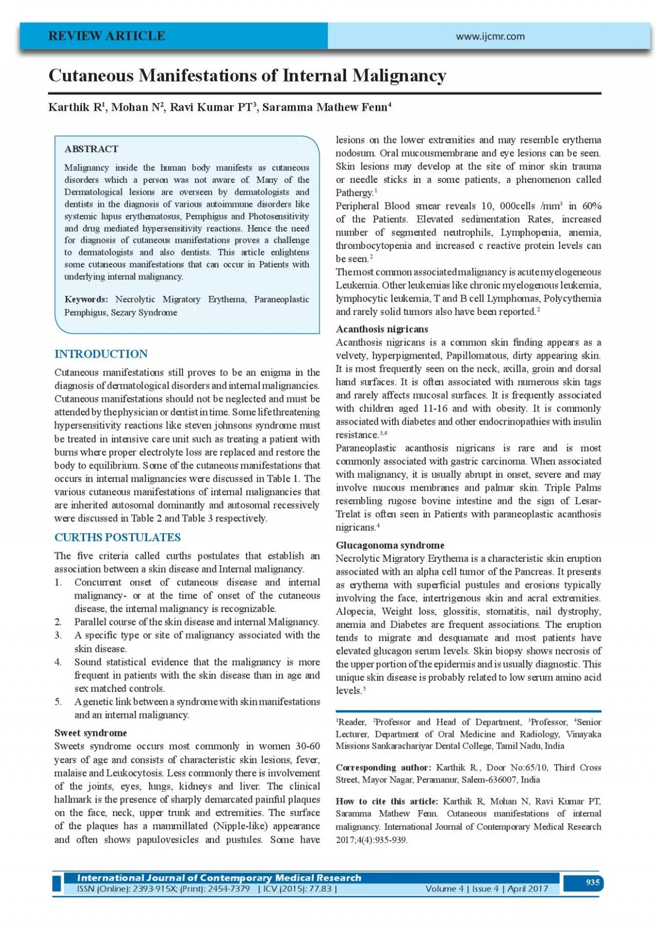
935 935 Karthik R 1 Mohan N 2 Ravi Kumar PT 3 Saramma Mathew Fenn 4 REVIEW ARTICLE Malignancy inside the human body manifests as cutaneous disorders which a person was not aware of Many of the ID: 941953

skin syndrome cutaneous malignancy syndrome skin malignancy cutaneous disease patients internal lesions x00660069 carcinoma manifestations 2017 erythema lymphoma cell

Share:

Link:

Embed:

Download Presentation from below link

Download Pdf The PPT/PDF document "Cutaneous Manifestations of Internal Mal..." is the property of its rightful owner. Permission is granted to download and print the materials on this web site for personal, non-commercial use only, and to display it on your personal computer provided you do not modify the materials and that you retain all copyright notices contained in the materials. By downloading content from our website, you accept the terms of this agreement.


Presentation Transcript

935 935 Cutaneous Manifestations of Internal Malignancy Karthik R 1 , Mohan N 2 , Ravi Kumar PT 3 , Saramma Mathew Fenn 4 REVIEW ARTICLE Malignancy inside the human body manifests as cutaneous disorders which a person was not aware of. Many of the Dermatological lesions are overseen by dermatologists and dentists in the diagnosis of various autoimmune disorders like and drug mediated hypersensitivity reactions. Hence the need for diagnosis of cutaneous manifestations proves a challenge to dermatologists and also dentists. This article enlightens some cutaneous manifestations that can occur in Patients with underlying internal malignancy. INTRODUCTION Cutaneous manifestations still proves to be an enigma in the diagnosis of dermatological disorders and internal malignancies. Cutaneous manifestations should not be neglected and must be attended by the physician or dentist in time. Some life threatening hypersensitivity reactions like steven johnsons syndrome must be treated in intensive care unit such as treating a patient with burns where proper electrolyte loss are replaced and restore the body to equilibrium. Some of the cutaneous manifestations that occurs in internal malignancies were discussed in Table 1 . The various cutaneous manifestations of internal malignancies that are inherited autosomal dominantly and autosomal recessively were discussed in Table 2 and Table 3 respectively. CURTHS POSTULATES The �ve criteria called curths postulates that establish an association between a skin disease and Internal malignancy. Concurrent onset of cutaneous disease and internal malignancy- or at the time of onset of the cutaneous disease, the internal malignancy is recognizable. Parallel course of the skin disease and internal Malignancy. A speci�c type or site of malignancy associated with the skin disease. 4. frequent in patients with the skin disease than in age and sex matched controls. A genetic link between a syndrome with skin manifestations and an internal malignancy. Sweet syndrome Sweets syndrome occurs most commonly in women 30-60 years of age and consists of characteristic skin lesions, fever, malaise and Leukocytosis. Less commonly there is involvement of the joints, eyes, lungs, kidneys and liver. The clinical hallmark is the presence of sharply demarcated painful plaques on the face, neck, upper trunk and extremities. The surface of the plaques has a mammillated (Nipple-like) appearance and often shows papulovesicles and pustules. Some have lesions on the lower extremities and may resemble erythema nodosum. Oral mucousmembrane and eye lesions can be seen. or needle sticks in a some patients, a phenomenon called Pathergy. 1 Peripheral Blood smear reveals 10, 000cells /mm 3 in 60% of the Patients. Elevated sedimentation Rates, increased number of segmented neutrophils, Lymphopenia, anemia, thrombocytopenia and increased c reactive protein levels can be seen. 2 The most common associated malignancy is acute myelogeneous Leukemia. Other leukemias like chronic myelogenous leukemia, lymphocytic leukemia, T and B cell Lymphomas, Polycythemia and rarely solid tumors also have been reported. 2 Acanthosis nigricans velvety, hyperpigmented, Papillomatous, dirty appearing skin. It is most frequently seen on the neck, axilla, groin and dorsal hand surfaces. It is often associated with numerous skin tags and rarely affects mucosal surfaces. It is frequently associated with children aged 11-16 and with obesity. It is commonly associated with diabetes and other endocrinopathies with insulin resistance. 3,4 Paraneoplastic acanthosis nigricans is rare and is most commonly associated with gastric carcinoma. When associated with malignancy, it is usually abrupt in onset, severe and may involve mucous membranes and palmar skin. Triple Palms resembling rugose bovine intestine and the sign of Lesar- Trelat is often seen in Patients with paraneoplastic acanthosis nigricans. 4 Glucagonoma syndrome Necrolytic Migratory Erythema is a characteristic skin eruption associated with an alpha cell tumor of the Pancreas. It presents as erythema with super�cial pustules and erosions typically involvi

ng the face, intertrigenous skin and acral extremities. Alopecia, Weight loss, glossitis, stomatitis, nail dystrophy, anemia and Diabetes are frequent associations. The eruption tends to migrate and desquamate and most patients have elevated glucagon serum levels. Skin biopsy shows necrosis of the upper portion of the epidermis and is usually diagnostic. This unique skin disease is probably related to low serum amino acid levels. 5 1 www.ijcmr.com International Journal of Contemporary Medical Research ISSN (Online): 2393-915X; (Print): 2454-7379 | ICV (2015): 77.83 |Volume 4 | Issue 4 | April 2017 www.ijcmr.com International Journal of Contemporary Medical Research ISSN (Online): 2393-915X; (Print): 2454-7379 | ICV (2015): 77.83 |Volume 4 | Issue 4 | April 2017 2 Professor and Head of Department, 3 Professor, 4 Senior Lecturer, Department of Oral Medicine and Radiology, Vinayaka Missions Sankarachariyar Dental College, Tamil Nadu, India Corresponding author: Karthik R., Door No:65/10, Third Cross Street, Mayor Nagar, Peramanur, Salem-636007, India How to cite this article: Karthik R, Mohan N, Ravi Kumar PT, Saramma Mathew Fenn. Cutaneous manifestations of internal malignancy. International Journal of Contemporary Medical Research 2017;4(4):935-939. 936 Hypertrichosis Lanuginosa Hypertrichosis Lanuginosa (malignant down) is an acquired excessive growth of Lanugo Hair. It usually begins on the face, neck and ears and eventually can involve most hair bearing skin. Glossitis is frequently an associated �nding. The most common associated cancers are lung, Breast, Gastrointestinal and carcinoid. This rare cutaneous �nding has also been reported in Patients with anorexia nervosa. 6 Trousseaus sign Trousseaus sign consists of recurrent and migratory super�cial thrombophlebitis, affecting both large and small cutaneous veins which is associated with an internal cancer, crops of oval to linear, erythematous, tender skin lesions are seen mostly on the arms, legs, �anks and abdomen. Thrombosis of internal veins can also occur and lead to a variety of symptoms. Men are more commonly affected. The most common associated carcinomas are lung and Pancreatic carcinoma. 7 Dermatomyositis The classic eruption of Dermatomyositis is a reddish purple erythema involving the face, typically the eyelids (Heliotrope sign). The rash may be faint or quite in�amed and edematous. In addition to the facial rash, lesions on the scalp, neck, upper trunk and extensor extremities are common. As the lesions mature, scaling and atrophy may develop. The erythema on the hands occurs over the knuckles rather than over the phalanges. Cuticular telengiectasis seen in Dermatomyositis. Flat topped red to violaceous Papules develop over the knuckles of Patients with Dermatomyositis. The skin lesions may precede clinical or laboratory evidence by weeks, months or years. A few patients may never develop muscle dysfunction. The skin lesions are notoriously resistant to topical steroid therapy. The cancer may occur before, during or after the development of dermatomyositis. A slight increase in the incidence of ovarian carcinoma is noted in patients newly diagnosed as Dermatomyositis and female Disease Internal malignancy Sweets syndrome Acute Myelogenous Leukemia Paraneoplastic Acanthosis Nigricans Gastric carcinoma Hypertrichosis Lanuginosa Lung, Breast, Gastrointestinal, carcinoid tumors Dermatomyositis Ovarian carcinoma Cutaneous erythema Lymphoma Paraneoplastic Pemphigus Lymphoma Erythema gyratum repens Breast cancer Basex syndrome Squamous cell carcinoma of upper aerodigestive tract Necrolytic Migratory erythema Alpha cell tumor of Pancreas (Glucagonoma) Sezary syndrome Lymphoma Pagets Disease of the Breast Ductal carcinoma Cowdens syndrome Breast cancer Multiple mucosal neuroma syndrome Medullary carcinoma of thyroid Acquired icthyosis Hodgkins Disease Pruritis Hodgkins Lymphoma Pyoderma Gangrenosum Myelocytic and Myelomonocytic Leukemias Table-1: Diseases associated with Internal Malignancies. Disorder Affected Gene Cancer association Clinical Features (Rare) Lisch Nodules Seizures Deafness Caf

é-au-lait macules Multiple Mucosal Neuro - ma syndrome RET Proto oncogene Medullary Thyroidcar - cinoma Neuromas of Lip, tongue and oral mucosa Neuromas of lip, tongue and oral mucosa Peutz Jeghers syndrome STK11 (Serine Threonine Kinase) Instestine Intestinal Polyps Pigmented macules on Vermilon border of lip, mucosa, face, acral extremities Gardners syndrome APC Adenomatosis Polyposis coli Gene Colon Osteomas Colon Polyps Desmoids Abnormal dentition Epidermoid cysts Cowdens syndrome PTEN (Phosphate and tension homologue delet - ed on chromosome 10) Breast Thyroid Mucosal papules Fibrocystic disease of the Breast Keratotic facial Papules Acral Keratosis Torres syndrome MSH2 (Melanocyte stim - ulating Hormone) MLH1 (Micronuclear Linker Histone) Colon Colon Polps Sebaceous tumors Keratoacanthomas Table-2: Autosomal dominant diseases associated with skin changes and malignancy 937 patients must be screened for ovarian carcinoma. 8 Sezary syndrome Sezary syndrome represents a triad of �ndings cutaneous erythema, lymphadenopathy and 10-15% atypical mononuclear cells in peripheral blood. This syndrome is a subset of cutaneous T Cell Lymphoma. Patients with sezary syndrome frequently have intolerable itching often to the point that they are suicidal. Lymphadenopathy, nail dystrophy and Hair loss are common associated features. The diagnosis is established by skin biopsy showing cutaneous T Cell Lymphoma, the presence of atleast 15% atypical mononuclear cells in Peripheral blood and the typical clinical picture. Approximately 10-15% of patients with erythroderma will have an associated lymphoma or more rarely leukemia. 9 Paraneoplastic Pemphigus Paraneoplastic Pemphigus is a blistering skin disease with a reported association with Lymphoma, although a few cases reported with solid tumors. The clinical picture resembles pemphigus vulgaris, bullous pemphigoid or Erythema multiforme Major (Stevens-Johnsons syndrome) with signi�cant oral mucous membrane involvement. The disease poorly responds to immunosuppressive therapy and is frequently fatal. 10 Skin and oral mucous membrane biopsy reveals epidermal acantholysis, epidermal spongiosis, suprabasilar clefts, basal cell vacuolar changes and dyskeratotic keratinocytes. Direct immuno�uorescenct examination reveals IgG and less commonly IgA with or without complement in the intracellular spaces and C3, IgG or IgM at the Basement membrane zone. 11 Antibodies have been demonstrated against desmoplakins, proteins in keratinocyte attachment plaques(desmosomes) and a 230kd protein in the Basement membrane zone. Rat bladder is a useful substrate for indirect immuno�uorescent examinationand shows positive staining with serum from patients with Paraneoplastic Pemphigus. 11 Erythema Gyratum repens It is a rare skin eruption that is characterised by widespread, ever changing pattern of skinlesions resembling wood grain. The erythematous circinate lesions may have a �ne scale and move upto 1cm a day. Almost all patients with this unique dermatosis have an associated malignancy. The most commonly associated malignancy with this unique dermatological �nding is Breast carcinoma. This can also be seen in lung, Bladder, cervical and Prostate cancers. The skin lesions clear within a few weeks after the removal of the malignancy and usually recur if the cancer returns. 12 Pyoderma Gangrenosum Pyoderma gangrenosum is an ulcerative skin disease ofunknown etiology characterised by painful, rapidly enlarging ulcers with an erythematous orviolaceous undermined border with a necrotic centre associated with internal malignancy in 7.2% of the Patients. Other reported hematologic cancers are multiplemyeloma, Polycythemia vera and Lymphoma. 13 Erythromelalgia Erthromelalgia is a rare skin disease characterised by erythematous, painful, burning of the feet, ankles and lower extremities. This disease is aggravated by Heat exposure and relieved by cooling. Many Patients �nd relief only by soaking their legs in ice water. The incidence of associated malignancy is variable between 3% and 65% of the cases. The associated haemato

logical malignancy are polycythemia or essential thrombocythemia. 14 Acquired icthyosis Acquired icthyosis was �rst reported to be associated with Hodgkins disease in 1940s. It is also reported in T- cell lymphoma, Kaposis sarcoma, Malignant Histiocytosis, Leiomyosarcoma, Multiple myeloma. 15 Pruritis: Generalised pruritis without skin lesions has been reported as a sign of internal malignancy. Pruritis is reported as an initial symptom of Hodgkins disease in 5- 10% of Patients and in 3% of those with non-hodgkins lymphoma. 16 Extramammary Pagets disease Extramammary Pagets disease occurson the axilla, breast, groin or anofenital region. It begins as a small eczematous patch on the nipple that gradually spreads to the areola and eventually to Disorder Affected Gene Inheritance Clinical Findings Cancer Ataxia telangiectasia (Louis Bar syndrome) ATM Autosomal recessive Progressive cerebellar ataxia Telengiectasia Recurrent sinus and Pul - monary infections Decreased or absent serum IgA Lymphomas Blooms syndrome RecQ3 Autosomal recessive Photosensitivity Telengiectasia of sun exposed skin Short stature Decreased serum Igs Recurrent infections Lymphomas Leukemias Dyskeratosis congenita DKC1 TERC X-Linked recessive Autosomal Dominant Skin atrophy and Hyper - pigmentation Nail dystrophy Oral Precancerous Leuko - keratosis Oral cancers Other malignancies Table-3: 938 the skin of the breast. It is often associated with an underlying adnexal carcinoma and about 20% of cases have carcinoma of the rectum or genitourinary tract. 17 Primary systemic Amyloidosis The cause of this disease ia plasma cell dyscrasia. The most common associated skin lesions are pruritic or ecchymoses that are seen most frequently on the skin areas like eyelids, neck, groin, axilla, umbilicus or oral mucosa. The haemmorhagic lesions may occur on areas of clinically normal skin or in skin having waxy papules, Plaques, nodules or tumors. The intracellular bleeding is due to in�ltration of blood vessel walls with amyloid protein. Other less common skin lesions include alopecia, ail dystrophies, Scleroderma like lesions, Macroglossia, cutis verticis gyrate, bullous lesions and dyspigmentation. 18 Basex syndrome (Acrokeratosis Paraneoplastica): Basex syndrome begins with acral violaceous erythema on the ears, nose, hands and feet. Early lesions may show small vesicles. As the lesionsprogress, they become hyperkeratotic and psoriasiform, especially on the hands and Feet. Paronychia and naildystrophy are common. Later the eruption may generalize and lesions on the face may appear dermatitis or lupus like. This syndrome is more common in men and is associated with squamous cell carcinoma of the upper aerodigestive tract. An another variant of Basex syndrome inherited as an autosomal dominant disease is characterised by acral follicular atrophoderma, early development of Multiple facial Basal cell carcinoma and in some hypohidrosis. 19 Multiple mucosa Neuroma syndrome This syndrome is characterised by the presence of multiple �esh coloured papules on the tongue, lips and occasionally other mucosal surfaces early in life. These patients have a characteristic thick prominent lips and a marfanoid habitus. 90% of these patients develop medullarythyroid carcinoma and phaeochromocytoma that is often multifocal or bilateral. 20 Gardners syndrome The cutaneous Hall mark of Gardners syndrome is epidermoid cysts which often appear before puberty, frequently on the extremities. These cysts may be many or few. The syndrome is also characterised by osteomas (typically on facial bones), �brous and desmoid tumours, abnormal dentition, lipomas, hypertrophy of renal pigmented epithelium and leiomyomas of the gastrointestinal tract. The syndrome is characterized by the early onset of colonic polyposis and has a very high incidence of colon cancer. 20 Peutz Jeghers syndrome Brown to Blue –black macules (lentigines) are present at birth or early infancy on the lips, oralmucosa, nasal mucosa, palms, soles, dorsal hand surfaces, central face and elbows. Polyps of the small intestine develops in 90% of the Patients. Polpys also occu

r in the stomach, colon and rectum. The average age of death is 36 years intussusception occurs in about 50% of the cases. 20 Torres syndrome This syndrome includes cutaneous sebaceous neoplasia and a high incidence of low grade colon cancer. The sebaceous tumors includes sebaceous adenomas, epitheliomas, carcinomas. About one-third of patients develop keratoacanthomas. The sebaceous skin tumours may be few or many but even one sebaceous adenoma should alert the clinician that patient may have this syndrome. 20 Carcinoid syndrome Carcinoid syndrome is a systemic manifestation of neuroendocrine carcinoid tumors that most commonly manifest by �ushing that progress to persistent telangiectasia and diarrhaea. Less common �nding include bronchospasm, cardiovascular dysfunction and Pellagra like skin changes (Photo disturbed Dermatitis). Development of this syndrome has prognostic signi�cance as liver metastases underlie most 21 CONCLUSION Cutaneous manifestations provide a challenge to the clinicians and dentists for treatment. A proper diagnosis of such cutaneous manifestations in a body is essential to treat the diseases early and betterment of the patients. REFERENCES 1. Cohen PR, Sweet’s syndrome – a comprehensive review of an acute febrile neutrophilic dermatosis, Orphanet J Rare Dis. 2007;26:2:34. 2. Von den Driesch P: Sweets syndrome (Acute Febrile neutrophilic Dermatosis). J Am Acad Dermatol. 1994; 31:535-556. 3. Yin WB, Gao Y, Lu Y, Gastric cancer associated with malignant acanthosis nigricans, Am J Med Sci. 2017;353:416. 4. Schwartz RA: Acanthosis nigricans. J Am Acad Dermatol. 1994;31:1-19. 5. Mountjoy L, Kollmorgen D, Glucagonoma associated rash, N Engl J Med. 2017;376:e18. 6. Hovenden AL: Acquired lanuginose acquisita associated with malignancy.Clin Dermatol. 1993;11:99-106. 7. Lesher JL Jr: Thrombophlebitis and thromboembolic Problems in Malignancy.Clin Dermatol. 1993;11:159- 163. 8. Callen JP: Dermatomyositis. Lancet. 2000;355:53-57. 9. Foss F: Mycosis Fungoides and the sezary syndrome, Curr opin oncol. 2004;5:421-428. 10. Anhalt GJ, KimSC, Stanley JR et al. Paraneoplastic Pemphigus. N Engl J Med. 1990;323:1729-1735. 11. Kasperkiewicz M et al, Pemphigus, Nat Rev Dis Primers. 2017;11:3:17026. 12. Tchernerv G et al. Erythema Gyratum repens: a pathogenetic mystery and therapeutic challenge, Wien Med Wochenschr. 2017;167:117-119. 13. Duguid CM, O Loughlin S, Otridge B, Powell FC. Paraneoplastic pyoderma gangrenosum.Aust J Dermatol. 1993;34:17-22. 14. Hou JL, Onajin O, Gangat N, Davis MD, Wolanskyj AP. 18, Erythromelalgia in patients with essential thrombocythemia and polycythemia vera, Leuk Lymphoma. 2017;58:715- 717. 15. Rizos E, Milinois HJ, Pavlidis N, Elisaf MS. Acquired icthyosis: A Paraneoplastic skin manifestation of Hodgkins disease.Lancet oncol. 2002;3:727. 16. Lober CW: Pruritis and Malignancy. Clin Dermatol. 1993; 11:125-128. 17. Bhatia P, Ahuja A, Dey P, Suri V. Vulval intraepithelial neoplasia with extra mammary Pagets disease: a rare association. J Clin Pathol . 2007;60:110-2. 939 18. Gonzalez – Ramos J et al. Relapsing bullous amyloidosis of the oral mucosa and acquired cutis laxa in a patient with multiple myeloma: a rare triple association. Clin Exp Dermatol. 2017;1. 19. Bolognia JL. Basex syndrome. Clin Dermatol. 1993; 11:37-42. 20. Koh KJ, Park HN, Kim KA. Gardner syndrome associated with multiple osteomas, intestinal polyposis, and epidermoid cysts. Imaging Sci Dent . 2016;46:267-272. 21. Owen CE, cutaneous manifestations of lung cancer, Semin Oncol . 2016;43:366-9. Source of Support: Nil; Con�ict of Interest: None Submitted: 05-04-2017; Accepted: 11-05-2017; Published: 17-05-2017 Karthik, et al.Cutaneous Manifestations of Internal Malignancy International Journal of Contemporary Medical Research Volume 4 | Issue 4 | April 2017 | ICV (2015): 77.83 |ISSN (Online): 2393-915X; (Print): 2454-7379 Karthik, et al.Cutaneous Manifestations of Internal Malignancy International Journal of Contemporary Medical Research ISSN (Online): 2393-915X; (Print): 2454-7379 | ICV (2015): 77.83 |Volume 4 | Issue 4 | April 2017