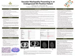
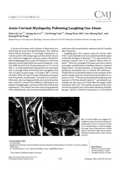
/
Thoracic myelopathy can be caused by various pathologies including thoracic disc herniation, Thoracic myelopathy can be caused by various pathologies including thoracic disc herniation,

Thoracic myelopathy can be caused by various pathologies including thoracic disc herniation, - PowerPoint Presentation

joy
joy . @joy
Follow
342 views
Uploaded On 2022-04-07

Thoracic myelopathy can be caused by various pathologies including thoracic disc herniation, - PPT Presentation

ligamentum flavum As the ligaments become ossified it causes narrowing of thoracic canal and eventually compression of spinal cord Fluoride is an important factor in bone mineralisation However ID: 910444

spine fluorosis opll thoracic fluorosis spine thoracic opll myelopathy skeletal fluoride ligamentum ossified flavum showing ray laminectomy ossification limb

Share:

Link:

Embed:

Download Presentation from below link

Download Presentation The PPT/PDF document "Thoracic myelopathy can be caused by var..." is the property of its rightful owner. Permission is granted to download and print the materials on this web site for personal, non-commercial use only, and to display it on your personal computer provided you do not modify the materials and that you retain all copyright notices contained in the materials. By downloading content from our website, you accept the terms of this agreement.


Presentation Transcript

Slide1

Thoracic myelopathy can be caused by various pathologies including thoracic disc herniation, ossified posterior longitudinal ligament (OPLL) and ossified ligamentum flavumAs the ligaments become ossified, it causes narrowing of thoracic canal and eventually compression of spinal cord.Fluoride is an important factor in bone mineralisation. However, excessive fluoride intake may cause intoxication – Fluorosis. Fluorosis is known to cause ossification in body tissues such as interosseous membranes, ligaments and tendons. When this ossification occurs in posterior longitudinal ligament or ligamentum flavum or duramater, compressive myelopathy occurs.

35 year old male, labourer, from Agra (U.P) presented with complaint of inability to walk , paraesthesia in bilateral lower limb and generalised body ache. Symptoms started one year back that gradually progressed to the current stage. Clinical examination revealed spasticity of both lower limb, brisk deep tendon reflexes, bilateral plantar extensor and patellar and ankle clonus. Loss of position and vibration sense in both lower limb. Diminished touch sensation below D6 dermatome. Bowel and bladder sensation intact. Motor and sensory examination in upper limb was normal.Radiograph of D-L spine shows diffuse densification of bone. CT scan shows ossified ligamentum flavum from D4 to D10 vertebra and OPLL from D5 TO L1 vertebra.in cervical spine also OPLL was present.MRI of thoracic spine shows OLF between D4 to D10 and OPLL from D5 to L1 vertebra.X-ray of both forearm showed significant calcification of interosseous membrane of forearm. X-ray pelvis both hip showed diffuse densification of bone.Based on radiograph, CT and MRI, patient was suspected of fluorosis. For confirmation serum ,urine and drinking water fluoride level were measured which was high.Radiographs of father and wife showed similar changes.After confirming diagnosis and proper consent, surgery was planned. Posterior decompression with en-bloc laminectomy done from D4 to D10 vertebra with the help of ultrasonic scalpel. No instrumentation done.Immediate post operative deterioration of neurology occurred which recovered over a period of 6 weeks. Patient has reduced spasticity and grade 3 muscle power in both lower limb after 2 months.

Case report

Introduction

Discussion

Fluoride is one of the necessary minor element in human and its daily requirement is 0.05 -0.07 mg/kg body weight

.

Main source of excess fluoride intake is from drinking water. In high fluoride areas where its concentration in water is more than 5-8 mg/L , dental fluorosis, skeletal fluorosis and even systemic fluorosis develops. Fluorosis is endemic disease in many countries of Asia especially India, China and Japan.

Neurological involvement in fluorosis occurs i

n late

stage. Spinal fluorosis causes compressive myelopathy mainly affecting the cervical and dorsal spine. Causes of compressive myelopathy in fluorosis are 1. OPLL, 2.

Ligamentum

flavum

ossification, 3. ossification within

duramater, 4. aggravation of pre-existing stenosis. Skeletal fluorosis is a rare cause of OLF. Mostly involve mid and lower thoracic region. OPLL most commonly involves cervical spine.Diagnosis of fluorosis is a great challenge as no definite diagnostic tests are available. In our study it was confirmed by skeletal survey, CT and MRI, urine serum and drinking water fluroide level.Since spinal fluorosis is a progressive disease, surgical decompression is warranted once a patient becomes symptomatic. Posterior decompression via laminectomy and en-bloc resection is most effective treatment. Despite adequate surgical decompression, the results are not always satisfactory.

Conclusion

1. Gupta MC, Bridwell KH. Textbook of spinal surgery. 4th ed. Philadelphia: Wolters Kluwer;2020. Chapter 52: treatment of thoracic myelopathy; p.516-522.2. Wang W , Kong L, Zhao H, Dong R, Li J, Jia Z , Ning Ji et al. Thoracic ossification of ligamentum flavum caused by skeletal fluorosis. Eur Spine J. 2007 Aug; 16(8): 1119–1128.3. Modi JV, Tankshali KV, Patel ZM, Shah BH, Gol AK. Management of Acquired Compressive Myelopathy due to Spinal Fluorosis. Indian J Orthop. 2019;53(2):324-332.4. Osman NS, Cheung ZB, Hussain AK, et al. Outcomes and Complications Following Laminectomy Alone for Thoracic Myelopathy due to Ossified Ligamentum Flavum: A Systematic Review and Meta-Analysis. Spine 2018;43(14):E842-E848. 5. Reddy DR. Neurology of endemic skeletal fluorosis. Neurol India .2009;57:7–12.

References

Skeletal fluorosis can cause thoracic myelopathy and can be diagnosed on basis of epidemic history, clinical symptoms, medical imaging and urinalysis. En-bloc laminectomy decompression is the treatment of choice.

Dental fluorosis

CT scan showing OPLL and ossified ligamentum flavum

CT scan of cervical spine showing OPLL

MRI scan showing OLF between D4 to D10 and OPLL from D5 to L1 vertebra

X-ray D-L spine showing densification of bone

X-ray forearm showing ossified interosseous membranes

Intraoperative fused lamina from D5 to D10

Specimen of En-bloc laminectomy

X-ray pelvis showing diffuse densification of bone

Serum, urine and drinking water fluoride level

Thoracic compressive myelopathy secondary to skeletal fluorosis

Dr. Ramesh Kumar, Dr.

Sumit

Kumar, Dr. T.

boruah

, Dr.

Atul

Sareen

, Dr. Ravi

Shreenivasan

, Dr.

Mohit

Patralekh

Department of Spine Surgery, CIO, VMMC &

Safdarjung

Hospital, New Delhi