/
Starter: Microscopes Block 1A - Cell structure 2.1.1 Starter: Microscopes Block 1A - Cell structure 2.1.1

Starter: Microscopes Block 1A - Cell structure 2.1.1 - PowerPoint Presentation

joyce
joyce . @joyce
Follow
0 views
Uploaded On 2024-03-13

Starter: Microscopes Block 1A - Cell structure 2.1.1 - PPT Presentation

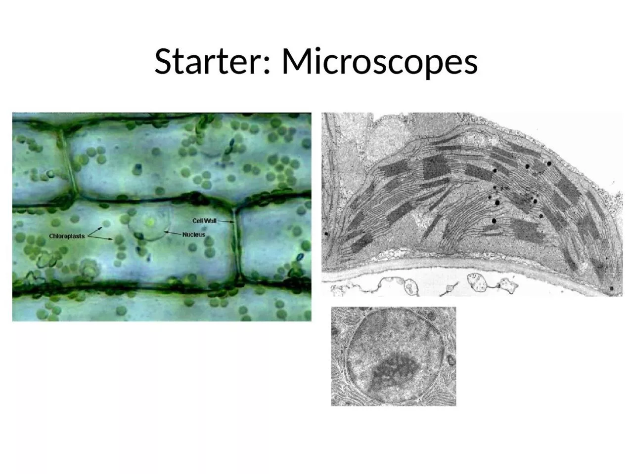
Microscopy Foundations in Biology SPEC Objectives and Success Criteria Objectives Compare and contrast different types of microscopes Describe the preparation of specimens for observation using microscopes ID: 1046894

electron light optical microscope light electron microscope optical image times grade scanning objects sample living laser cell cells resolution

Share:

Link:

Embed:

Download Presentation from below link

Download Presentation The PPT/PDF document "Starter: Microscopes Block 1A - Cell str..." is the property of its rightful owner. Permission is granted to download and print the materials on this web site for personal, non-commercial use only, and to display it on your personal computer provided you do not modify the materials and that you retain all copyright notices contained in the materials. By downloading content from our website, you accept the terms of this agreement.


Presentation Transcript

1. Starter: Microscopes

2. Block 1A - Cell structure 2.1.1MicroscopyFoundations in Biology

3. SPEC

4. Objectives and Success CriteriaObjectivesCompare and contrast different types of microscopesDescribe the preparation of specimens for observation using microscopesSuccess criteriaIdentify the differences between Optical, Electron and Laser scanning microscope (grade E)Describe how a specimen can be prepared for observation and used to identify structures (grade C)Compare a light microscope with an electron microscope (grade A)

5. Cell TheoryAll living things consist of cellsNew cells are formed only by the division of pre-existing cellsThe cell contains information that acts as the instructions for growth. This information can be passed to new cells

6. Use the table to complete the notes comparing microscopes Light microscopeLaser scanning microscopeElectron microscopeTEMSEMHow do they work?     Magnification    Resolution    Advantages     Disadvantage     Compare a light microscope with an electron microscope (grade A)

7. The light microscopeUses a number of lenses to view an image through the eye pieceLight passes through the condenser lens and then through the specimenBeam of light is focused through the objective lens and then through the eye piece lensMagnification: How many times a structure is enlargedResolution: The ability to see two objects that are close together as separate objects. Objects that are close together can only be distinguished if light waves can pass through them, allowing you to see detail

8. Optical (Light) MicroscopeRelatively cheapEasy to usePortableCan study whole living specimens

9. Key TermsResolution: the ability to distinguish two separate points as distinct from each other. Increases the clarity of the image.Magnification: the number of times greater an image is than the original object x 100 = 100 times wider and 100 times longer

10. Optical (Light) MicroscopeMagnification available on light microscopex40x100x400x1500 or x2000Resolution – (limited) maximum of 200nm. (0.2mm) This is due to the wavelength of visible light (400-700nm)Ribosomes have a diameter of 20nm Can you see them using an optical microscope?Hint: In optical microscopy objects that are closer than half the wavelength of light cannot be seen separately.TASK – Label microscopes on worksheet

11. Laser scanning (confocal) microscopesLaser light used to scan the image point by point. It can focus at specific depths. Computer assembles information into one image.High resolution, high contrast, depth sensitivityFluorescent dye can be used to allow more specific targeting of features to be studied.Can be used for whole cells and living organismsUse in diagnosis of disease (eg eye diseases) and in medical research

12. Electron MicroscopeEM generates beam of electrons (0.004nm width) Wavelength is 125 000x shorter than visual light so increases resolution.Electron beam passes through very thin prepared sample.Resolution is 0.5nmMagnification can be up to 500 000xExpensive, can only be used in a controlled environmentVacuum needed for sample preparationlargePreparing slides is complexSkill and training needed

13. Transmission Electron MicroscopeElectrons pass through thin part of sample less easily so create contrast. Sample dehydrated and stained with metal saltsElectrons focused on photographic plate (or screen)Resulting 2D grey-scale image is an Electron micrographMagnification up to 2 million times so smaller organelles can be seen, such as ribosomes(future- 50 million x)What might be a disadvantage of Electron microscopy?

14. Scanning Electron MicroscopeElectrons are reflected off the surface of a metal-salt-stained sample3D shape can be seen so greater field of view, grayscale, but computer programmes can add false colourYou can see surface features in detailMagnification x 15-200 000

15. EMAdvantages of EMDisadvantages of EMResolution is x2000 more than LMSamples have to be placed in a vacuumProduces detailed imagesVery expensiveSEM produces 3D imagesNeed to be highly skilled to create samples

16. Comparing LM and EM15002000)x 200 000x 2 000 000Identify the differences between Optical, Electron and Laser scanning microscope (grade E)