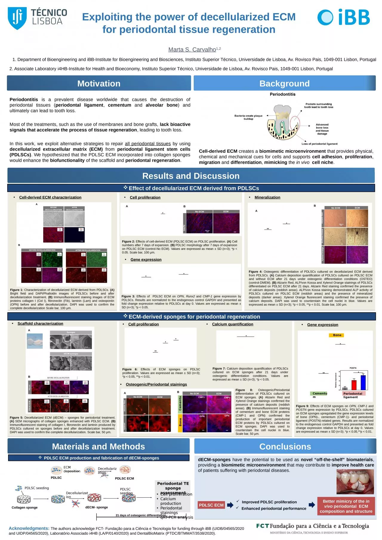
/
Periodontitis  is a prevalent disease worldwide that causes the destruction of periodontal Periodontitis  is a prevalent disease worldwide that causes the destruction of periodontal

Periodontitis is a prevalent disease worldwide that causes the destruction of periodontal - PowerPoint Presentation

julia
julia . @julia
Follow
27 views
Uploaded On 2024-02-03

Periodontitis is a prevalent disease worldwide that causes the destruction of periodontal - PPT Presentation

periodontal ligament cementum and alveolar bone and ultimately can lead to tooth loss Most of the treatments such as the use of membranes and bone grafts lack bioactive signals that accelerate the process of tissue regeneration ID: 1044311

pdlscs ecm pdlsc periodontal ecm pdlscs periodontal pdlsc sponges cultured cell derived osteogenic calcium days expressed decellularized values staining

Share:

Link:

Embed:

Download Presentation from below link

Download Presentation The PPT/PDF document "Periodontitis is a prevalent disease wo..." is the property of its rightful owner. Permission is granted to download and print the materials on this web site for personal, non-commercial use only, and to display it on your personal computer provided you do not modify the materials and that you retain all copyright notices contained in the materials. By downloading content from our website, you accept the terms of this agreement.


Presentation Transcript

1. Periodontitis is a prevalent disease worldwide that causes the destruction of periodontal tissues (periodontal ligament, cementum and alveolar bone) and ultimately can lead to tooth loss. Most of the treatments, such as the use of membranes and bone grafts, lack bioactive signals that accelerate the process of tissue regeneration, leading to tooth loss.In this work, we exploit alternative strategies to repair all periodontal tissues by using decellularized extracellular matrix (ECM) from periodontal ligament stem cells (PDLSCs). We hypothesized that the PDLSC ECM incorporated into collagen sponges would enhance the biofunctionality of the scaffold and periodontal regeneration.Exploiting the power of decellularized ECM for periodontal tissue regenerationMarta S. Carvalho1,21. Department of Bioengineering and iBB-Institute for Bioengineering and Biosciences, Instituto Superior Técnico, Universidade de Lisboa, Av. Rovisco Pais, 1049-001 Lisbon, Portugal 2. Associate Laboratory i4HB-Institute for Health and Bioeconomy, Instituto Superior Técnico, Universidade de Lisboa, Av. Rovisco Pais, 1049-001 Lisbon, PortugalMotivationResults and DiscussionBackgroundAcknowledgments: The authors acknowledge FCT- Fundação para a Ciência e Tecnologia for funding through iBB (UIDB/04565/2020 and UIDP/04565/2020), Laboratório Associado i4HB (LA/P/0140/2020) and DentalBioMatrix (PTDC/BTMMAT/3538/2020).Materials and MethodsConclusionsFigure 1: Characterization of decellularized ECM derived from PDLSCs. (A) Bright field and DAPI/Phalloidin images of PDLSCs before and after decellularization treatment. (B) Immunofluorescent staining images of ECM proteins collagen I (Col I), fibronectin (Fib), laminin (Lam) and osteopontin (OPN) before and after decellularization. DAPI was used to confirm the complete decellularization Scale bar, 100 μm.Cell proliferationGene expressionFigure 3: Effects of PDLSC ECM on OPN, Runx2 and CMP-1 gene expression by PDLSCs. Results are normalized to the endogenous control GAPDH and presented as fold change expression relative to PDLSCs at day 0. Values are expressed as mean ± SD (n=3); *p < 0.05.Scaffold characterizationMineralizationFigure 4: Osteogenic differentiation of PDLSCs cultured on decellularized ECM derived from PDLSCs. (A) Calcium deposition quantification of PDLSCs cultured on PDLSC ECM and without ECM after 21 days under osteogenic differentiation conditions (OSTEO) (control-DMEM). (B) Alizarin Red, ALP/von Kossa and Xylenol Orange stainings of PDLSCs differentiated on PDLSC ECM after 21 days. Alizarin Red staining confirmed the presence of calcium deposits (reddish areas). ALP/von Kossa staining demonstrated ALP activity of PDLSCs cultured on PDLSC ECM (reddish areas) and the presence of mineralized deposits (darker areas). Xylenol Orange fluorescent staining confirmed the presence of calcium deposits. DAPI was used to counterstain the cell nuclei in blue. Values are expressed as mean ± SD (n=3); *p < 0.05, **p < 0.01. Scale bar, 100 μm.ECM-derived sponges for periodontal regenerationFigure 5: Decellularized ECM (dECM) – sponges for periodontal treatment. (A) SEM micrographs of collagen sponges enhanced with PDLSC ECM. (B) Immunofluorescent staining of collagen I, fibronectin and laminin produced by PDLSCs cultured on sponges before and after decellularization treatment. DAPI was used to confirm the complete decellularization. Scale bar, 50 μm. Figure 2: Effects of cell-derived ECM (PDLSC ECM) on PDLSC proliferation. (A) Cell numbers after 7 days of expansion. (B) PDLSC morphology after 7 days of expansion on PDLSC ECM (control-No ECM). Values are expressed as mean ± SD (n=3); *p < 0.05. Scale bar, 100 μm.Effect of decellularized ECM derived from PDLSCsCollagen spongePDLSC seedingDecellularizationdECM- spongePDLSC seedingPeriodontal TE sponge assessmentCell proliferationCalcium productionPeriodontal stainingsqRT-PCR analysis21 days of osteogenic differentiationCell-derived ECM characterizationAlizarin RedNo ECM      ALP/von KossaXylenol OrangeECM100 µm*A***BDAPI/PhalloidinNo ECM  ECM100 µm**Figure 9: Effects of ECM sponges on OPN, CMP-1 and POSTN gene expression by PDLSCs. PDLSCs cultured on ECM sponges upregulated the gene expression levels of bone (OPN)-, cementum (CMP-1)- and periodontal ligament (POSTN)-related genes. Results are normalized to the endogenous control GAPDH and presented as fold change expression relative to PDLSCs at day 0. Values are expressed as mean ± SD (n=3); *p < 0.05,**p < 0.01.Gene expressionCell proliferationFigure 6: Effects of ECM sponges on PDLSC proliferation. Values are expressed as mean ± SD (n=3); *p < 0.05, **p < 0.01.****Calcium quantificationFigure 7: Calcium deposition quantification of PDLSCs cultured on ECM sponges after 21 days under osteogenic differentiation conditions. Values are expressed as mean ± SD (n=3); *p < 0.05.Osteogenic/Periodontal stainingsFigure 8: Osteogenic/Periodontal differentiation of PDLSCs cultured on ECM sponges. (A) Alizarin Red and Xylenol Orange stainings confirmed the presence of calcium deposits (reddish areas). (B) Immunofluorescent staining of cementum and bone ECM proteins (CMP-1 and OPN) confirmed the production of important periodontal ECM proteins by PDLSCs cultured on ECM sponges. DAPI was used to counterstain the cell nuclei in blue. Scale bar, 50 μm.Alizarin RedNo ECMECMXylenol Orange No ECMECMCMP1OPNOPNCMP1BoneCementum 50 µm50 µm100 µmPDLSCECM depositionDecellularizationPDLSC ECMBPDLSC ECM production and fabrication of dECM-spongesCell-derived ECM creates a biomimetic microenvironment that provides physical, chemical and mechanical cues for cells and supports cell adhesion, proliferation, migration and differentiation, mimicking the in vivo cell niche.AdECM-sponges have the potential to be used as novel “off-the-shelf” biomaterials, providing a biomimetic microenvironment that may contribute to improve health care of patients suffering with periodontal diseases.PDLSC ECMImproved PDLSC proliferation Enhanced periodontal performanceBetter mimicry of the in vivo periodontal ECM composition and structure*BAABBoneCementumPeriodontal ligament****