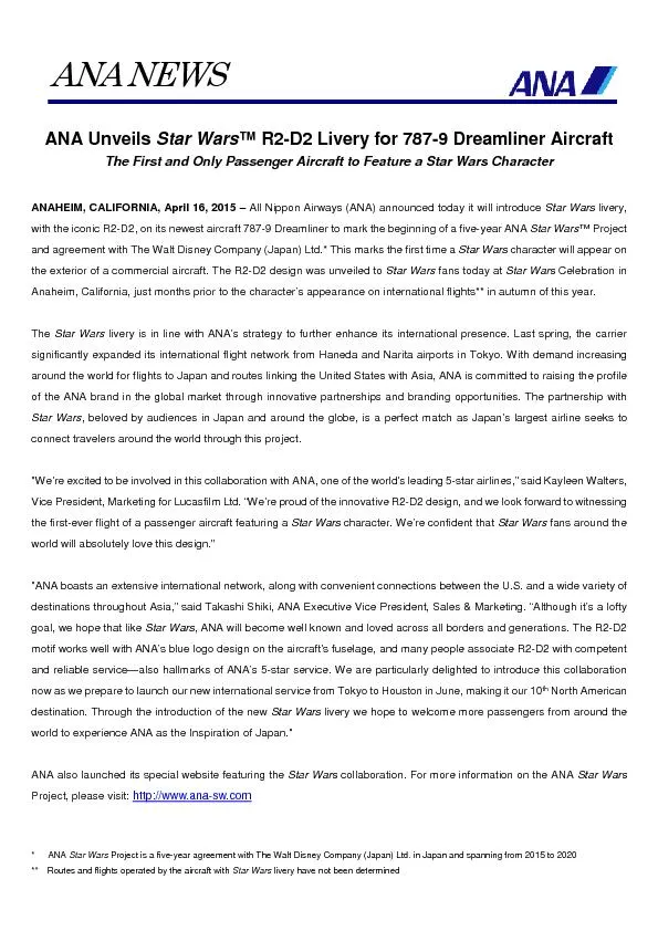
Megan O Nakashima Eric D Hsi Department of Clinical Pathology Cleveland Clinic Foundation Clinical History Previously healthy 53 year old man Three day history of a generalized petechial rash hematuria and rectal bleeding following a flulike illness ID: 815917
Download The PPT/PDF document "Case 291 Ana L. Ruano, Juraj Bodo," is the property of its rightful owner. Permission is granted to download and print the materials on this web site for personal, non-commercial use only, and to display it on your personal computer provided you do not modify the materials and that you retain all copyright notices contained in the materials. By downloading content from our website, you accept the terms of this agreement.
Slide1
Case 291
Ana L. Ruano, Juraj Bodo,
Megan O. Nakashima, Eric D. Hsi
Department of Clinical Pathology
Cleveland Clinic Foundation
Slide2Clinical History
Previously healthy 53 year old man.
Three day history of a generalized petechial rash, hematuria and rectal bleeding following a flu-like illness.
WBC = 8.85 K/UL;
Hgb
=12.5 g/
dL
;
Plt
= 40 K/UL.
Laboratory studies showed evidence of disseminated intravascular coagulation.
Slide3Peripheral blood
, 1000x
Bone marrow aspirate
, 1000x
Peripheral blood
, 1000x
Bone marrow aspirate
, 1000x
Slide4Cytochemical Stains
Myeloperoxidase
α
-
naphthyl butyrate esterase
Slide5Bone marrow biopsy
, 100x
1000x
Slide6Slide7Interphase
FISH
for
PML-RARA
(dual fusion probes):
Positive
Karyotype: 47,XY,+8,t(15;17)(q24;q21)[20]
Slide8Proposed Diagnosis
Acute promyelocytic leukemia with (t15;17)(q22;q12)/
PML-RARA
.
CONSENSUS DX: Acute promyelocytic leukemia
with t(15;17)(q22;q12)
PML-RARA
Slide9Clinical History (cont.)
Initially treated with systemic corticosteroids, vitamin K, platelets, fresh frozen plasma and cryoprecipitate.
Upon diagnosis of acute promyelocytic leukemia, ATRA was initiated.
Slide10Increasing shortness of breath, low SaO
2
, development of altered mental status and left hemiplegia. Head CT showed intraparenchymal hemorrhage.
Bilateral multifocal opacities on CXR; systemic corticosteroids initiated for apparent “differentiation syndrome”.
Clinical condition rapidly deteriorated. Brain death declared; patient expired shortly after removal from ventilator.
Slide11Acute Promyelocytic Leukemia
Time to diagnosis
prognosis
Expedient methods of diagnosis may significantly improve patient outcomes
Slide12Patient sample
Positive control (NB4)
Negative control (HL60)
Proximity Ligation Assay:
PML and RARA primary antibodies
for detection of PML-RARA fusion protein
Slide13Proximity Ligation Assay
Method for co-localizing any two biologic features that can be targeted by antibodies
Formats
Homogenous mixtures
Solid phase
Localized (
in situ)
Slide14Applications
Detection of post-translational protein modifications (eg. phosphorylation, mucin glycoforms)
A
B
Detect and quantify single protein expression
Duolink Brightfield II User Manual
Olink Bioscience Uppsala, Sweden www.olink.com
Detect and quantify protein interactions
Slide15Procedure
1
2
3
Primary antibodies
PLA probes
Ligation solution
Duolink Brightfield II User Manual
Olink Bioscience Uppsala, Sweden
www.olink.com
Slide164
5
Rolling circle amplification (RCA)
Detection -HRP
Duolink Brightfield II User Manual
Olink Bioscience Uppsala, Sweden www.olink.com
Slide17Patient sample
Positive control (NB4)
Negative control (HL60)
Proximity Ligation Assay:
PML and RARA primary antibodies
for detection of PML-RARA fusion protein
Slide18Advantages
Amenable to bright field microscopy
Fast - Results in approximately 6 hours
Easy to quantify – use of image analysis
Single cell analysis possible
Independent of PML gene break point
Slide19Conclusion
The case illustrates the application of a an alternate technology to test for fusion proteins via an easily interpretable bright field method potentially adaptable to the clinical laboratory.
Slide20References
Bodo J, Lin JJ,
Maciejewski
JP,
Schade
AE, Hsi ED. Detection of PML/RARA Fusion Protein in APL Using Proximity Ligation Assay as an Alterative to FISH Testing. 2013 Annual meeting of United States and Canadian Academy of Pathology, Baltimore, MD.
Duolink Brightfield II User Manual Olink Bioscience Uppsala, Sweden www.olink.comGustafsdottir SM et al. Proximity Ligation Assays for Sensitive and Specific Protein Analysis. 2005 Anal Biochem. 345(1):2-9.Zieba A et al. Bright-field microscopy visualization of proteins and protein complexes by in situ proximity ligation with peroxidase detection. 2010 Clin Chem 56(1):99-110
Gullberg M et al. A sense of closeness: protein detection by proximity ligation. 2003
Curr Opin
Biotechnol 14(1):82-6.Koos B et al. Analysis of Protein Interactions in situ by Proximity Ligation Assays
Current Topics in Microbiology and Immunology Springer-Verlag Berlin Heidelberg 2013.Figueiredo J et al. The importance of E-cadherin binding partners to evaluate the pathogenicity of E-cadherin missense mutations associated to HDGC. 2013 Eur J Hum Genet;21(3):301-9.Pinto R et al. Identification of new cancer biomarkers based on aberrant mucin glycoforms by in situ proximity ligation. 2012 J Cell Mol Med Jul;16(7):1474-84.Leuchowius
KJ et al. High content screening for inhibitors of protein interactions and post-translational modifications in primary cells by proximity ligation. 2010 Mol Cell Proteomics Jan;9(1):178-83.Chen TC et al. Protein phosphorylation profiling using an in situ proximity ligation assay: phosphorylation of AURKA-elicited EGFR-Thr654 and EGFR-Ser1046 in lung cancer cells
. 2013 PLoS One ;8(3):
e55657.Aubele M et al. In situ quantification of HER2-protein tyrosine kinase 6 (PTK6) protein-protein complexes in paraffin sections from breast cancer tissues. 2010 Br J Cancer 103(5):663-7.