/
Histology of Salivary Glands Histology of Salivary Glands

Histology of Salivary Glands - PowerPoint Presentation

kittie-lecroy
kittie-lecroy . @kittie-lecroy
Follow
581 views
Uploaded On 2016-04-27

Histology of Salivary Glands - PPT Presentation

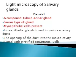
What is a gland Gland is an organ of secretion made up of specialized secretory cells derived from the surface epithelium on which it opens General Features Epithelial in origin Present as discrete organs or in layers ID: 295829

glands gland salivary cells gland glands cells salivary secretion compound secretory mucous serous alveolar tubulo simple ducts duct based

Share:

Link:

Embed:

Download Presentation from below link

Download Presentation The PPT/PDF document "Histology of Salivary Glands" is the property of its rightful owner. Permission is granted to download and print the materials on this web site for personal, non-commercial use only, and to display it on your personal computer provided you do not modify the materials and that you retain all copyright notices contained in the materials. By downloading content from our website, you accept the terms of this agreement.


Presentation Transcript

Slide1

Histology of Salivary GlandsSlide2
Slide3
Slide4

What is a gland?

Gland is an organ of secretion made up of specialized

secretory

cells derived from the surface epithelium on which it opens.Slide5

General Features

Epithelial in origin

Present as discrete organs or in layers.

Secretory

cells form functional units called

secretory

end pieces- may

be flask (

Acinus)or cylindrical (Tubular)shapedSlide6

Types of Secretory

unitsSlide7

General Features

Fluid secreted may be enzymes, hormones or mucus.

Secretion is modulated by nervous and hormonal influences.

Myoepithelial

cells-

star shaped, contractile, lie between the

secretory

cells and the basement membraneSlide8

Mixed Salivary GlandSlide9

Development

Develop as

invagination

of the epithelium

into the underlying vascular connective tissue.

Distal part

forms glandular or

Secretory end Piece

– functionally an active portion.Proximal part-Excretory Duct-opens on the surface of the epitheliumSome cells get detached

from the epithelial surface- Ductless or endocrine glandsSlide10

Development of GlandSlide11

Glandular EpitheliumSlide12
Slide13
Slide14
Slide15

Classification of Glands

Based on the site of Secretion

Exocrine Gland

Endocrine Gland

Paracrine

Gland-

secretes its products into the local extracellular space affecting the surrounding cells

e.g. enteroendocrine cells of gastrointestinal tract (GIT)Slide16
Slide17

Classification of Glands

Based on the Number of cells

Unicellular Gland-

goblet

cells

in

the respiratory and intestinal tracts

Multicellular Gland-

all glands other than goblet cellsSlide18

Classification of Glands

Based on the Number of Ducts and the shape of

secretory

end piece

Simple Gland-

one duct

Compound Gland-

has minor and major ducts

Both the types are further subdivided intoTubulo, Alveolar/Acinar

orTubulo-alveolar/acinousSlide19
Slide20

Multicellular GlandsSlide21

Compound Tubulo

-alveolarSlide22

Compound Tubulo

-alveolarSlide23

Compound Glands

Simple Alveola

r-Penile urethra

Simple Branched

alveolar-Sebaceous gland

Compound Alveolar-

Pancreas, Parotid, Mammary gland and glands of Respiratory tract.

Simple Tubular-Crypts of Leiberkuhn

Simple branched tubular-Uterine glands,Pyloric and fundic glandsCompound Tubular

-Brunner’s Gland, Cardiac glandsSimple coiled tubular-Sweat glandCompound Tubulo-alveolar- Submandibular

& Sublingual salivary glandsSlide24

Mixed Salivary GlandSlide25

Classification of Glands

Based on the Mode of Secretion

Merocrine

Gland-

No loss of Cytoplasm-e.g. most of the

compound

glands e.g. Pancreas

Also known as

Eccrine or Epicrine

Apocrine Gland- Partial loss of cytoplasm-e.g. lactating mammary gland, sweat glands in the axilla

and external genitalia

Holocrine

Gland-

Complete loss of cytoplasm e.g.

sebaceous and tarsal gland

Cytocrine

Gland-

Cells are released as secretion

. e.g

.

Testis (spermatozoa

)Slide26

Modes of SecretionSlide27

Classification of Glands

Based on the Nature of Secretion

Serous Gland-

thin, watery secretion rich in enzymes e.g. Parotid gland

Mucous Gland-

thick, viscous secretion for protection and lubrication. e.g. Sublingual salivary gland

Mixed Gland (

seromucous

)- both watery and viscous material.e.g. Submandibular salivary glandSlide28

Difference between Serous & Mucous

Acini

Serous

Thin, watery

Proteinaceous

secretion

Zymogen

granules in cyto

Central rounded NucleusSmall LumenIndistinct cell bondaries

Darkly stainedEnzymatic actionParotid Gland

Mucous

Thick, viscous

Mucopolysaccharides

Mucigen

droplets

Nucleus-flat & peripheral

Large Lumen

Distinct cell boundaries

Lighly

stained

Protection & lubrication

Sublingual glandSlide29

Mixed salivary gland

Serous

Acini

Mucous

Acini

Seromucous

Acini

- having Serous

demilunesSlide30
Slide31
Slide32

General Architecture of a Compound Gland

Gland may be divided into lobes and lobules.

Parenchyma-

Secretory

end pieces-

Acini

/tubules /

tubulo-acinar)Ducts- Intralobular, interlobular

, main excretory duct Stroma

CapsuleSepta (interlobular, interlobar)Loose

intralobular

connective tissue

supporting the parenchymaSlide33

Clinical

ADENOMA:

Benign tumors arising in the gland

ADENOCARCINOMA:

Malignant growth in the glandSlide34
Slide35

Serous AcinusSlide36

DuctsSlide37
Slide38

Striated Duct

Larger than the

acinus

Eosinophilic

columnar cells

Central nuclei

Vertical

Cytoplasmic stiations due to

foldings in the basal plasma membranesSlide39

Parotid GlandSlide40
Slide41

Parotid GlandSlide42

Intra-glandular adipose tissue in parotid glandSlide43

Submandibular GlandSlide44

Submandibular GlandSlide45

Mixed Salivary GlandSlide46
Slide47

Mucous AciniSlide48
Slide49

Sublingual-purely mucous glandSlide50

SublingualSlide51

Minor salivary glands of Palate

Aggregations of Mucous

acini

No striated ductSlide52

Minor salivary glands of Palate(High Power)

Mucous

acini

with central Lumen

Large Pyramidal cells with granular cytoplasm

Nucleus towards the basement membraneSlide53

The minor salivary glands are small aggregates of

unencapsulated

mucous or serous glands. In the tongue they are in intimate contact with the striated muscle tissue.Slide54

Keratin cocktail stains intercalated, striated and interlobular ducts, but

acinar

and

myoepithelial

cells are mostly negative.Slide55

MCQ

The serous gland can be identified by the presence of serous

acinus

with

A) Small Lumen

B)Large Lumen

C)Flat peripheral Nuclei

D)Mucigen dropletsSlide56

MCQ

When there is a complete loss of cytoplasm resulting in cell death of the

secretory

cell during the process of secretion, the gland is said to be

A)

Merocrine

B)

ApocrineC) HolocrineD)

CytocrineSlide57

MCQ

Sebaceous gland is an example of

Holocrine

gland

Apocrine

gland

Merocrine

glandUnicellular glandSlide58

MCQ

Mucous

Acinus

A) Secretes thin watery fluid

B) Has flat, peripheral nucleus

C) Has a small lumen

D) Contains

zymogen granulesSlide59

MCQ

Sweat glands in the

axilla

are an example of

A)

Merocrine

gland

B) Apocrine glandC) Holocrine glandD) Cytocrine

gland