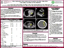
/
 Tracheo  esophageal fistula  Tracheo  esophageal fistula

Tracheo esophageal fistula - PowerPoint Presentation

kittie-lecroy
kittie-lecroy . @kittie-lecroy
Follow
397 views
Uploaded On 2020-04-03

Tracheo esophageal fistula - PPT Presentation

Dr S Parthasarathy MD DA DNB MD Acu Dip Diab DCA Dip Software statistics PhDphysiology Mahatma Gandhi Medical College and Research Institute Puducherry India What is it ID: 774906

fistula tube surgeon aspiration fistula tube surgeon aspiration ventilation problems caudal left intra spontaneous lung trachea esophagus gastrostomy post

Share:

Link:

Embed:

Download Presentation from below link

Download Presentation The PPT/PDF document " Tracheo esophageal fistula " is the property of its rightful owner. Permission is granted to download and print the materials on this web site for personal, non-commercial use only, and to display it on your personal computer provided you do not modify the materials and that you retain all copyright notices contained in the materials. By downloading content from our website, you accept the terms of this agreement.


Presentation Transcript

Slide1

Tracheo esophageal fistula

Dr. S. Parthasarathy

MD., DA., DNB, MD (

Acu

), Dip.

Diab

. DCA,

Dip. Software statistics, PhD(physiology)

Mahatma Gandhi Medical College and Research Institute,

Puducherry

, India

Slide2

What is it ??

There is a connection between trachea and esophagus

Congenital

Sometimes plain esophageal

atresia

is talked with fistula

Slide3

GROSS classification

Slide4

Why does it happen ??

The embryogenesis

- not

completely

defined

.

The trachea and esophagus develop from a common site, the foregut, in the first 4 to 5 weeks of

gestation.

Both the esophagus and the trachea originate from the median ventral diverticulum of the primitive foregut.

The

TEF lesion results from failure of the two structures to separate during division of the endoderm.

Slide5

Slide6

1 in 4000 --- C A E

C

A

E

Slide7

Slide8

Diagnosis

antenatal polyhydramnios Excessive salivation choking, coughing,aspiration pneumonia, cyanosis. Attempts at feeding - met with explosive vomiting passing an oral (nasal) gastric tube is impossible.A chest radiograph of a coiled oral gastric tube in the cervical esophageal pouch is diagnostic.No contrast please

Slide9

Slide10

VACTERL

A common association is the VACTERL complex, consisting of vertebral,

anorectal

, cardiac,

tracheoesophageal

, renal, and limb defects

VATER, VACTER and VACTERL

30 % may have !!

But the ligation of a TEF is urgent.

Slide11

Preoperative management --FUSA

F

eedings

– NO

U

pright

positioning

S

uctioning

intermittent

A

ntibiotic

administration

Dehydration and acid base to be corrected

Should we

intubate

preop

?? Does this prevent aspiration

Slide12

Two ways of aspiration

If significant aspiration pneumoniadefinitive corrective surgery ??decompressing gastrostomy local or caudal anesthesia

Slide13

Waterson prognostic criteria

Weight - > 2.5 , 1.8 – 2.5 , <1.8

Associated anomalies

Pneumonia

A, B or C

Slide14

Preoperative work up

Echocardiography ( routine + right sided aortic arch)

X ray chest

Blood gases

Lumbar ultrasound –

Xray

spines – caudal ??

USG abdomen

Rigid

bronchoscopy

(airway +

fogarty

)

Tracheoscopy

flexible (

Zurisch

et al)

1 unit of packed red blood cells should be type and crossed matched

Slide15

Surgery

Repair primary repair involves isolation and ligation of the fistula followed by primary anastamosis of the esophagus. A staged repair is possible in sick neonates Posterior thoracotomy Thoracoscopic Bronchoscopic clipping of HSometimes stabilizing and do it in 7 – 10 daysFUSA remains

Slide16

Gastrostomy

Slide17

Sometimes

During gastrostomy, they may clamp the fistula with esophagostomy and do the surgery after a few months also. Esophagostomy done from the neck to remove the secretionsDescribed

Slide18

Routine monitors

Precordial

steth

in the left

axilla

Adequate IV access

Radial artery

cannulation

(Lt) or umbilical

0.15 mg atropine IV

Temperature , IV fluids

Urine output – 1 ml/kg /hour

Slide19

Anaesthetic

management

Slide20

Awake intubation

Inhalation Induction Intubation in sitting posture Proper suctioning Circuits, scopes, cuffed ETT,Neonatal ventilators

Slide21

Fish mouth fixation

Slide22

Principle

Intubate purposely Rt main bronchus Withdraw slightly to get bilateral air entry Reverse the curve of the tube Reverse the side of murphy eye

Gas leak

Ventilate lungs and not the fistula

Slide23

Ventilation - adequate ??

Gastric distension

Gastrostomy

to water – to ETCO2

Tube kink

Anomaly – different

Even OLV

Slide24

Ventilate to the stomach – fistula – no ventilation

Tight bag with occlusion of the fistula

Slide25

To prevent leak

Slide26

Its easier to describe spontaneous –

but open chest with surgeon pushing the right lung its difficult to maintain with spontaneous

appropriate positioning of ETT is mandatory.

Slide27

Spontaneous or controlled

Are we sure – we are ventilating the lungs and not the fistula ??

Allow spontaneous

& Give caudal

0.5–1

mL

/ kg of 0.25%

bupivacaine

with epinephrine

(5 μ

g/

mL

)

Try threading far up .. But guarantee ?? USG

Fentanyl

Relaxants after clamping the fistula

Slide28

No caudal ??

Avoidance of regional anesthesia with its corresponding decrease in systemic vascular resistance is warranted in patients with coexisting congenital heart disease such as

hypoplastic

left heart syndrome (HLHS).

Cautious caudal in CVS diseases

Slide29

Intra op problems

left lateral

decubitus

position.

During the surgical repair, the right lung is compressed and packed away, which may result in hypoxia.

the trachea and/or

endotracheal

tube is compressed and occluded by the surgeon.

Alternatively, the

endotracheal

tube can become obstructed by blood clots or may migrate into the fistula tract.

Slide30

Intra op problems

During localisation of the fistula, an anaesthesiologist can help the surgeon by applying traction to the wire loop.

Some routinely use

100% oxygen

during these anesthetics, even in premature infants who are at risk for developing the ROP.

Class C -- bad lungs HFO used - reports !!

Extubate

in healthy infants -- class A

Otherwise ??

Slide31

Intra op problems

The surgeon will get hold of the fistula and pull it.

It will dislodge the position of ETT.

The rt. Lung is collapsed by the surgeon. The tube becomes RT.

endobronchial

. ??

No ventilation – that’s why keep the

steth

Left

axilla

and watch for breath sounds ..

Slide32

Prognosis

Slide33

Post op problems

Surgical postoperative complications include anastomotic leak, stricture, gastro esophageal reflux, tracheomalacia, recurrent TEF. flush ligation is a must. Otherwise - Diverticulum –stasis, infection and giving way

Slide34

Thoracoscopic approach

The advantages

Reduction of musculoskeletal

sequelae

that often develop following open

thoracotomy

in the newborn period. These have been well described as “winged” scapula, asymmetry of thoracic wall and thoracic scoliosis.

superior visualization of fistula and surrounding structures including

vagus

nerve with the

thoracoscopic

approach

.

Slide35

Post op giving way

Don’t extend Neck of neonate

anastomosis

will stretch and give way as there is always a gap between the two ends of

oesophagus

, which surgeon has

mobilised

to bring together

Slide36

Post operative problems

Need for ventilation arises secondary to

Compression of lung for several hours

Pre-existing aspiration pneumonia

Is always preferred in the backdrop of other coexistent congenital anomalies

Slide37

Summary

Incidence

Types

Commonest

Clinical features

FUSA

Intubation and positioning , techniques

Intra op hypoxemia

Waterson

,

spitz

, post op ventilation

Slide38

Thank you all