PDF-(BOOS)-Fundamentals of Clinical Hematology
Author : kylakomar98 | Published Date : 2022-06-23
This reference presents the fundamentals of hematology including erythrocytes leukocytes thrombocytes and coagulation and briefly discusses disease states in a concise
Presentation Embed Code
Download Presentation
Download Presentation The PPT/PDF document "(BOOS)-Fundamentals of Clinical Hematolo..." is the property of its rightful owner. Permission is granted to download and print the materials on this website for personal, non-commercial use only, and to display it on your personal computer provided you do not modify the materials and that you retain all copyright notices contained in the materials. By downloading content from our website, you accept the terms of this agreement.
(BOOS)-Fundamentals of Clinical Hematology: Transcript
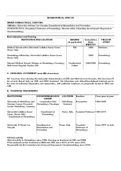
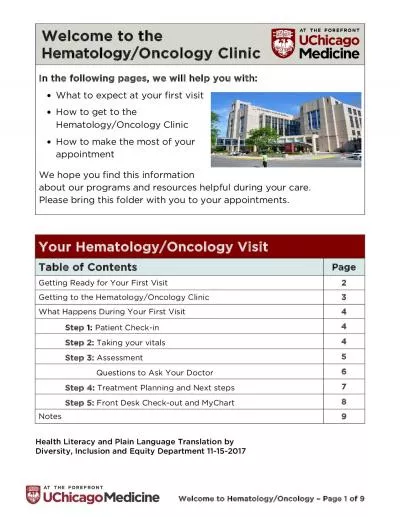
This reference presents the fundamentals of hematology including erythrocytes leukocytes thrombocytes and coagulation and briefly discusses disease states in a concise yet comprehensive manner This readerfriendly text features outlines objectives study questions bibliographies Do It Now application exercises special emphasis statements and Fast Facts summaries Demonstrates the integration collaboration balance and wholeness of quality clinical laboratory practices by introducing related areas and their procedures. Current Controversies in Hematology and Oncology he cost of cancer drugs is at an all-time high, with several lifesaving agents costing more than $100,000 a year. Much of the discussion related to hea Targeted Cancer Therapies. Dr . Ongóndi. .M. KNH-Dept. Internal Medicine. Hemato. -Oncology. Objectives. Traditional chemotherapy. Targeted chemotherapy. Country status. Challenge !!!. Administer sufficiently high dose of . MARIATERESAVOSO,13/04/1965 Affiliation:UniversityRomeTorVergata, DepartmentBiomedicinePrevention POSITIONTITLE:AssociatedProfessorHematology, DirectortheLaboratoryforadvanceddiagnosisOncohematology Be Specializing in the diagnosis and management of hematological disorders the Hematology practice at Valley Children146s provides consultations 24 hours a day We provide care for a large pediatric patie Dr. . Versha. Prasad. Anticoagulants Used In the Hematology Laboratory. Anticoagulants are defined as substances which prevent blood clotting / coagulation, and allow separation of the blood into cellular and liquid (plasma) components. Generally plasma contains coagulation factors. . , RN. Evy Warmbier, MSN, RN, CNE. Objectives. To Identify the Basic Hematological Components. To Understand the Clotting Cascade. To Relate the Fibrinolytic Systems Regarding Medication Administration. ARC Jour Volume 4 , Issue 2, 2019, PP 1 - 4 www.arcjournals.org ARC Journal of Hematology Page | 1 Difficulties Diagnostic of Amegacaryocytic Thrombocytopenic Purpura in Developing Country: A Case 65 Rotation Name Peds/Onc at Michigan State University Educational Purpose of the Rotation It is expected that residents rotating through the Hematology/Oncology experience will successfully master sp Description: The pediatric hematologyoncology division sees a wide spectrum of pediatric disease including but not limited to leukemia, hemophilia, solid tumors, ITP, and other blood dyscrasias. The Welcome to the Oncology Clinic In the following pages, we will help you with: hat to expect at your first visit ow to get to the Hematology/Oncology Clinicow to make the most of your appointmentWe - Oncology The Tata Group has always believed in returning wealth to the Society it serves. It has established philanthropic trusts which have created National Institutions in Science and Technology Get complete detail on IT Audit Fundamentals exam guide to crack ISACA IT Audit Fundamentals. You can collect all information on IT Audit Fundamentals tutorial, practice test, books, study material, exam questions, and syllabus. Firm your knowledge on ISACA IT Audit Fundamentals and get ready to crack IT Audit Fundamentals certification. Explore all information on IT Audit Fundamentals exam with number of questions, passing percentage and time duration to complete test. Get complete detail on Data Science Fundamentals exam guide. You can collect all information on ISACA Data Science Fundamentals tutorial, practice test, books, study material, exam questions, and syllabus. Firm your knowledge on Data Science Fundamentals and get ready to crack ISACA Data Science Fundamentals certification. Explore all information on Data Science Fundamentals exam with number of questions, passing percentage and time duration to complete test. Get complete detail on ISACA Blockchain Fundamentals exam guide to crack the certification exam. You can collect all information on ISACA Blockchain Fundamentals tutorial, practice test, books, study material, exam questions, and syllabus. Firm your knowledge on Blockchain Fundamentals and get ready to crack ISACA Blockchain Fundamentals certification. Explore all information on ISACA Blockchain Fundamentals exam with number of questions, passing percentage and time duration to complete test.
Download Document
Here is the link to download the presentation.
"(BOOS)-Fundamentals of Clinical Hematology"The content belongs to its owner. You may download and print it for personal use, without modification, and keep all copyright notices. By downloading, you agree to these terms.
Related Documents