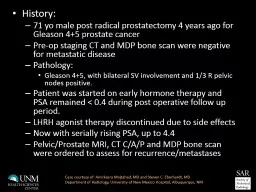
/
History:  71  yo  male post radical prostatectomy 4 years ago for Gleason 4+5 prostate History:  71  yo  male post radical prostatectomy 4 years ago for Gleason 4+5 prostate

History: 71 yo male post radical prostatectomy 4 years ago for Gleason 4+5 prostate - PowerPoint Presentation

laxreffa
laxreffa . @laxreffa
Follow
343 views
Uploaded On 2020-06-17

History: 71 yo male post radical prostatectomy 4 years ago for Gleason 4+5 prostate - PPT Presentation

Preop staging CT and MDP bone scan were negative for metastatic disease Pathology Gleason 45 with bilateral SV involvement and 13 R pelvic nodes positive Patient was started on early hormone therapy and PSA remained lt 04 during post operative follow up period ID: 780711

recurrence bone radiology scan bone recurrence scan radiology mri courtesy prostatectomy eberhardt steven prostate amirkasra mdp unm lesion psa

Share:

Link:

Embed:

Download Presentation from below link

Download The PPT/PDF document "History: 71 yo male post radical pros..." is the property of its rightful owner. Permission is granted to download and print the materials on this web site for personal, non-commercial use only, and to display it on your personal computer provided you do not modify the materials and that you retain all copyright notices contained in the materials. By downloading content from our website, you accept the terms of this agreement.


Presentation Transcript

Slide1

History: 71 yo male post radical prostatectomy 4 years ago for Gleason 4+5 prostate cancerPre-op staging CT and MDP bone scan were negative for metastatic diseasePathology: Gleason 4+5, with bilateral SV involvement and 1/3 R pelvic nodes positive. Patient was started on early hormone therapy and PSA remained < 0.4 during post operative follow up period.LHRH agonist therapy discontinued due to side effectsNow with serially rising PSA, up to 4.4Pelvic/Prostate MRI, CT C/A/P and MDP bone scan were ordered to assess for recurrence/metastases

Case courtesy of: Amirkasra Mojtahed, MD and Steven C. Eberhardt, MDDepartment of Radiology, University of New Mexico Hospital, Albuquerque, NM

Slide2

Initial MDP bone scan (A) demonstrates marked radiopharmaceutical uptake within the right acetabulum anteriorly, which correlates with sclerotic lesion on CT (B) performed 2 weeks later. B

ACase courtesy of: Amirkasra Mojthed, MD, Steven C. Eberhardt MD, UNM Radiology

Slide3

MRI Pelvis/Prostate (3T, no ER coil) performed 2 days after CT: Axial (A) and coronal (B) T2WI of the prostatectomy bed show no local recurrence. Large FOV axial T1WI (C) demonstrates a hypointense lesion within the anterior right acetabulum/pubic root, correlating with lesion on preceding CT and bone scan.B

ABC

Case courtesy of: Amirkasra Mojthed, MD, Steven C. Eberhardt MD, UNM Radiology

A

Slide4

Teaching pointIn cases of biochemical failure following RRP, bone scan, CT and MRI can be useful in finding a cause for the rising PSA. Large field of view sequences on MRI can be helpful in assessing for regional osseous metastases or adenopathy, in addition to small field of view sequences for recurrence in the operative bed. CT and MDP bone scan can be used to provide a more broad surveyCase courtesy of: Amirkasra Mojthed, MD, Steven C. Eberhardt MD, UNM Radiology

Slide5

Useful referencesMay EJ, Viers LD et al.  Prostate cancer post-treatment follow-up and recurrence evaluation.  Abdominal Radiology, May 2016, Volume 41, Issue 5, pp 862-876Sella, Tamar, et al. "Suspected Local Recurrence after Radical Prostatectomy: Endorectal Coil MR Imaging 1."Radiology 231.2 (2004): 379-385.Casciani, Emanuele, et al. "Endorectal and dynamic contrast-enhanced MRI for detection of local recurrence after radical prostatectomy." 

American Journal of Roentgenology 190.5 (2008): 1187-1192.