PDF-What Is an Arteriovenous Malformation AVM
Author : layla | Published Date : 2022-08-16
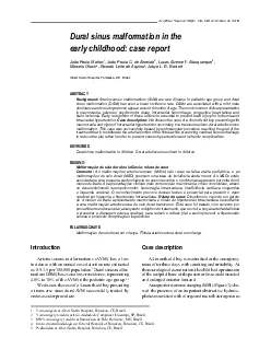
From the Cerebrovascular Imaging and Intervention Committee of the American Heart Association Cardiovascular Council Randall T Higashida MD Chair What is a brain
Presentation Embed Code
Download Presentation
Download Presentation The PPT/PDF document "What Is an Arteriovenous Malformation AV..." is the property of its rightful owner. Permission is granted to download and print the materials on this website for personal, non-commercial use only, and to display it on your personal computer provided you do not modify the materials and that you retain all copyright notices contained in the materials. By downloading content from our website, you accept the terms of this agreement.
What Is an Arteriovenous Malformation AVM: Transcript
Download Rules Of Document
"What Is an Arteriovenous Malformation AVM"The content belongs to its owner. You may download and print it for personal use, without modification, and keep all copyright notices. By downloading, you agree to these terms.
Related Documents