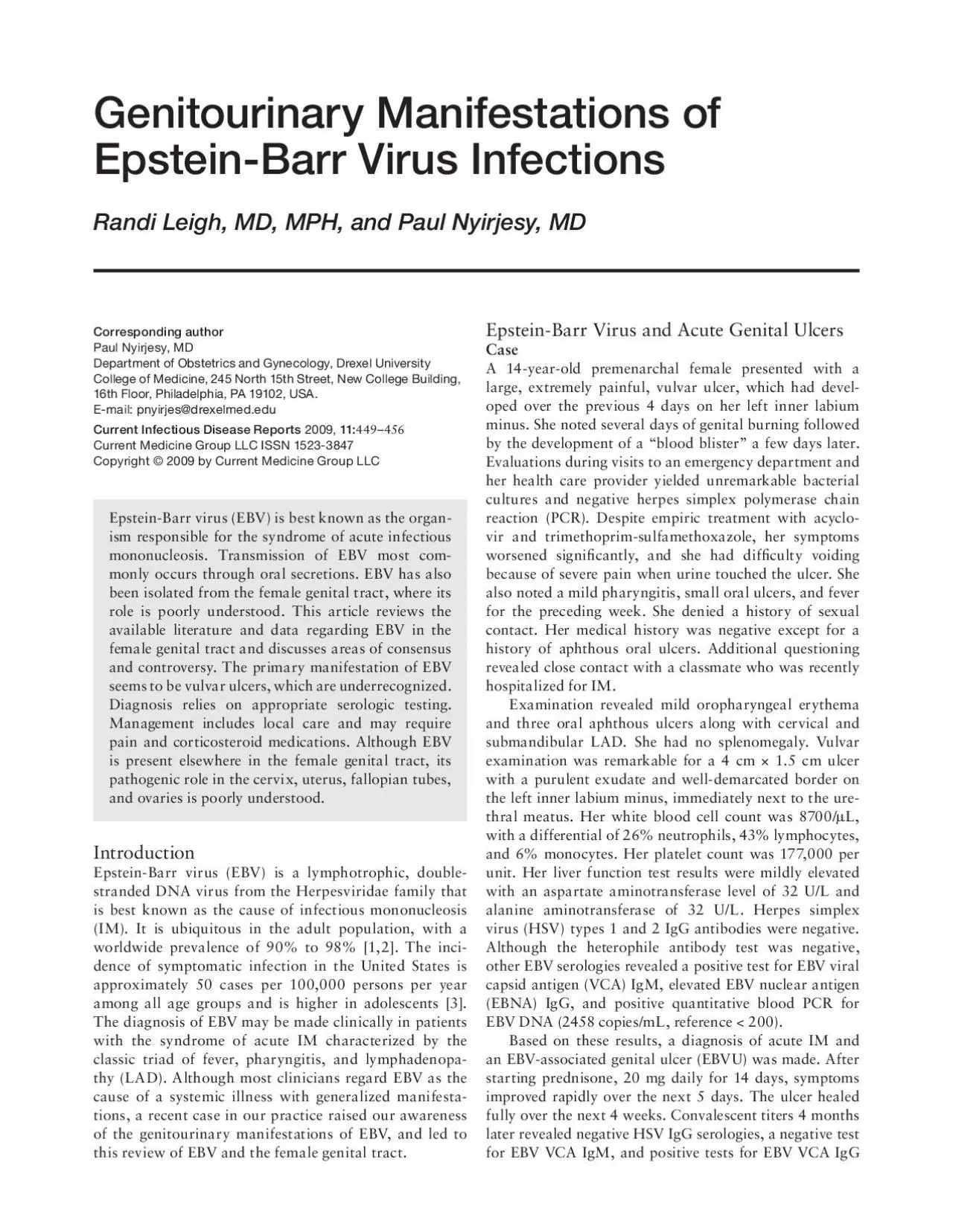
/
Genitourinary Manifestations of EpsteinBarr Virus InfectionsRandi Lei Genitourinary Manifestations of EpsteinBarr Virus InfectionsRandi Lei

Genitourinary Manifestations of EpsteinBarr Virus InfectionsRandi Lei - PDF document

leah
leah . @leah
Follow
342 views
Uploaded On 2022-09-07

Genitourinary Manifestations of EpsteinBarr Virus InfectionsRandi Lei - PPT Presentation

450 Urinary Tract Infectionsand EBNA One year after initial presentation the patient had a recurrence of her ulcer which responded within a few days to a 7day course of prednisone Clinical pr ID: 952495

virus ebv ebvu genital ebv virus genital ebvu epstein barr infection ulcers patients cases acute women cervical ulcer tract

Share:

Link:

Embed:

Download Presentation from below link

Download Pdf The PPT/PDF document "Genitourinary Manifestations of EpsteinB..." is the property of its rightful owner. Permission is granted to download and print the materials on this web site for personal, non-commercial use only, and to display it on your personal computer provided you do not modify the materials and that you retain all copyright notices contained in the materials. By downloading content from our website, you accept the terms of this agreement.


Presentation Transcript

Genitourinary Manifestations of Epstein-Barr Virus InfectionsRandi Leigh, MD, MPH, and Paul Nyirjesy, MD Corresponding authorPaul Nyirjesy, MDDepartment of Obstetrics and Gynecology, Drexel University College of Medicine, 245 North 15th Street, New College Building, 16th Floor, Philadelphia, PA 19102, USA. E-mail: pnyirjes@drexelmed.eduCurrent Infectious Disease Reports 2009, 11:449456Current Medicine Group LLC ISSN 1523-3847 450 Urinary Tract Infectionsand EBNA. One year after initial presentation, the patient had a recurrence of her ulcer, which responded within a few days to a 7-day course of prednisone. Clinical presentationOur review identi ed 39 cases of EBVU published between 1966 and 2009, 34 in women between the ages of 10 and 23. Table 1 summarizes the pertinent clinical features of EBVU. Five patients were premenarchal, 7 had reached menarche, and 27 had no documentation of pubertal status. Of 29 patients for whom sexual histories were documented, 72% denied sexual activity. Associated symptoms were often prodromal and ranged from 1 day to several weeks before ulcers appeared. Although one third developed the classic syndrome of IM, only two developed the syndrome before the appearance of a vulvar lesion. In most patients, EBVU demonstrated a preference for the labia minora. Lesions tended to be large (0.3…4 cm) and well circumscribed, with violaceous margins. In the nine bilateral cases,  ve had a symmetric or kissingŽ pattern. Modes of transmissionEBV is thought to be transmitted to oropharyngeal epithelial cells and circulating B cells via infected oral secretions. Once B-cell infection is established, EBV migrates into the lymphoid tissues and establishes latency in memory B cells. Prolonged viral shedding of EBV from epithelial cells may occur for up to 18 months after the resolution of symptomatic infection [4€€,5]. In a large prospective study, 90% of healthy seropositive partici-pants demonstrated viral shedding at least once, whereas 25% demonstrated viral shedding on multiple tests over 15 months [6]. Furthermore, in highly concentrated sali-vary samples, the virus can be identi ed in up to 100% of healthy seropositive individuals, suggesting that the virus is never truly latent [7]. In the genital tract, EBV DNA was isolated by PCR from samples taken from the cervix and vagina of women, the urethra and anal mucosa of men and women, and the coronal sulcus of uncircumcised males [8]. In 1994, Taylor et al. [9] isolated EBV DNA by PCR from cervical Table 1. Signs and symptoms reported in 39 cases of Epstein-Barr virus…induced vulvar ulcersSigns and symptomsPresentTotal documentedAmong cases with documentation, Reported symptomFever3233 97Sore throat1821 86Headache 7 7100Fatigue1010100Arthralgia/myalgia 6 6100Leukorrhea 3 3100Dysuria1416 88Genital prodrome (pain and/or pruritus) 4 4100HistoryHistory of recurrent oral ulcers 3 4 75Physical examination “ ndingsCervical lymphadenopathy2228 79Inguinal lymphadenopathy1017 59Pharyngitis/tonsillitis1215 80Organomegaly 216 13Oral ulcers 4 4100Description of ulcerSize of dominant ulcer  1 cm1638 42Solitary ulcer 229 7Two or more ulcers2729 93Bilateral 914 64Labia minora only2534 74Necrotic/exudative appearance2020100Data from Cheng et al. [1], Barnes et al. [4€€], Portnoy et al. [10], Halvorsen et al. [11€€], Pelletier et al. [12], Taylor et al. [13], Farhi et al. [14€€], Svedman et al. [15], Llanos et al. [18], Hernandez-Nunez et al. [20], Brown and Stenchever [23], Hudson and Perlman [26], Johnson and Landry [27], Logeart et al. [28], Nicolas et al. [30], Groulier et al. [33], Lorenzo and Robertson [34], Tenorio et al. [36], McKenna et al. [37], Sisson and Glick [48], DeKlotz and Frieden [49].) Genitourinary Manifestations of Epstein-Barr Virus Infections I Leigh and Nyirjesy samples in 42% of women with normal Pap smears and 36% of women with abnormal  ndings. A possible expla-nation for these results is that the virus might be present in cell-free form, in lymphocytes, or in cervical squamous epithelial cells [9]. Among the cases we reviewed, EBV was isolated by culture ( = 1) [10], by PCR of vulvar biopsies ( = 3) [11€€,12] or ulcer swab ( = 1) [13], and by in situ hybridization ( = 2) [13,14€€]. Although other attempts to isolate EBV using the same methods failed [4€€,13,14€€,15], it is clear that EBV virus is present in the male and female genital tracts, including sites of EBVU, during acute and latent infec

tion. It is unknown whether genital epithelial cells are infected by direct inoculation, circulating B lymphocytes, hematogenous spread, another route, or some combination thereof. The presence of EBV in the genital tract, along with the knowledge that EBV infects epithelial cells at other body sites, led to the hypothesis that EBV may be sexually trans-mitted. One study of EBV transmission in HIV-infected men who have sex with men provides convincing evidence that sexual transmission of EBV is possible among this group [16]. Studies failed to provide substantial evidence to either refute or con rm heterosexual transmission of EBV. However, sexual transmission does not appear to be a necessary prerequisite for genital or gynecologic mani-festations of EBV. In fact, most cases of EBVU occur in young women who deny a history of any sexual activity.Although the question of whether EBV can be trans-mitted through vaginal intercourse is important, an equally relevant question is whether it can infect genital epithelium via contact with salivary secretions. Free EBV virus, infected oropharyngeal epithelial cells shed into salivary secretions, or infected B lymphocytes in saliva may all serve as possible vectors. Thus, orogenital trans-mission and self-inoculation via salivary secretions are potential routes of EBV infection that cannot be ruled out based on available data. Most investigations of EBV in the genital tract failed to explore patients sexual histories in suf cient detail to answer this question with certainty [17€€]. Only seven of the cases (18%) reviewed speci cally addressed whether a patient who was not sexually activeŽ had ever engaged in oral sex, and only three patients had done so. An even smaller number of patients speci cally reported on other sexual behaviors and practices, includ-ing digital-genital stimulation and masturbation. A better understanding of how EBV enters the female genital tract is desirable because this information has the potential to affect understanding of EBVU epidemiology and the role of EBV in genitourinary and gynecologic pathology. Epstein-Barr virus…associated ulcers: underrecognized disorder vs increasing incidence With clinicians largely unaware of the possibility that EBV can cause acute genital ulcers (AGUs), it is well accepted that EBVU is an underrecognized disorder [4€€]. Presumptive diagnoses of herpes likely contribute to missed cases of EBVU. Furthermore, as demonstrated by at least two cases, a history of con rmed herpes does not exclude the possibility of EBVU [14€€,18]. More than a dozen reports describe women with AGUs in whom the possibility of EBVU was not explored, including a small number of women in whom exhaustive workups failed to identify any cause [19€€,20,21]. It remains unknown if EBVU can occur as a result of reactivated latent infection. In multiple cases of women with AGU of unknown etiol-ogy, testing revealed an otherwise negative workup and serologies consistent with past EBV infection (anti-VCA IgG or anti-EBNA IgG positive), although no clear linkage was offered for these  ndings [19€€,20,21,22]. Excluding our case and that of a 19-year-old sexually active patient with con rmed EBVU who reported suffering a similar episode of AGU at age 12, before coitarche [19€€], reports of EBVU fail to address recurrence. Although the growing number of case reports of EBVU may re ect a greater awareness of the condition, it may also be the result of increasing incidence. Adolescents and young adults are now engaging in oral sex more fre-quently and at younger ages than in the past [23]. If direct inoculation of the genital epithelium via infected salivary secretions occurs, then changes in sexual behavior trends among the group most susceptible to symptomatic EBV infection may be increasing genital manifestations of EBV. However, it is also possible that the increase in EBVU cases may re ect an overall increase in the number of complicated cases of EBV infection over the past three decades [24,25]. Pathophysiology of acute genital ulcers secondary to Epstein-Barr virusTwo hypotheses are proposed to explain the pathophysiol-ogy of EBVU [5,10]. The virus itself may cause the ulcers. According to this theory, infected cells undergo cytolysis and cause epithelial erosion and eventually ulceration. A more likely explanation is that the ulcers are caused by the host immune response, in which immune complex deposition results

in complement  xation and reactive localized in ammation, then cell lysis. Consistent with this theory, biopsies collected from 11 patients demon-strated pathologic  ndings of vasculitis, thrombosis of super cial blood vessels, and nonspeci c mixed dermal in ltration [1,4€€,11€€,12,14€€,18,26…29]. EBVUs may simply be one of many mucocutaneous manifestations of EBV infection. The large number of possible sites of involvement (including the skin and nasal, oral, esopha-geal, and gastric mucosa) supports the idea that the host response to infection causes the ulcer. Of note, our literature review revealed only one case of a penile ulcer attributed to EBV [30]. The most likely reason for this gender discrepancy is that less keratinized tissue (eg, the labia minora, where most EBVUs occur), is more vulnerable to ulceration than the more keratinized epithelium of the male genitalia. It is also possible that female sex hormones serve as catalysts in ulcer formation. Exogenous estrogen and progesterone are associated with 452 Urinary Tract Infectionsother, nonulcerating cutaneous lesions including the vas-culitic panniculitis erythema nodosum, and vulvar ulcers secondary to progesterone autoimmune dermatitis and estrogen hypersensitivity were observed [31]. DiagnosisA thorough history in patients presenting with AGU is important and should include a detailed sexual history, use of soaps and detergents, current and recent medications, travel history, and sick contacts. The differential diagnosis is extensive and beyond the scope of this review. A sum-mary of clinical clues that may help distinguish EBVU from other key diagnoses is provided in Table 2. Notably, the timing of signs and symptoms associated with EBVU may vary, with many appearing days after the presentation of the ulcer. Rash may be a particularly important sign in patients with EBVU. Although 3% to 19% of all patients with acute EBV infection develop a maculopapular rash [7], this sign is observed in more than 70% of patients with acute EBV who receive antibiotics, particularly amoxicil-lin [5]. EBVU may be distinguished from HSV ulcers by their rapid enlargement despite treatment with antiherpetic agents. EBVU also tend to be larger, deeper, and more necrotic than ulcers caused by herpes. Numerous diagnostic tests can help clinicians dis-tinguish among EBVU, HSV, and other causes of AGU. Patients with EBVU demonstrate laboratory  ndings typi-cal for acute IM, including atypical lymphocytosis, mild thrombocytopenia, and mild elevations in liver transami-nases [7]. Culture or PCR of the lesion for HSV can help distinguish EBVU from HSV, and provide guidance for counseling sexually active patients. HSV and cytomegalo-virus (CMV) serology during the acute and convalescent periods also should be considered. Selection of other tests should be based on  ndings from the history and physical examination, and may include tests for syphilis and HIV, and for rare causes of AGU including adenovirus, in u-enza and coxsackie viruses, typhoid and paratyphoid, toxoplasmosis, tuberculosis, Mycoplasma pneumoniaeand others [4€€,20,32].EBV serology should be performed in the acute andconvalescent phases of illness. Wide variation may occur in the types of antibodies produced and the timing of their appearance during primary EBV infection [6]. The pres-ence of IgM antibodies against EBV VCA in the absence of antibodies against EBNA is diagnostic of acute EBV infection. Anti-VCA IgM typically forms during the  rst 2 weeks of illness and peaks several weeks later. However, it is worth noting that the ulcer preceded anti-VCA IgM antibodies in three cases [11€€,33,34]. Anti-VCA IgG and anti-EBNA antibodies typically form during the third to sixth week after the onset of infection and may persist for life. Nonspeci c heterophile antibodies may also form; however, as in our case, they can give a false-negative result during the  rst 2 weeks of infection. Moreover, 70% to 80% of patients develop anti…early antigen (EA) antibodies during the acute phase of infection. Persistence of anti-EA antibodies for more than 6 months is unusual and may be a marker of an aberrant immune response. A rapid test for EBV and serum PCR for EBV DNA are newer methods being used with increasing frequency [7]. Because of varia-tions in the timing and type of antibody production during primary EBV infection and the possibility of false-positive and false-negative tes

t results, convalescent serology per-formed at least 10 to 14 days after the initial presentation and repeated several months after the acute phase are help-ful in con rming the diagnosis.Culture of ulcers for EBVU was attempted, but was successful in only one of  ve cases. Also useful is PCR for EBV DNA from the ulcer, or immunohistochemistry and indirect immuno uorescence from a biopsy. Nucleic acid hybridization techniques are the only methods that dif-ferentiate between the two known strains of EBV. These latter methods are not currently used but may become more important with improved understanding of the dif-ferences between EBV strains. Biopsy  ndings are often nonspeci c and nondiagnostic, but provided clinically useful information in 36% (4/11) of the cases reviewed, with EBV DNA isolated in three specimens by PCR [9,10,35] and with one in which the pathologist noted a prominent starry sky patternŽ similar to that observed in the EBV-induced cancer Burkitts lymphoma [29]. Management EBVU is typically a self-limited condition. However, among the 20 case reports that described healing time, the mean time to complete resolution of the ulcer was approximately 20 days, with one lasting 10 weeks [4€€]. Although supportive treatment of EBVU is the norm, a range of management options is available.Topical therapy and pain managementA moisturizing barrier (eg, zinc oxide ointment) may pro-vide local relief. Patients with severe dysuria may need to urinate while directing a stream of water onto the vulva or while in a sitz bath. Low-potency topical steroids, silver sulfadiazine, topical lidocaine, fusidic acid, and estrogen cream have been used in the treatment of EBVU with variable bene t [18,26]. A broad range of analgesics, including narcotics, were used in the cases reviewed. AntimicrobialsEighteen patients in the case reports reviewed had negative bacterial cultures of ulcers, but one grew Pseudomonas aeruginosa [36] and another Staphylococcus aureus [37]. Although bacterial superinfection appears rare, antibiotic therapy may be considered in patients with markedly necrotic or purulent ulcers. Broad-spectrum agents with coverage for genitourinary pathogens should be selected. Antiherpetic agents for acute EBV infection appear to have little clinical ef cacy. Although acyclovir therapy reduces oropharyngeal EBV shedding, the quantity of virus and rate of viral shedding return to pretherapeutic levels soon after patients stop taking the medication [4€€,17€€]. Genitourinary Manifestations of Epstein-Barr Virus Infections I Leigh and Nyirjesy Table 2. Distinguishing Epstein-Barr virus…associated ulcers from other common causes of acute genital ulcersFeaturesEBVUHSVSyphilisChancroidLymphogranuloma venereumHistoryMean age, 14…17Seroprevalence increases with age; sharpest increase at ages 19 and 2925…29 35 UnknownIncubation period0 d…3 wk (mean, 8 d)2…7 d1…12 wk1…14 d3 d…6 wkFeverYesYesRare in primary infectionOccasionally mild feverYesPain/dysuriaYesYesRarelyYesOccasionallyFavored locationLabia minoraLabia majora, labia minora, perianal, vaginal walls, cervixNo preference; can appear on vagina, cervix, perineum, anusLabia majora, labia minora, perianal; rarely affects vagina or cervixCommonly causes rectal ulcers; iso-lated genital/vaginal ulceration rareOther+ Sick contacts; + history of oral ulcers in many casesPrimary infec-tion may be asymptomaticChancre is associated with primary syphilisEndemic in Africa, Asia, Caribbean; rare in United States, most outbreaks in citiesVery rare in United States; associated with proctocolitis (ie, rectal bleeding or purulent dis-charge, tenesmus)Physical examinationAppearance of primary lesionLarge, vesicular, punched-outŽ ulcer; often necrotic or pseudo-membrane formingSmall (but multi-ple small ulcers may coalesce), vesicularPapular chancrePapular (occa-sionally pustule forming)Papular or vesicular (occasionally pustule forming)Number of lesionsSolitary or multiple; commonly one dominant and sev-eral satellite ulcersMultiple, occasionally coalescingUsually solitaryUsually multiple, occasionally coalescingUsually solitaryDiameter, 0.1…4.00.1…0.20.5…1.50.2…2.00.2…1.0DepthDeepSuper“ cialSuper“ cial or deepDeepSuper“ cial or deepColor/appearanceViolaceous/purplish, irregular borders, often with central necrosisReddish blisters small ulcersIndurated, nontender, reddish-brown, elevated, sharply demarcated; may coalesce and form plaquesPurulent

base, irregular bordersVesicle or papule painless ulcer; severe cases may result in vulvar elephantitisLADCervica�l inguinal; unilateral or bilateralFirm, tender, bilateral, inguinalFirm, tender, bilateralTender, usually unilateral, occasionally suppurativeTender, loculated, usually unilateral, occasionally suppurativeLaboratoryDiagnostic gold standardEBV VCA IgM+ and EBNA IgG-; + serum PCR for EBV DNA+ HSV culture or PCR (serum or from ulcer swab)+ RPR or VDRL plus + FTA-ABS or TP-PA or TPHA; + dark-“ eld exam+ Culture for Haemophilus ducreyi + Culture for Chlamydia tracho-matis followed by genotypingEBNA„Epstein-Barr virus nuclear antigen; EBV„Epstein-Barr virus; EBVU„Epstein-Barr virus…associated ulcer; FTA-ABS„” uorescent treponemal antibody absorption; HSV„herpes simplex virus; LAD„lymphadenopathy; PCR„polymerase chain reaction; RPR„rapid plasma reagin; TPHA„Treponema pallidum hemagglutination assay; TP-PA„Treponema pallidum particle agglutination; VCA„viral capsid antigen; VDRL„Venereal Disease Research Laboratory test.Adapted from Sisson and Glick [48] and Taylor et al. [50].) 454 Urinary Tract InfectionsAntiviral therapy has limited impact on the numbers of circulating EBV-infected B lymphocytes, and, most impor-tantly, antiviral medications do not provide relief from EBV symptoms [7]. They are not recommended for acute EBV infection, nor do data exist to support their use for EBVU.Systemic corticosteroidsThe use of corticosteroids in treating acute EBV infec-tion is controversial. In patients with severe pharyngitis, steroids reduce swelling and pain within 24 hours of initiating therapy [38€€]. A recent Cochrane review of seven randomized clinical trials found strong evidence to suggest that steroids may shorten the length of stay in hospitalized patients with EBV, decrease the time until resumption of normal activities, and reduce the severity of sore throat pain [2,38€€]. Current guidelines recommend steroids primarily to treat complicated EBV infection, de ned as airway compromise, thrombocytopenia, and certain neurologic and cardiac complications [7]. Steroid therapy in mild EBV infection is linked to septicemia and to neurologic and other complications [7]. Concerns also exist that muting the normal host immune response to EBV may predispose some patients to EBV-linked cancers (eg, Burkitts lymphoma) [38€€,39€€]. The effect of sys-temic corticosteroids on EBV relapse is unknown.No apparent difference in average healing times of the ulcer(s) was observed between patients who did or did not receive steroid therapy. Although steroids may not reduce healing time, they appear to signi cantly reduce the severity and duration of pain from EBVU; this is consis-tent with  ndings in patients with severe EBV infection, who typically report marked symptomatic improvement within 24 hours of their  rst steroid dose [38€€]. Among the cases we reviewed, one patient who received steroids experienced a recurrence and one patient remained asymptomatic and free of recurrence 1 year after EBVU diagnosis. No information about follow-up was provided for the remaining  ve patients who received steroids. The ef cacy of steroids in EBVU is dif cult to assess because of variations in dose, duration of treatment, and timing of therapy initiation. That EBVU might constitute a new cri-terion for complicated EBV is worth considering, because 21% of cases required hospitalization during the course of the illness. With regard to recommended doses, Jenson [2] suggests oral prednisone, 40 mg/d for 1 to 3 days. The studies included in the Cochrane review describe starting doses of steroids between 5 and 25 mg/d [38€€]. Epstein-Barr Virus in the Lower Female Genital Tract: Vaginal, Cervical, and Uterine ManifestationsSeveral studies con rm the presence of EBV in the cervi-cal secretions of healthy women [8,13,17€€,31,35,39€€,40]. Shedding of EBV from the female genital tract is more com-mon among EBV-immune women who are sexually active, particularly those with sexually transmitted infections [8,31,35,40]. Apart from ulcers, data are sparse regarding clinical and pathologic signi cance of EBV in the lower female genital tract. Vaginal ulceration from EBV infection is extremely rare. Our review revealed a case of a 21-year-old patient with EBVU who had multiple super cial vaginal ulcer-ations 48 hours after her initial presentation [27]. Because speculum examinations are usually deferred

in premen-archal girls, the actual frequency of vaginal ulceration in patients with EBVU may be higher. Although other herpes viruses (eg, HSV and CMV) are known to cause cervicitis, the data for EBV are more tenuous [17€€]. EBV was found in the cervical secretions of 6 of 15 women (40%) with acetowhite lesions who showed signs of nonspeci c in ammation (cervicitis) on colposcopy [41]. Young et al. [29] describe three women with cervical and uterine pathology suspicious for EBV involvement, including one woman with a chief complaint of vaginal bleeding who had cervical involvement with a con rmed diagnosis of acute EBV. In addition, signi cant debate occurs regarding the role of EBV in cervical car-cinogenesis. Although the human papillomavirus (HPV) was identi ed as the main causative agent in cervical cancer, most patients with HPV-induced dysplasias do not develop cancer. That other factors play a role in cervical carcinogenesis is widely accepted, and EBV is considered a possible cofactor. Several studies explored the possible correlation between HPV and EBV infection in women with cervical dysplasia. In one study, EBV DNA was isolated from 55% of samples taken from women with cervical cancer and only 26% of samples from women with no known cervical pathology [39€€]. Thoe et al. [42] isolated EBV DNA from 63% of tissue samples taken from women with a diagnosis of cervical cancer and none from healthy controls. Other studies fail to link coinfec-tion with EBV and HPV to changes suggestive of cervical carcinogenesis [43]. Even where evidence was identi ed for a role for EBV in cervical dysplasia, the question per-sists whether the relationship between EBV and cervical cancer is causal. In summary, not enough evidence exists to prove or exclude that EBV acts as a cofactor in the development of gynecologic malignancies.Finally, a few cases of EBV-induced malignancies arising in the lower female genital tract were reported. These include a recent case report of extranodal lymphomatoid granuloma-tosis initially involving the uterine cervix [44], and a report by Goker et al. [45] of an HIV-negative patient with multiple gynecologic malignancies including endometrial adenocar-cinoma and vulvar intraepithelial neoplasia. An extensive immunologic workup of the patient in the Goker et al. [45] report revealed only a positive test for EBV VCA IgG. Upper Genital TractThe role of EBV in upper genital tract pathology is even less well-de ned. Most notable is EBVs association with primary Burkitts lymphoma of the ovary, of which more Genitourinary Manifestations of Epstein-Barr Virus Infections I Leigh and Nyirjesy than a dozen case reports exist. Many of these cases were discovered during pregnancy, and in a few, the primary ovarian tumor was recognized before other, extraovarian manifestations of disease [46]. Based on epidemiologic data [47], some researchers speculate that EBV plays a role in the development of ovarian epithelial cell tumors. A recent preliminary study evaluating the risk of epithe-lial ovarian cancer in relationship to age at IM diagnosis found that women with elevated IgG anti-VCA antibod-ies had a 5.3-fold increased risk of ovarian cancer. The study also found an association between ovarian cancer risk and EA antibody titers. Because the presence of EA antibody is considered suggestive of an atypical response to EBV, this  nding may explain why disease develops in some, but not all, EBV-exposed individuals [47…50].ConclusionsEBV remains an underappreciated cause of AGU, and prompt recognition can lead to more appropriate patient counseling and care. Although controversial, systemic corticosteroids may help decrease the severity of symp-toms and should be considered for large, painful ulcers. Despite the ubiquity of the organism in the global popula-tion, its role in causing EBVU and possibly other disease in the female genital tract remains relatively unstudied. Important questions remain about mode of transmission and the role of host factors in causing genital disease. AcknowledgmentsThe authors thank Clifton Burt, Stephanie Doupnik, and Benjamin Kummer for their help in translating medical articles for this review.DisclosureNo potential con icts of interest relevant to this article were reported.References and Recommended ReadingPapers of particular interest, published recently, have been highlighted as:€ Of importance€€ Of major importance1. Cheng SX, Chapman

MS, Margesson LJ, et al.: Genital ulcers caused by Epstein-Barr virus. J Am Acad Dermatol 2004, 51:824…826.2. Jenson HB: Acute complications of Epstein-Barr virus infec-tious mononucleosis. Curr Opin Pediatr 2000, 263…268.3. Auwaerter PG: Infectious mononucleosis in middle age. JAMA 1999, 281:454…459.4.€€ Barnes CJ, Alió AB, Cunningham BB, Friedlander SF: Epstein-Barr virus-associated genital ulcers: an under-recognized disorder. Pediatr Dermatol 2007, 130…134.This article is a case report that includes thorough discussions of pos-sible mechanisms of ulceration in EBVU and treatment considerations.5. Ikediobi NI, Tyring SK: Cutaneous manifestations of Epstein-Barr virus infection. Dermatol Clin 2002, 20:283…289.6. Hollier LM, Grissom H: Human herpes viruses in pregnancy: cytomegalovirus, Epstein-Barr virus, and varicella zoster virus. Clin Perinatol 2005, 32:671…696.7. Katz BZ, Miller G: Epstein-Barr virus infections. In Krugmans Infectious Diseases of Children, edn 11. Edited by Gershon AA, Hotez PJ, Katz SL. Philadelphia: Elsevier; 2004. Available at http://www.MDconsult.com. Accessed June 2009.8. Naher H, Gissmann L, Freese UK, et al.: Subclinical Epstein-Barr virus infection of both the male and female genital tract„indication for sexual transmission. J Invest Dermatol 1992, 791…793.9. Taylor Y, Melvin WT, Sewell HF, et al.: Prevalence of Epstein-Barr virus in the cervix. J Clin Pathol 1994, 47:92…93.10. Portnoy J, Ahronheim GA, Ghibu F, et al.: Recovery of Epstein-Barr virus from Genital Ulcers. N Engl J Med 1984, 966…967.11.€€ Halvorsen JA, Brevig T, Aas T, et al.: Genital ulcers as initial manifestation of Epstein-Barr virus infection: two new cases and a review of the literature. Acta Derm-Venereol 2006, 86:439…442.This article reviews relevant data regarding some symptoms associated with EBVU and healing time in multiple cases. 12. Pelletier F, Leblanc L, Estavoyer JM, et al.: Ulcere de Lipshutz au cours dune primo-infection a virus Epstein-Barr [in French]. Ann Dermatol Venereol 2002, 904…907.13. Taylor S, Drake SM, Dedicoat M, Wood MJ: Genital ulcers associated with acute Epstein-Barr virus infection. Sex Transm Infect 1998, 74:296…297.14.€€ Farhi D, Wendling J, Molinari E, et al.: Non-sexually related acute genital ulcers in 13 pubertal girls. Arch Dermatol 2009, 145:38…45.This article is a descriptive prospective study of 13 immunocompe-tent adolescent females with AGUs, four of whom were diagnosed with EBVU. The article includes a detailed review of 24 cases of EBVU. The prospective nature of the study supports the notion that the incidence of EBVU is likely higher than was once thought.15. Svedman C, Holst R, Johnsson A: Ulcus vulvae acutum, a rare diagnosis to keep in mind. Eur J Obstet Gynecol Reprod Biol 2003, 115:104…105.16. van Baarle D, Hovencamp E, Dukers NH, et al.: High prevalence of Epstein-Barr virus type 2 among homosexual men is caused by sexual transmission. J Infect Dis 2000, 181:2045…2049.17.€€ Pagano JS: Is Epstein-Barr virus transmitted sexually? J Infect Dis 2007, 195:469…470.This article discusses the controversy about whether EBV is sexually transmitted and explores the data supporting this theory.18. Llanos AN, Climent VD, Eguinoa JE, Lllave EP: Epstein-Barr virus primary infection: a poorly known cause of acute genital ulcers [in Spanish]. Revista Clinica Espanola 1996, 196:570…571.19.€€ Huppert JS, Gerber MA, Deitch HR, et al.: Vulvar ulcers in young females: a manifestation of aphthosis. J Pediatr Adolesc Gynecol 2006, 19:195…204.This article describes a prospective study of young women with vulvar ulcers. The authors explore the etiology and discusse important treatment considerations, including corticosteroids, in the setting of AGU.20. Hernandez-Nunez A, Cordoba S, Romero-Mate A, et al.: Lipchutz ulcers„four cases. Pediatric Dermatology 2008, 25:364…367.21. Deitch HR, Huppert J, Hillard PJA: Unusual Vulvar Ulcerations in Young Adolescent Females. J Pediatr Adolesc Gynecol 2004, 13…16.22. Pardo A, Figueroa JP: Acute vulvar ulcer or Lipschutz disease [in Spanish]. Revista Chilena de Obstetricia Gynecologico y Ginecologica 1966, 31:219…221.23. Brown ZA, Stenchever MA: Genital ulceration and infectious mononucleosis: a report of a case. Am J Obstet Gynecol 1977, 127:673…674. 456 Urinary Tract Infections24. Tattevin P, Tulzo YL, Minjolle S, et al.: Increasing incidence of severe Epstein-Barr virus-related infectious mononucleosis: surveillance s

tudy. J Clin Microbiol 2006, 44:1873…1874.25. Morris MC, Edmunds EJ: The changing epidemiology of infectious mononucleosis? J Infect 2002, 107…108.26. Hudson LB, Perlman SE: Necrotizing genital ulcerations in a premenarcheal female with mononucleosis. Obstet Gynecol 1998, 92:642…644.27. Johnson TR, Landry MS: College blues and hues. Obstet Gynecol 2007, 109:1447…1448, 1461…1462.28. Logeart I, Blondon H, Caulin C: Acute ulceration of the vulva during a primary Epstein-Barr virus infection [in French]. Presse Medicale 1998, 27:571…572.29. Young RH, Harris NL, Scully RE: Lymphoma-like lesions of the lower female genital tract: a report of 16 cases. Int J Gynecol Pathol 289…299.30. Nicolas X, Ansart S, Jaffuel S, et al.: Genital ulcerations due to Epstein-Barr virus primary infection [in French]. Revue de Medecine Interne 2005, 26:913…916.31. Anderrson-Ellstrom A, Bergstrom T, Svennerholm B, Milsom I: Epstein-Barr virus DNA in the uterine cervix of teenage girls. Obstet Gynecol Scand 1997, 76:779…783.32. Lampert A, Assier-Bonnet H, Chevallier B, et al.: Lip-schutzs genital ulceration: a manifestation of Epstein-Barr virus primary infection. Br J Dermatol 1996, 135:663…665.33. Groulier JL, Pellegrin JL, Marchal C et al.: Genital ulceration in infectious mononucleosis [in French]. Presse Medicale 1986, 15:2167.34. Lorenzo CV, Robertson WS: Genital ulcerations as presenting symptom of infectious mononucleosis. J Am Board Fam Pract 2005, 18:67…68.35. Kantakamalakul W, Kanyok R, Naksawat P, Puthavathana P: Prevalence of type speci c Epstein-Barr virus in the genital tract of genital herpes suspected patients. J Med Assoc Thai 1999, 82:263…267.36. Tenorio V, Parra C, Uriz S, et al.: Febre I ulcers genitals en una nena de 2 anys [in Catalan/Spanish]. Pediatria Catalana 2004, 64:88…89.37. McKenna G, Edwards S, Cleland H: Genital ulceration secondary to Epstein Barr virus infection. Genitourin Med 1994, 70:356…357.38.€€ Candy B, Hotopf M: Steroids for symptom control in infec-tious mononucleosis. Cochrane Database Syst Rev 2006, CD004402.This article is a comprehensive review of the data available on use of corticosteroids in acute EBV infection and the data in support of and against this treatment modality.39.€€ Santos NBM, Villanova FE, Andrade PM, et al.: Epstein-Barr virus detection in invasive and pre-invasive lesions of the uterine cervix. Oncol Rep 2008, 21:403…405.The authors report compelling data to support a link between Epstein-Barr virus and cervical carcinogenesis.40. Gradlione A, Vercillo R, Napolitano M, et al.: Prevalence of human papillomavirus, cytomegalovirus, and Epstein-Barr virus in the cervix of healthy women. J Med Virol 1996, 50:1…4.41. Voog E, Ricksten A, Stenglein M, et al.: Are acetowhite lesions of the cervix correlated to the presence of Epstein-Barr virus DNA? Int J STD AIDS 1997, 432…436.42. Thoe SYS, Wong KK, Pathmanathan R, et al.: Elevated secretory IgA antibodies to Epstein-Barr virus (EBV) and presence of EBV DNA and EBV receptors in patients with cervical carcinoma. Gynecol Oncol 1992, 50:168…172.43. Lattario F, Furtado YL, Silveira FA, et al.: Evaluation of DAPK gene methylation and HPV and EBV infection in cervical cells from patients with normal cytology and colposcopy. Arch Gynecol Obstet 2008, 277:505…509.44. Melegh Z, Sutak J, Whiteway A, et al.: Lymphomatoid granulomatosis of the uterine cervix. Pathol Res Pract 2009, 305:371…374.45. Goker BO, Bese T, Ilvan S, et al.: A case with multiple gynecological malignancies. Int J Gynecol Cancer 2005, 15:372…376.46. Magliore LK, Pettker CM, Buhimschi CS, Funai EF: Burkitts lymphoma of the ovary in pregnancy. Obstet Gynecol 2006, 108:743…745.47. Littman AJ, Rossing MA, Madeleine MM, et al.: Asso-ciation between late age at infectious mononucleosis, Epstein-Barr virus antibodies, and ovarian cancer risk. Scand J Infect Dis 2003, 35:728…735.48. Sisson BA, Glick L: Genital ulceration as a presenting mani-festation of infectious mononucleosis. J Pediatr Adolesc Gynecol 1998, 11:185…187.49. DeKlotz CMC, Frieden IJ: Picture of the month quiz case. Vulvar ulcerations resulting from acute Epstein-Barr virus infection. Arch Pediatr Adolesc Med 2008, 162:86…87.50. Taylor SN, Martin DH, Leone P, et al.: Chancroid, genital herpes, lymphogranuloma venereum and syphilis. In Current Diagnosis and Treatment of Sexually Transmitted Diseases. Edited by Klausner JD, Hook EW. New York: McGraw-Hill; 2007:69…75, 84…92, 108…116, 119…142