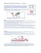
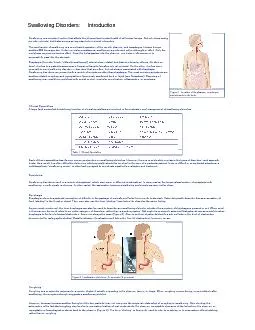
/
Figure 1. Location of the Figure 1. Location of the

Figure 1. Location of the - PDF document

liane-varnes
liane-varnes . @liane-varnes
Follow
392 views
Uploaded On 2015-11-08

Figure 1. Location of the - PPT Presentation

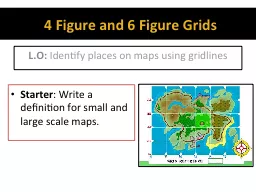
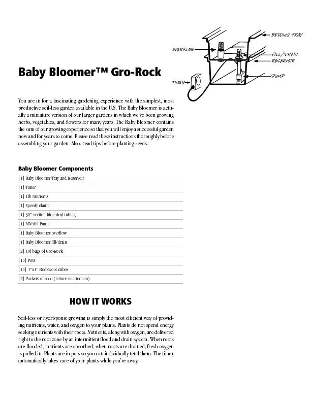
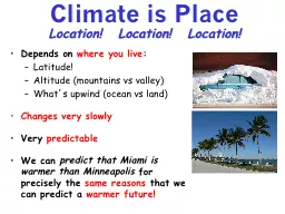
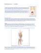
pharynx esophagusand stomach in the body Swallowing Disorders Introduction Swallowing is a complex function that affects the physical and mental health of all human beings Not only does ID: 187190

pharynx esophagus and stomach the

Share:

Link:

Embed:

Download Presentation from below link

Download Pdf The PPT/PDF document "Figure 1. Location of the" is the property of its rightful owner. Permission is granted to download and print the materials on this web site for personal, non-commercial use only, and to display it on your personal computer provided you do not modify the materials and that you retain all copyright notices contained in the materials. By downloading content from our website, you accept the terms of this agreement.


Presentation Transcript

Figure 1. Location of the pharynx , esophagus,and stomach in the body. Swallowing Disorders: Introduction Swallowing is a complex function that affects the physical and mental health of all human beings. Not only does eatingprovide nutrients, but it also serves as important role in social interaction.The mechanism of swallowing is a coordinated operation of the mouth, pharynx bolus passes into the pharynx , involuntary reflexes serve tosuccessfully pass it to the stomach. Dysphagia (from the Greek, "difficulty swallowing") refers to two related, but distinct, clinical problems. On the onehand, it refers to a patient's awareness of impaired transit of swallowed oral contents. On the other, dysphagia Swallowing disorders can present with a variety of symptoms other than dysphagia . The most common symptoms areswallow-related coughing and regurgitation of previously swallowed food or liquid (see Symptoms). Disorders ofswallowing may result from problems with neural control, muscular coordination, inflammation, or neoplasia. Clinical Specialties Table 1. Clinical Specialties Each of these specialties has it’s own unique perspective on swallowing disorders. However, there is considerable overlap in the types of disorders each specialtytreats. As a result, it is often difficult to determine which specialty should be involved in the care of a particular patient. In more difficult or complicated situations, amultidisciplinary "swallowing center" is often best Figure 2. Localization of stricture; A, accurate; B, proximal. CoughingCoughing is a nonspecific response to a variety of stimuli usually originating in the pharynx, larynx, or lungs. When coughing occurs during, or immediately afterswallowing, the symptom strongly suggests a swallowing problem.However, because humans swallow throughout the day, patients may not recognize the temporal relationsh Figure 3. A food bolus penetrating the airway. ChokingAs mentioned above (see Dysphagia and Coughing), "choking" is a term frequently used by patients (and occasionally by physicians) to describe the feeling of foodsticking in the esophagus or coughing. Although both symptoms may occur in patients with swallowing disorders, they imply somewhat different mechanisms ofdysfunction. In analyzing symptoms, it is therefore important to determine exactly what the patient means by the term.RegurgitationThe swallowing mechanism is designed to assure unidirectional movement of the swallowed bolus. "Regurgitation" refers to the return of food or liquid back to themouth or pharynx, after it has apparently successfully passed out of either region (Figure 4). Figure 4. Regurgitation of a food bolus. With regurgitation, material returns effortlessly to the mouth or throat. This contrasts with vomiting, during which nausea and retching are frequently present andcontraction of the abdominal muscles and diaphragm play an important role. When patients suggest that the regurgitated material tastes like ingested food, aswallowing disorder is usually present. Regurgitation of sour or bitter-tasting food or liquid suggests that at least some of the regurgitated material had reached thestomach. When sour or bitter regurgitation is present, the problem may fall within the spectrum of gastroesophageal reflux disease (GERD), rather than a swallowingdisorder.Nasal RegurgitationThe nasopharynx closes through a combination of soft palate elevation and contraction of the upper pharyngeal constrictor muscles (the superior pharyngealconstrictors). Failure of this closure mechanism, pharyngeal retention, or esophagopharyngeal regurgitation can result in nasal regurgitation (Figure 5). Figure 5. Nasal regurgitation of a food bolus. Other SymptomsDepending on the type of swallowing disorder, patients may present with a sore throat, hoarseness, shortness of breath, and chest discomfort or pain. Therelationship between swallowing and these symptoms may not be obvious. All of these symptoms may arise from a variety of other sources and none are specific toswallowing disorders. © Copyright 2001-2013 | All Rights Reserved.600 North Wolfe Street, Baltimore, Maryland 21287 Swallowing Disorders: Anatomy Physiology of SwallowingNormal swallowing requires the coordinated activity of the oral cavity, pharynx, and esophagus. A properly functioning swallowing mechanism provides efficient,unidirectional flow of the ingested bolus, while avoiding undesired diversion into the nasal cavity or respiratory tree.Between swallows, the pharynx and esophagus are at rest. The nasal cavity and larynx are in open communication with the pharynx permitting the individual tobreathe freely. The entrance to the esophagus remains closed by the upper esophageal sphincter (UES). This muscle serves to prevent the esophagus from filling withair during inspiration. At the lower end of the esophagus, the lower esophageal sphincter (LES) separates the esophagus from the stomach. The LES prevents refluxof irritating gastric contents into the esophagus. The mouth prepares the bolus for swallowing. The lips prevent drooling. The tongue positions all or part of the oral contents on its upper surface. The back of thetongue, palatine arch, and soft palate prevent premature spillage of the bolus into the pharynx. At the beginning of a swallow, as the tongue forces the bolus upagainst the hard palate and then back into the pharynx, the soft palate elevates and the posterior wall of the nasopharynx contracts to prevent nasal regurgitation.Penetration of the airway is prevented through a combination of events: inhibition of respiration, elevation of the larynx, approximation of vocal cords, and deflection ofthe epiglottis over the larynx. The presence of multiple mechanisms to prevent airway blockage suggests the importance of this function to the organism's well being.The failure of any one mechanism, such as the surgical removal of the epiglottis (epiglottectomy), usually does not result in aspiration of the swallowed bolus.Propulsion of the swallowed bolus is accomplished through a combination of a piston-like compression of the back of the tongue against the posterior pharyngeal wall,and a sequential wave of pharyngeal muscles (pharyngeal peristalsis) (Figures 6 and 7).As the swallowed bolus enters the pharynx, inhibition of neural stimulation results in the relaxation of the UES. This permits the swallowed bolus to enter theesophagus unimpeded. The peristaltic wave continues the length of the esophagus (esophageal peristalsis). Before the bolus arrives in the distal esophagus,cessation of firing of excitatory nerves and activation of inhibitory nerves relax the LES. This permits easy passage of the bolus into the stomach.The initiation of swallowing by the oral cavity is under voluntary control, whereas control of the pharynx and esophagus are involuntary. This means that once the initialsignal is received from the brain, the pharyngeal and esophageal phases of swallowing are carried out automatically. Initiation of swallowing is directed by thebrainstem, which integrates sensory information from the swallowing channel with information from the other areas of the brain. Integration signals are then sent backto the swallowing channel to initiate the act of swallowing (Figure 8). Figure 8. Central nervous system control of the pharyngeal phase of swallowing. Once initiated, the esophageal phase of swallowing can continue without central nervous system involvement, with the brain serving to modify esophageal function. © Copyright 2001-2013 | All Rights Reserved.600 North Wolfe Street, Baltimore, Maryland 21287 Swallowing Disorders: Causes OverviewThe mechanism of swallowing disorders may be either structural or motor. Structural disorders include luminal stenosis and diverticula. Motor disorders includeparesis (muscle weakness), sphincteric dysfunction, and spastic disorders. Structural Disorders Luminal StenosisLuminal stenosis occurs as a result of mechanical narrowing of the esophageal lumen in patients with esophageal strictures (Figure 9). Symptoms arise when theswallowed food is too large to pass. The typical symptom in a patient with an esophageal stricture is dysphagia for solid food, often followed by regurgitation ofundigested material. Figure 9. A, Esophageal stricture showing obstruction of food bolus; B, barium swallow. In general, patients can swallow with little difficulty. This remains true until the swallowing channel is narrowed by about 50%. Even beyond this point, frequency andseverity of symptoms may vary substantially, depending on the degree of narrowing, the presence of any associated motor dysfunction, and the choice andpreparation of food.DiverticulumA diverticulum is a pouch extending out from the normal wall of the swallowing channel. Diverticula (the plural of diverticulum) can develop in either the pharynx oresophagus (Figure 10). Although small diverticula may not cause symptoms, larger diverticula can cause dysphagia for liquids and solids. Regurgitation of undigestedfood, often hours after ingestion is a characteristic symptom of patients with diverticula. Figure 10. A, Zenker’s diverticulum; B, esophageal diverticulum. Motor Disorders ParesisPharyngeal weakness is commonly associated with neurological conditions, such as a cerebral vascular accident, amyotrophic lateral sclerosis (ALS), head and necktrauma, or brain surgery. Severe esophageal weakness is relatively rare. When it does exist, it is characteristically found in patients with certain types of collagenvascular disorders, especially scleroderma and mixed connective tissue disease. It is also found in patients with certain neurological and muscle conditions that canaffect the function of the smooth muscle of the GI tract.This condition may be asymptomatic. However, when the patient is symptomatic, dysphagia with liquids and solids usually occurs, often associated with regurgitation.Coughing during swallowing is common with pharyngeal weakness, because of aspiration due to pharyngeal retention or impaired laryngeal closure. Mild degrees ofweakness are not uncommon in the elderly, in patients with neurological conditions, and in patients with gastroesophageal reflux disease .Sphincteric DysfunctionThe upper esophageal sphincter (UES) and lower esophageal sphincter (LES) assure unidirectional flow of the swallowed bolus. However, they must relax duringswallowing so as not to pose a barrier to flow. The best-described disorder of sphincteric function is achalasia. This is a condition in which the failure of LES relaxation, associated with severe esophageal weakness, combines to retain food in the esophagus. Patients with sphincteric dysfunction typically complain ofdysphagia for liquids and solids (Figure 11). Figure 11. A, Manometric tracings in a normal patient; B, manometric tracing in a patient with achalasia Spastic DisordersWhereas spastic disorders of the pharynx are rare, those of the esophagus are common. A certain amount of motor dysfunction may be seen in normal individuals.Esophageal dysmotility may range from mild, infrequent events that are within normal limits, to profound incardination associated with every swallow. Figure 12. A, Diffuse esophageal spasm; B, barium swallow x-ray. Diffuse esophageal spasm (Figure 12) represents one extreme of esophageal dysmotility. However, the term is often used loosely to refer to any degree of dysmotilitynoted by barium or manometric studies (Figure 13). Figure 13. Manometric studies comparing normal and spastic tracings; A, normal; B, spastic. Neurogenic DysphagiaNeurogenic dysphagia is a general term used to describe any neurological or muscular condition that affects oral or pharyngeal motor function. Common causes ofneurogenic dysphagia include stroke (cerebral vascular accidents), amyotrophic lateral sclerosis (ALS), brain injury due to trauma or previous surgery, and local softtissue and nerve damage due to head and neck surgery. However, many disorders of the nervous system and muscles can cause neurogenic dysphagia. In somepatients, neurogenic-type radiographic abnormalities occur even when no diagnosable neurological or muscular disorder is present.Symptoms of neurogenic dysphagia generally include dysphagia and swallow-induced coughing. Nasal regurgitation, if present, strongly suggests a neurogenicprocess. Some patients fail to recognize the presence or severity of aspiration, possibly because of associated cognitive problems or impaired sensation. Patientsmay experience recurrent bouts of pneumonia due to swallowed saliva, liquid, and food entering the airway. The prognosis depends on the severity of the swallowingimpairment and the treatability of the underlying condition. Among the more treatable causes of neurogenic dysphagia are myasthenia gravis, polymyositis, and bothhyper- and hypothyroidism. Isolated UES Dysfunction Upper esophageal sphincter (UES) dysfunction refers to an impaired opening of the cricopharyngeal segment, which is also called the pharyngoesophageal segment(PE). This is a 1–2 cm region where the UES is located. Incomplete opening of the cricopharyngeal segment is a common radiographic finding on videoradiography(Figure 14). Figure 14. Radiographic image of cricopharyngeal dysfunction. Because the UES protects against esophagopharyngeal regurgitation, an incomplete opening may be a sign of the body’s attempt to prevent regurgitation in the faceof downstream obstruction due to esophageal disorders. Alternatively, impairment of the PE opening may reflect inadequate force of bolus propulsion due topharyngeal weakness in neurogenic dysphagia. Therefore, radiographic evidence of a "tight" PE, may point to pharyngeal or esophageal dysfunction. UESdysfunction must be interpreted with caution. Isolated UES dysfunction (referred to as cricopharyngeal spasm or cricopharyngeal achalasia) refers to failure of the PE opening without evidence of pharyngealparesis or evidence of esophageal disease. In patients with this condition, dysphagia is caused by obstruction at the level of the PE, due to either fibrosis (scarring) ofthe PE or UES dysfunction. Failure of UES relaxation may be documented by manometry. Zenker’s and Esophageal DiverticulumA diverticulum is a sac-like protrusion found in the normal swallowing channel. Diverticula may result from inflammatory or neoplastic processes outside the channel.As these processes evolve, they pull on the adherent wall of the channel, forming the diverticulum. This type of diverticulum is referred to as a traction diverticulum.Traction diverticula most often occur in the midesophagus as a result of inflammation in the mediastinum. Today, most diverticula are due to a different pathogenesis.They appear to result from gradual weakening of the wall of the swallowing channel as a result of abnormally increased intraluminal pressure (pulsion diverticula). Figure 15. A, Zenker’s diverticulum; B, barium x-ray study. Zenker's (hypopharyngeal) diverticulum is an abnormal, sac-like protrusion on the wall of the lower pharynx (the hypopharynx) (Figure 15). It develops along theposterior pharyngeal wall, just above the upper esophageal sphincter. This area of the pharynx is a region of relative weakness because of the absence of anycovering muscle layer. However, this area of weakness is a normal feature of human anatomy and does not in and of itself account for the development of thediverticulum. The primary cause of a Zenker's diverticulum appears to be abnormal relaxation of the upper esophageal sphincter. As a result, the pharyngeal pressuregenerated during swallowing is increased. Over time, the pharyngeal wall begins to bulge with each swallow until a diverticulum develops and persists betweenswallows.Pulsion diverticula may be asymptomatic when small. When they grow, however, they retain liquid, food, and regurgitated matter. A patient may wake up coughing,only to find food, recognizable as portions of the meal eaten hours before, in the mouth or on the pillow.Diverticula may occur in the esophagus as well as the pharynx (Figure 16). Most esophageal diverticula result from abnormal intraluminal pressure as a consequenceof increased downstream resistance. This may be due to a stricture, esophageal spasm, or abnormal lower esophageal sphincter function. Figure 16. A, Esophageal diverticulum; B, barium swallow x-ray; C, endoscopic view Esophageal StenosisA stenosis refers to any point of narrowing of a channel that results from a structural abnormality, as opposed to a motor disorder. Although the term stricture is oftenused to describe the same thing, stricture more specifically refers to a narrowing that results from a thickening of the wall of the swallowing channel. Rings, webs,inflammatory strictures, or tumors may also narrow the lumen. Stenosis tends to cause more difficulty in swallowing solids than liquids, but dysphagia for solids only isstrongly suggestive of a stenosis. The major difference among the different types of stenotic lesions is the rate of progression of symptoms.Esophageal rings or webs refer to a short, ring-like band protruding into the lumen. These rings or webs are typically composed of a fold of the lining of the esophaguswithout any muscle tissue. They may be congenital or acquired, and may occur anywhere in the pharynx or esophagus. The most common is the Schatzki's ring(Figure 17), which by definition occurs at the junction of the esophagus and stomach. Named for the radiologist who described it, Schatzki’s ring is very common andonly causes symptoms when it narrows the channel by about 50%. A ring-like band located elsewhere in the pharynx or esophagus is often called a web; thedistinction is relatively arbitrary. Webs and rings typically cause intermittent dysphagia for solids, which may remain stable or progress slowly over time. Figure 17. A, Schatzki’s ring; B, barium swallow x-ray; C, endoscopic Typically the patient describes intermittent dysphagia of sudden onset, separated by symptom-free periods (without swallowing difficulty). Symptoms are oftengreatest with tough anddifficult-to-chew foods. Patients may describe particular problems with foods generally considered to be soft, such as pasta or bread. Symptoms are more likely tooccur when the patient is eating hurriedly, is offered a limited choice of foods (e.g., when dining out), or is distracted by conversation.Gastroesophageal reflux disease (GERD) is the most common cause of inflammatory strictures. Inflammation resulting from prolonged contact with irritating gastriccontents causes scarring. Thickening of the esophageal wall narrows the channel, producing the dysphagia for solids characteristic of an esophageal stenosis.Esophageal strictures tend to be longer than rings or webs (Figure 18). Unless the cause of inflammation has been removed, the surface of the stricture may appearinflamed and even ulcerated in some cases. Figure 18. A, B, Normal esophagus compared with erosive esophagitis; A’, B’, endoscopic views Unlike webs and rings, dysphagia from an inflammatory stricture tends to grow steadily worse over time. The intervals between symptomatic episodes becomeshorter and the variety of foods that cause symptoms increases over a period that can often be measured in months rather than years. Symptoms may improve whenthe cause of inflammation is removed (e.g., by effective treatment of reflux disease) and as the swelling associated with inflammation decreases. However, even witheffective treatment, symptoms may continue to progress as the scarring increases. With complete eradication of inflammation, scarring gradually stops and the size ofthe channel stabilizes.A final cause of esophageal stricture is infiltration of the esophageal wall by a tumor. A tumor may also grow into the channel, further narrowing the lumen. Comparedwith an inflammatory stricture, malignant strictures tend to progress rapidly, often causing dysphagia for liquids as well as solids within a few months of onset of symptoms (Figure 19). Rapidly progressive dysphagia for solids is a cause of concern and requires early evaluation. Figure 19. A, esophageal cancer; B, barium swallow x-ray; C, endoscopic view. Diffuse Esophageal SpasmEsophageal dysmotility refers to any abnormality in the coordination or strength of esophageal contractions. Diffuse esophageal spasm (Figure 20) refers to severedegrees of esophageal dysmotility that may produce symptoms of dysphagia and chest pain. Figure 20. A, Barium swallow x-ray showing diffuse esophageal. stricture; B, manometric tracing. Dysphagia typically occurs with both liquids and solids. Chest pain — which may simulate cardiac pain — may be swallow-induced, but may also be unrelated toswallowing. Even when severe, esophageal dysmotility, does not always produce symptoms.Esophageal spasm is often diagnosed on the basis of clinical symptoms alone. Documentation of abnormal motility may be difficult to obtain because esophagealspasm can be intermittent and therefore may not be detected on short duration studies such as barium x-ray or esophageal manometry. Some degree of abnormalmotility occurs in normal asymptomatic volunteers, and the line between esophageal spasm and abnormal contractions that can occur in normal individuals is poorlydefined.Esophageal dysmotility (including spasm) may result from a variety of causes including downstream obstruction, esophageal irritation, or neurological diseasesaffecting the autonomic nervous system, which controls much of the gastrointestinal function. When no cause has been determined, the condition is referred to asidiopathic esophageal spasm, but this is a relatively rare phenomenon. The most common cause of esophageal spasm is gastroesophageal reflux disease (GERD).It is important to look for reflux and a stenotic lesion in anyone with symptoms suggestive of esophageal spasm because the treatment of these secondary spasticconditions is different from that of idiopathic spasm. Benign esophageal stenosis is usually treated by esophageal dilation. Reflux is treated with dietary measuresdesigned to avoid stimulation of gastric acid and to decrease the volume of gastroesophageal regurgitation. These measures are usually combined with drugs thatdecrease acid secretion and improve gastric emptying. Idiopathic spasm, however, may be treated with drugs (smooth muscle relaxants) that decrease the strength ofesophageal contractions. Unfortunately, smooth muscle relaxants are often ineffective for the treatment of esophageal spasm.AchalasiaAchalasia is a motor disorder of the esophagus without an obvious etiology. In achalasia, nerve cells (myenteric plexus) located between the esophageal musclelayers are damaged (degenerate). The result is a complete loss of coordinated esophageal contractions (peristalsis) and failure of lower esophageal sphincter (LES)relaxation (Figure 21). This combination produces obstruction at the esophagogastric junction and loss of effective propulsion. Figure 21. A, Anatomic findings in achalasia; B, endoscopic image; B, radiographic image. The mean age for the onset of achalasia ranges between 30–60 years, with a peak in the 40s. The prevalence appears to be about 8–12 per 100,000 population. Thedisorder is more prevalent in men than women with a ratio of about 2:1.The initial clinical presentation of achalasia is characterized by solid food dysphagia. Patients complain of fullness in the chest while eating, with a filling or overflowsensation as the meal progresses. Food is unable to pass effectively into the stomach and piles up above the LES, resulting in the gradual dilation of the esophagus.This causes dysphagia for liquids and solids and regurgitation of recognizable food, often many hours after ingestion. This form of regurgitation is almost alwaysfound in patients with hypopharyngeal or esophageal diverticulum or achalasia. Patients may also complain of substernal chest pain and weight loss.In most instances, the esophagus becomes dilated, and sometimes tortuous probably as a result of neuropathic changes in the esophagus and the pressure exertedon the esophageal wall by retained food. endoscopic ultrasound (EUS) studies reveal a thickened lower esophageal sphincter (31 mm as compared 22 mm) andthickened muscularis propria. © Copyright 2001-2013 | All Rights Reserved.600 North Wolfe Street, Baltimore, Maryland 21287 Swallowing Disorders: Diagnosis Objective TestsThe clinical examination, particularly the patient's description of the nature and history of their symptoms, should always be the initial step in the evaluation ofswallowing disorders. The patient's self-report often suggests both the type of disorder most likely responsible for the complaints and the objective tests required todetermine, or verify, the specific cause. Minor abnormalities of the swallowing mechanism are frequent, especially in older patients. They must not beover-interpreted. The clinical examination offers a check against objective findings and allows for comparison and interpretation.The following objective tests are commonly used in the evaluation of patients with symptoms of swallowing disorders: Barium Radiography, Modified Barium Swallow,Radionuclide Clearance Studies, Pharyngoscopy, Esophagoscopy, Manometric Studies, and Continuous (24 Hour) pH Monitor. Barium RadiographyExcept when re-evaluating a patient with an established diagnosis, barium x-ray studies are usually the first step in the evaluation of swallowing disorders. Thesestudies permit evaluation of the entire swallowing channel (mouth, pharynx, and esophagus). Even when the location of the abnormality is suggested by the clinicalhistory, barium radiography allows assessment of both the structure and function of the swallowing mechanism. When symptoms suggest an esophageal condition, astandard barium swallow (often performed as part of an upper GI [UGI] series) is generally adequate. However, the flow of barium through the pharynx is too rapid forthe radiologist to record important details of swallowing. Therefore, when a pharyngeal condition is suspected, a video-barium study (videopharyngoesophagram,during which the flow of barium through the pharynx and esophagus is recorded on a videotape recorder) is essentialModified Barium SwallowA modified barium swallow is a variant of the video-barium study in which the effect of various maneuvers on the efficiency of swallowing is evaluated. Maneuversmay include modifications in the type of swallowed bolus, in head or body position, and in the timing of swallowing in relationship to respiration. Primarily designed toevaluate the effect of therapeutic maneuvers on patients with established neurological or post surgical disorders of oral and pharyngeal function, the modified bariumswallow is sometimes used as the first test in patients in whom these disorders are strongly suspected. However, those who specialize in this procedure (most oftenspeech language pathologists with special training in the field of swallowing) may have limited familiarity with structural disorders of the pharynx and even less withradiological abnormalities of the esophagus. In general, the modified barium swallow should be used only after a detailed diagnostic video-barium study has beenperformed under the supervision of an experienced radiologist, or as a combined study in which the swallowing therapist and radiologist collaborate.Radionuclide Clearance StudiesDuring esophageal clearance studies a small amount of radioactive material is mixed with liquid or solids. These studies provide important information about thephysiology of swallowing and reflux disease; however, their use is limited in clinical practice. The major clinical application of radionuclide clearance studies is forachalasia, in which esophageal weakness combines with failure of the lower esophageal sphincter relaxation, causing retention of food in the esophagus. Theesophageal clearance study is used to quantify the severity of retention and to determine the extent of improvement after treatment (Figure 22). Figure 22. Esophageal cornflake study in a patient with achalasia; A, before treatment; B, aftertreatment. PharyngoscopyPharyngoscopy is a standard component of the otolaryngologist's (head and neck physician) swallowing evaluation and is usually performed without sedation during aclinic visit. Pharyngoscopy involves the visual examination of pharynx and larynx ( but not the esophagus), using either a specially designed mirror (indirectpharyngoscopy) or a flexible telescope (flexible pharyngoscope). Pharyngoscopy can detect inflammation, tumors, or abnormal function of the pharynx or larynx(Figure 23).  \n A, A’, Normal vocal cords; B, B’, nodular vocal cords; A’,B’, endoscopic views. EsophagoscopyEsophagoscopy involves visual examination of the esophagus, using either a rigid or flexible endoscope. Because rigid esophagoscopy requires anesthesia andinvolves more discomfort and risk, flexible endoscopes are generally used for swallowing evaluations except in specific instances. Flexible endoscopes allowexamination of the stomach and first part of the small intestines (duodenum) as well as the esophagus. Endoscopy is the best test for the evaluation of esophagealmucosal abnormalities, such as esophagitis (Figure 24). It is also a good way to evaluate conditions that narrow the esophagus (esophageal strictures), although mildto moderate strictures may be missed. Endoscopy is a poor means of evaluating esophageal function, but it does play a supplemental role in assessing specificconditions that may cause such motor abnormalities. Figure 24. A, A’, Normal esophagus; B, B’, erosive esophagitis; A’,B’, endoscopic views Endoscopy is the primary means of obtaining tissue for microscopic analysis. Although the endoscopist may examine the pharynx during intubation, endoscopes usedfor upper endoscopy are somewhat thicker and less flexible than those developed for the pharynx. Additionally, gastroenterologists perform most upper endoscopies;generally their training and experience with pharyngeal disease is limited. Therefore, upper endoscopy does not substitute for pharyngoscopy when the latter isindicated. Manometric StudiesManometric studies evaluate pressure changes that occur during swallowing. The test involves use of a thin, flexible catheter that is passed through the nose ormouth into the esophagus. The tube has a series of pressure sensors attached to, or imbedded into, specific locations throughout. These pressure sensors permitassessment of the strength and coordination of the pharyngeal and esophageal peristalsis wave and the strength and relaxation function of the upper and loweresophageal sphincters (Figure 25). Figure 25. Esophageal peristalsis with corresponding manometric tracing. The test has been used extensively for the evaluation of esophageal motor disorders, particularly when barium studies are unremarkable or when x-ray findings raisequestions about motor dysfunction. Manometry is also used to properly position pH probes for the evaluation of gastroesophageal reflux disease (see below). Finally,it is used preoperatively in patients for whom antireflux surgery is planned to help determine the appropriateness and approach to surgery. A special variant of thetest, in which the pressure catheter is attached to a small computer or to record data, permits continuous recording of esophageal pressure for an extended period (upto 24 hours). This is occasionally used for patients with suspected esophageal spasm in whom standard manometric studies have proven unrevealing. Pharyngealmanometry can be used for the evaluation of pharyngeal and UES function. This requires special equipment because the speed of pharyngeal pressure changesduring swallowing exceeds the capacity of the usual equipment used for esophageal manometry. The equipment used for pharyngeal manometry is not available in allesophageal manometry laboratories. The role of manometry in the evaluation of pharyngeal dysfunction is not as well established as that for esophageal motordisorders. Continuous (24 Hour) pH MonitorpH monitoring permits evaluation of gastroesophageal reflux disease (GERD). pH monitoring involves the placement of a thin, flexible catheter with an acid-sensitivetip through the nose into the esophagus (Figure 28A). A short version of this test (20–30 minutes) has been available for decades. The procedure was later adaptedfor the evaluation of reflux prolonging the test over extended periods of time —up to 24 hours (24-hour, or continuous pH monitor study) (Figure 26 B,C). ContinuouspH monitoring has played a critical role in advancing our understanding of GERD. It is considered the best test for the evaluation of GERD and permits determinationof the presence and severity of reflux. Figure 26. A, pH monitor placement. Inset shows the catheter position in the esophagus; B, normal pHmonitor tracing; C, abnormal continuous pH monitor tracing. Continuous (24-hour) pH monitoring has demonstrated that most normal individuals (nonrefluxers) actually reflux on a daily basis. Therefore, gastroesophageal refluxdisease implies not just the presence of reflux, but reflux in excess of that experienced by nonrefluxers. © Copyright 2001-2013 | All Rights Reserved.600 North Wolfe Street, Baltimore, Maryland 21287 Swallowing Disorders: Therapy OverviewThe treatment of patients with swallowing disorders should be individualized. The symptoms along with the physical and emotional impact upon quality of life shouldbe assessed and considered in the development of a treatment strategy. To date, relatively small numbers of well-controlled clinical trials have been conducted;therefore, experience, rather than analysis of data, is often the basis for treatment.Reassurance / Psychological InterventionsAlthough swallowing disorders are almost always caused by structural abnormalities or motor dysfunction, the severity of symptoms occasionally appear to exceedwhat would be expected. Swallowing disorders are rarely a result of psychological factors alone; anxiety may play a role in exaggerating the severity of symptoms,especially if the patient is concerned about the possibility of cancer or heart disease (if chest pain is a prominent associated symptom). Clinical reassurance alone isgenerally not effective and is often ill advised if it takes the place of an appropriate evaluation. However, after the evaluation is performed, symptom severity mayimprove with reassurance. Formal psychological intervention, including counseling and biofeedback, may help patients who fail to respond adequately to reassurancealone. MedicationsMost causes of neurogenic dysphagia have no specific therapy. However, it is important to detect those for which drug therapy is available (e.g., myasthenia gravis,polymyositis, Parkinson's disease, and both hyper- and hypothyroidism).A number of different drug therapies, administered for varying lengths of time, for patients with non-achalasia motility disorders have been utilized with differingdegrees of success. However, very few of the studies have been well-controlled. Nor have they included large numbers of patients examined over time. Despite theseshortcomings, several drugs have shown utility in well-controlled studies.Short-acting nitrates have been shown to be effective in the relief of chest pain and dysphagia within minutes. The duration of action is approximately 30 minutes.Short-acting nitrates have been used in patients with diffuse esophageal spasm. These drugs are often used for rapid treatment of predictable patterns of symptoms.Anticholinergic drugs such as hyoscyamine, atropine, cimetropium bromide, and propantheline have been associated with a decrease in peristalsis. These drugs,when compared to placebo, have demonstrated a reduction in symptoms in patients with non-achalasia motility disorders. Further studies of this class of drugs arewarranted.Calcium channel blockers have demonstrated utility in reducing lower esophageal sphincter pressure and the amplitude of esophageal contractions. Nifedipine, indoses of 10–30 mg 3 times per day, has been used in esophageal motility disorders and has shown to decrease the frequency, amplitude, and duration of chest painattacks when compared to placebo. Diltiazem has also been shown to decrease the amplitude of pain, lower esophageal sphincter pressure, and impact the durationof esophageal contractions in patients in placebo-controlled trials.Experimentally induced stress can result in considerable changes in esophageal peristalsis. Sedatives and tranquilizers have been shown to be helpful in patientswith non-achalasia esophageal motility disorders accompanied by anxiety disorders and depression. Sedatives and tranquilizers such as trazodone, imipramine, andalprazolam may be useful in this group of patients with symptoms of chest pain and/or dysphagia. Swallowing RetrainingSwallowing therapy is commonly recommended for neurogenic dysphagia and postoperative pharyngeal dysfunction and is most often performed by a speech-language pathologist with expertise in this area. Swallowing therapy employs modifications of bolus volume and consistency, head and neck position, and swallowingrelated maneuvers to improve swallowing efficiency. There are also exercises that have been developed to help selected patients with these types of disorders. Themodified barium swallow is often used to determine the effect of potential interventions on the bolus transit and airway penetration. The information gathered from thisthe modified barium swallow may serve as the basis of treatment recommendations. Botulinum ToxinBotulinum toxin has been shown to improve symptoms in patients with achalasia. Success in this group has prompted clinical trials in patients with non-achalasiaesophageal motility disorders who were unresponsive to drug therapy. The clinical trial group demonstrated short-term relief with intrasphincteric injection of botulinumtoxin. DilationDilation utilizing any number of available dilator systems is used to increase the diameter of the swallowing channel. This procedure, usually performed at the time ofan endoscopy, is very successful for webs, rings, and benign strictures. Re-stenosis is relatively common, especially in inflammatory strictures. However, effectivetreatment of the underlying inflammation will decrease the need for repeat dilations (Figure 27, 28, 29). Figure 27. Endoscopic dilation. Technique for Savary dilation of an esophageal stricture. Figure 28. Endoscopic dilation. Technique for Maloney dilation of an esophageal stricture. Figure 29. Endoscopic dilation. Technique for through-the-scope (TTS) dilation of an esophagealstricture. In patients with achalasia, abrupt inflation of large diameter balloon dilators has been used for decades to weaken the lower esophageal sphincter and allow food topass by gravity. Dilation of the upper esophageal sphincter has been successful in patients with isolated upper esophageal dysfunction, and in selected patients withneurogenic dysphagia. Enteral FeedingSome patients with swallowing disorders are so impaired that they are unable to take adequate nutrition safely by mouth. This is especially true in patients withneurogenic or postoperative dysphagia. Enteral feeding, most often provided by an endoscopically placed gastrostomy tube, provides a means of assuring adequatenutrition and decreasing the severity of aspiration. At the same time, measures are taken to correct the swallowing problem.Enteral feeding should be considered for any patient who has lost substantial amounts of weight or has had recurrent aspiration pneumonia due to swallowingdisorders, unless the initial evaluation indicates that the problem can be substantially improved in a short period of time. Patients are often reluctant to accept agastrostomy. The physician must take the time to explain that malnutrition and excessively prolonged meals have a serious impact on the quality of a patient's life. Itmust also be explained that the gastrostomy tube does not prevent further efforts to treat the swallowing problem and it can be removed if and when the patient'sability to swallow, and consumption of an adequate number of calories, safely improves. Structural LesionsStructural lesions may be treated surgically and endoscopically. The goal of therapy is mechanical disruption. Myotomy is often used for sphincteric dysfunction.Myotomy reduces the resting sphincter tone by 50% without completely abolishing it by increasing sphincter opening capacity and reducing resistance totranssphincteric flow. Myotomy of the lower esophageal sphincter (Heller myotomy) is an effective alternative to dilation for achalasia. The major limitations are thediscomfort associated with the operation itself and a relatively high risk of postoperative gastroesophageal reflux. Many surgeons add an anti-reflux operation to themyotomy to prevent reflux. Recent techniques for laparoscopic and thoracoscopic myotomy have been developed and have become the preferred approach to thesurgical treatment of achalasia. Myotomy of the circopharyngeal muscle (the major component of the upper esophageal sphincter) is commonly used as part of thesurgical approach to diverticulectomy for Zenker's diverticula. It is also occasionally used for isolated upper esophageal sphincter dysfunction (a rare condition). Itsuse in patients with neurogenic dysphagia remains controversial. Esophageal StenosisAvoiding offending foods, choosing foods prepared in creamy sauces, and eating slowly with careful attention to small bites and chewing may minimize the frequencyof symptoms of esophageal stenosis.Dysphagia from an inflammatory stricture requires the source of inflammation to be removed (e.g., by effective treatment of reflux disease) thereby decreasingassociated swelling. With complete eradication of inflammation, scarring gradually stops and the size of the channel stabilizes.Methods of mechanical disruption such as myotomy or dilation (refer to Figures 27–29), are efficacious in the treatment of esophageal webs and stenoses wherethere is preserved contractility. Almost half of the patients who undergo dilation will require repeat procedures over the course of many years.Zenker’s DiverticulumSmall Zenker's diverticulum do not require treatment intervention other than periodic esophagrams. In cases where treatment is required, surgical and endoscopicalternatives are available (Figure 31). Figure 31. Posterior view of Zenker’s diverticulum. Treatment involves relieving the source of resistance by cutting the upper esophageal sphincter (cricopharyngeal myotomy) and removing the diverticulum(diverticulectomy). Diverticulectomy with (Figure 32) or without myotomy (Figure 33) is the traditional approach to the treatment of Zenker's diverticulum. A one-stagepharyngoesophageal diverticulectomy with or without myotomy has been shown to provide excellent results with low mortality (1.2%), morbidity (8%) and recurrencerates (3.6%). Figure 32. Technique for diverticulectomy with myotomy. Figure 33. Technique for diverticulectomy without myotomy. The surgery is performed through a left cervical incision. The diverticulum is retracted, dissected, and resected with sutured or stapled closure. Myotomy, dissection ofthe cricopharyngeal muscle fibers away from the mucosa, may be performed before or after diverticulectomy. Long-term follow-up studies are needed to assessphysiologic consequences of these procedures. Complications such as infection, vocal cord paralysis, fistulas, and aspiration may result from surgical intervention.Recently, techniques for the endoscopic approach to treatment of a Zenker's diverticulum have been developed. In these procedures, the wall between thediverticulum and the esophageal lumen is divided, thereby cutting the upper esophageal sphincter and allowing swallowed food and liquid to decant out of theunroofed pouch. Modifications of this technique using laser versus electrocoagulation have shown comparable results, though the laser procedure appeared toproduce less pain and faster recovery. Whether this represents a more efficacious procedure than the traditional transcutaneous diverticulectomy is still uncertain.Endoscopic management of Zenker's is not popular in the US. Most surgeons perform myotomy with diverticulectomy. Esophageal DiverticulumSmall diverticula are often asymptomatic and should prompt further evaluation for an alternative cause of symptoms. However, when symptoms do arise from adiverticulum, the condition is often treated by surgical removal (diverticulectomy). Diverticula are usually a consequence of chronic obstruction downstream from thediverticulum. Unless the obstructive lesion is found and treated, symptoms may not improve with diverticulectomy.Midesophageal diverticula are often asymptomatic and do not require treatment. However, if treatment does become necessary it is important to exclude esophagealmotility disorders before recommending surgery. Though surgery is rarely indicated, diverticulectomy with or without myotomy is the procedure of choice.Epiphrenic diverticula, arising near the diaphragm, usually do not require treatment. However, in symptomatic patients the goal of therapy should be aimed atremoving the cause of abnormal resistance to avoid further enlargement of the diverticulum. Diverticulectomy alone or with myotomy has shown favorable results insymptom relief, weight gain, and absence of recurrence.Neurogenic DysphagiaA multidisciplinary approach to the treatment of neurogenic dysphagia is often needed for optimal patient management. Since neurogenic dysphagia may be thepresenting or accompanying symptom of many systemic diseases, the strategy is to treat any underlying disease whenever possible. Structural, neuromyogenicmechanisms need to be identified by use of radiographic and endoscopic evaluation; treatment is dictated by these assessments. The risk of aspiration pneumoniaalso needs to be assessed. If this risk is high, gastrostomy feeding should be instituted. Local therapy should be considered after structural lesions, treatableunderlying disease, and the safety of oral feeding have been established.Modification of the diet, swallowing therapy, and surgery (singularly or in combination), constitute therapeutic options. The treatment for neurogenic dysphagiaemphasizes measures to improve the efficiency of swallowing to minimize aspiration. Swallowing retraining, most often under the direction of a specially trainedspeech-language pathologist or occupational therapist, is an important method of intervention. Swallowing technique is modified improving the speed and range ofmovement of oropharyngeal muscles. The mechanics involved in swallowing are selectively modified to facilitate flow and minimize aspiration. In select patients,when objective testing suggests that the upper esophageal sphincter is contributing to the severity of swallowing problems, therapy directed at weakening the upperesophageal sphincter (myotomy) might be helpful. When adequate nutrition cannot be maintained safely, alternative methods of feeding—most often a feeding tubeplaced directly into the stomach—may be advisable.Isolated UES DysfunctionThe severity of symptoms in isolated upper esophageal sphincter dysfunction often responds to treatment directed at weakening the lower esophageal sphincter(using, for example, dilation or myotomy). These measures work only occasionally in patients with neurogenic dysphagia, even when the cricopharyngeal segmentappears to open poorly. Diffuse Esophageal SpasmPatients with symptoms suggestive of esophageal spasm require assessment to determine if reflux or stenotic lesions are playing a role. These secondary spasticconditions differ from idiopathic spasm. Benign esophageal stenosis is usually treated by esophageal dilation. Reflux is treated by dietary measures designed to avoidthe stimulation of gastric acid and to decrease the volume of gastroesophageal regurgitation. These measures are usually combined with drugs that decrease acidsection and improve gastric emptying. Idiopathic spasm is treated with drugs (smooth muscle relaxants) that decrease the strength of esophageal contractions. Often,however, smooth muscle relaxants are ineffective for the treatment of esophageal spasm. Recent studies suggest that patients with symptoms of esophageal spasm,especially chest pain, have a sensory disorder in which the threshold for perceiving esophageal stimulation as a painful phenomenon is decreased. The implication ofthis sensory disorder for treatment of spasm-like chest pain remains unclear. AchalasiaDilation of the obstructed distal esophagus is the focus of achalasia treatment. Treatment involves decreasing the pressure in, and thereby the resistance to, flowthrough the lower esophageal sphincter.Dilation is probably the most common approach to treating the patient with achalasia. Pneumatic dilation is performed endoscopically (Figure 34). Patients aresedated with intravenous medications. Esophageal lavage and aspiration of gastric contents are performed prior to positioning the endoscope in the antrum. A stiffguide wire is advanced through the biopsy channel of the scope and positioned in the antrum (Figure 34A). The dilator is passed over the guide wire and advancedthrough the gastroesophageal junction. The balloon is slowly inflated and held in position. Gradual increase in the size of the dilator may be necessary (Figure 34B).The objective is to stretch the circular muscle of the lower esophageal sphincter to allow the passage of solids and liquids without subsequent reflux or perforation(Figure 34C,D). Figure 34. Technique for pneumatic dilation of achalasia. Controversy exists regarding the efficacy of using rapid, maximal, short duration stretches compared with rapid, submaximal long duration stretches. Most patients(65%–80%) report a significant improvement in their symptoms of dysphagia. Physiologically lower esophageal pressures decrease immediately after pneumaticdilation, but increase over time with a concomitant increase in symptoms.Recently, intrasphincteric injection of the potent neurotoxin botulinum toxin has been successfully used in patients with achalasia. Safe and effective in the majority ofpatients, it is particularly effective in the elderly and has earned a place in the management of patients considered poor candidates for dilation or myotomy. Theprocedure involves injection of the lower esophageal sphincter causing a chemical denervation of the sphincter. Twenty to 25 units of botulinum toxin are injected intoeach quadrant of the lower esophageal sphincter with a sclerotherapy needle passed through the endoscope (Figure 35). Although it is the safest of availabletechniques, botulinum toxin injection has a limited duration of effect, lasting on average one year. Repeat treatment is necessary to maintain the effect. Some patientsmay experience mild chest pain and there have been reports of skin rashes noted after treatment. Figure 35. Technique for intrasphincteric injection of botulinum toxin for achalasia. Minimally invasive surgery, using either a laparoscopic or a thorascoscopic technique has significantly decreased the morbidity associated with achalasia surgery. Asingle anterior lateral myotomy (Figure 36) or a modified Heller myotomy is typically the surgical procedure performed. A lower esophageal sphincter myotomy incisesenough muscle to relieve symptoms but not enough to result in gastroesophageal reflux. In many cases, an antireflux procedure is performed at the same time. Figure 36. Surgical technique for single anterior lateral myotomy. OverviewThe inability to obtain adequate nutrition results in weight loss and easy fatigability. Difficulty swallowing can lead to a voluntary decrease in food intake andalterations in diet that can further impair nutrition. Aspiration can lead to pneumonia, bronchiectasis, and life-threatening pneumonia (aspiration pneumonia). Theseproblems are correctable, either by treatment of swallowing disorders or by provision of adequate calories through an alternative route.Treatment may have its own complications. All drugs have the potential for producing side effects—some allergic and others dose-related. Dilation may result inperforation or less often, gastrointestinal bleeding. These complications are relatively rare with dilation for benign strictures, but are more common if the dilation isperformed for malignant strictures or achalasia. The risks of radiation therapy are discussed in the section on esophageal cancer. Surgery carries risks associatedwith anesthesia and the specific surgical procedure. © Copyright 2001-2013 | All Rights Reserved.600 North Wolfe Street, Baltimore, Maryland 21287