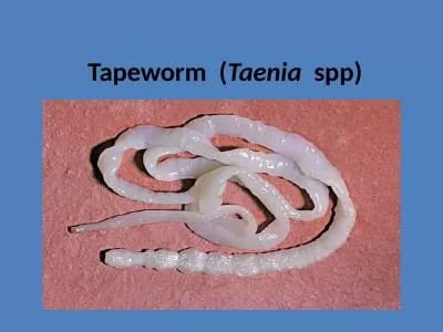
PGGCG11 Chandigarh Classification Tapeworm infestation is the infection of the digestive tract by adult parasitic flatworms called cestodes or tapeworms Live tapeworm larvae coenuri ID: 775318
Download Presentation The PPT/PDF document " Taenia Dr. Shivani Gupta," is the property of its rightful owner. Permission is granted to download and print the materials on this web site for personal, non-commercial use only, and to display it on your personal computer provided you do not modify the materials and that you retain all copyright notices contained in the materials. By downloading content from our website, you accept the terms of this agreement.
Slide1
Taenia
Dr.
Shivani
Gupta,
PGGCG-11,
Chandigarh
Slide2Classification
Tapeworm infestation
is the infection of the digestive tract by adult
parasitic
flatworms called
cestodes or
tapeworms.
Live tapeworm larvae (coenuri)
are
sometimes
ingested by
consuming
undercooked food. Once inside the
digestive
tract,
a larva can grow into a very large adult
tapeworm.
Additionally, many
tapeworm larvae cause
symptoms
in an
intermediate
host. For
example,
cysticerciosis is a disease of
humans
involving larval
tapeworms
in the
human
body
.
Kingdom: Animalia
Phylu
m
:P
l
a
t
yhe
l
m
in
t
hs
Class:
Cestoda
Sublass:
Eucestoda
Slide3Major
Attributes:
Endoparasitic.
Acoelomates
Body
is
covered
by
tegument.
Anterior
end
is
a
scolex.
Body
segments
called
proglottids.
Hermaphroditic.
Adult
tapeworm
in
intestine
Slide4Morphology
There are three
main
features to a
tapeworms
external
morphology:
the strobila
(proglottids)
the scolex and the
tegument.
1.Strobila/Proglottids
-unique
among the
metazoa
-a
linear
series of
sets
of reproductive organs
of
both sexs - each
is
referred to as a
genitalium
-the area around the genitalium is a
proglottid
-tapeworms
with
mulitple
proglottids
are
describes as being
polyzoic
-new
proglottids
are continuously
differentiated
near the anterior end in a process
called
strobilation - as each segment moves from the anterior end to the posterior end a new one takes it place-the new proglottids are produced in an undifferentiated zone between the scolex and the strobila, called the neck - this area contains stem cells which give rise to the new proglottids
Slide5Morphology
-
while the
segments
travel down
the
length of the worm they sexually
mature,
by the
time
they reach
the
end of the worm the
genitalia
have already copulated and
produced
eggs
-a proglottid can
mate
with
itself,
with others in the
same
worm
or
with another worm entirely (it depends on the species of
tapeworm)
-a
segment containing
eggs is
called
agravid-when the gravid reaches the end of the worm, it detaches and passes out of the host in the host feces-from here the eggs can be ingested by a new host, leading to tapeworm infection
Hooks of
tapeworm
Mature
Proglottids
Slide6Morphology
Mature
proglottid
Scolex
and anterior
strobila
Slide7Morphology
2.
Scolex:
-The tapeworm body consists of an anterior, head-like scolex and the trunk, or strobila, consisting of a linear series of
segments,
or proglottids. The scolex
is
wider than the
strobila
to which it joined by a narrow
neck
.
-The scolex attaches the worm to the gut wall of
the
host. For this purpose it
has
a
retractable
rostellum
armed with
two
rings of
rostellar
hooks
. Just
posterior to
the rostellum
is
a ring of four
suckers
; two
dorsolateral and
two
ventrolateral.
-
T
wo lateral nephridial canals may be visible on each side of the scolex. They connect with each other near the rostellum via a set of convoluted scoelcial nephridial canals and extend posteriorly through the strobila.
Slide8Morphology
3.
Body
Wall
:
-The body wall consists of
a
syncytial,
microvilliated,
absorptive
neodermis
,
a
basal
lamina,
and layers
of
circular and longitudinal
muscles.
The nuclei
of
the
syncytium are
submerged
below the basal lamina and muscle layers into the parenchyma. Inside the body wall is connective tissue consisting of the mesenchymal parenchyma in which the reproductive, excretory, and nervous systems are embedded.
Tegument
of
tapeworm
Slide9Excretory
System
-
The
excretory
system
consists of numerous
flame
bulb
protonephridia
scattered
throughout the parenchyma of each proglottid
but
they are
not
evident in
these
preparations. Individual
protonephridia
drain
into
an
elaborate system of nephridial canals which
ultimately
open to the exterior
at the posterior end of the
last
proglottid of
the
strobila. Dorsal
and
ventral
lateral
nephridial canals on each side of each proglottid extend the length
of
the
worm. In addition to the two nephridial canals, each side
possesses
a
longitudinal nerve cord
.
-The larger
ventral nephridial canal
is a pale, wide, longitudinal band
lateral
to the
testes.
The dorsal canal is
much smaller in diameter
and is located
medial to the
ventral canal, between it and
the
testis.
In each
proglottid
the
right and
left
ventral canals are connected by a
transverse nephridial canal
extending across the posterior edge of the proglottid just anterior to the
junction
with the
next
proglottid.
The
canals
may
be
easier
to
see
in
immature
proglottids.
Slide10Nervous
System
-The
right
and
left
lateral
longitudinal
nerve
cords
arise
from
nerve
rings
in
the
scolex
and
pass
post
e
riorl
y
in
the
s
i
des
o
f
t
h
e
proglottids
.
They
are
slender
longitudinal
lines lateral
to
ventral
nephridial
canals.
Slide11Reproductive
System-Male
-Most structures in the proglottid belong to the
reproductive
system.
The
male
and
female systems
share a
common
genital pore (= gonopore) and
genital atrium
but
are otherwise independent of
each
other.
The
common
genital pore
is a large aperture on
either
the right or
left
side of the
proglottid.
It opens into a shallow, cuplike
genital
atrium
.
The
male
and
female systems
both open into the atrium via
its
own
gonoduct.
-The two ducts join the
medial
border of the genital
atrium.
The anterior duct
is the thicker and is the
male
gonoduct.
The
gonoduct
is
regionally
specialized.
The wide portion of the gonoduct
attached
to the atrium is the
muscular
cirrus
sac
. Inside the sac is the convoluted,
eversible,
tubular
cirrus
,
which is the
intromittent
organ,
or
penis. During
copulation
the
cirrus is extended from the genital pore and inserted into the genital
atrium
and vagina of another
proglottid.
-The next region of the
male
gonoduct is the tubular
sperm duct
,
also
convoluted, which extends to
the
testes
. Its entire length is
not
visible.
The
testes
are numerous
small
spheres
scattered
throughout the
parenchyma. Each
is
drained by a tiny
tributary
of the sperm
duct,
but
these cannot
be
seen.
There is no
seminal
vesicle and autosperm
are
stored
in the coils of the sperm
duct.
Slide12Reproductive
System-Female
-The
smaller and more
posterior of the two ducts entering the genital atrium is
the
female
gonoduct, which is
also
regionally
specialized. The
first
region
is
the
vagina
. It receives the partner's cirrus and sperm
during
copulation.
The vagina extends
medially
and
posteriorly
to the
small
seminal receptacle
This is a
clear,
unstained, oval
chamber
where allosperm
received by the vagina
are
stored. It is usually
easily
visible. A
short
duct
exits the posterior end of the
seminal
receptacle and joins the
oviduct.
-The
germarium
(= ovary) is divided into large right and
left
lobes
lying
on
either
side of the
seminal
receptacle. It is the
site
of oogenesis and
produces
large numbers of
small,
yolkless
oocytes.
The two
lobes of
the
germarium
are connected across the
midline
by a short, wide, transverse
isthmus
.
-The
follicles
of the
germarium
open into
small
ducts which drain into the
isthmus. The
narrow oviduct arises from the
isthmus
and
extends
posteriorly
for a short distance before receiving the duct from the
seminal
receptacle .After receiving the duct from the
seminal
receptacle the
oviduct
continues posteriorly to
the
ootype.
Fertilization
occurs in the
oviduct.
Slide13Reproductive
System-Female
-Yolk
cells
are produced by the single
vitellarium
at the posterior end
of
the
proglottid.
A short
vitelline
duct exits the
vitellarium
and extends
anteriorly to join the oviduct at the ootype. Mehlis’s gland surrounds
the
ootype.
-A
small
uterine duct, usually
not
discernible,
extends from the ootype to
the
uterus
. Shelled eggs
move
from the ootype through
the
uterine
duct into
the
uterus. Within the
shell meiosis
is
completed,
a
zygote
forms,
and development proceeds to the oncosphere larval
stage.
The
uterus is a blind sac with
lateral
branches in which embryonated
“eggs”
are
stored.
The
size
and
visibility
of the uterus vary with
the
maturity
of the
proglottid.
-
As the proglottid ages the
accumulating
“eggs” cause the uterus to
become
larger, darker, and
more
visible.
They will
eventually fill
it,
distending
it so it occupies the entire
proglottid.
There is no opening of the uterus
to the exterior and eggs are released by rupture of the
proglottid.
Slide14Mature
Proglottid
(Showing
male and female reproductive system)
Slide15Fertilization
and
Development
-Tapeworms are hermaphroditic and
commonly self-fertilize,
a
convenience
for an
animal
that
lives
in a habitat where it
may
be
the
only
member
of
its
species.
Cross-fertilization
may
occur if
more
than
one
worm is present in
the
host.
-
Fertilization
of oocytes (from the
germarium)
by sperm (from the
seminal
receptacle) occurs in
the
oviduct. The fertilized oocyte (with
meiosis
still
in progress)
moves
posteriorly in the oviduct to the ootype where it
associates with a yolk
cell
from
the
vitellarium. The
role of
Mehlis's
gland
is unclear
but
it
may
secrete a thin,
delicate membrane
around the yolk
cells
but
it does
not
secrete the eggshell as was
formerly
thought.
-The eggshell is the product of a joint effort by the yolk
cell
and the
developing
embryo. Shelled
embryonated “eggs”, with
development
in
progress,
move
anteriorly into the uterus where they
accumulate
and are
stored. In the uterus
meiosis
is
completed,
a zygote is
formed,
cleavage
ens
u
es,
and
de
v
e
l
o
p
m
ent
ad
v
ances
to
the
o
n
cos
p
h
e
re
l
a
r
v
a
st
a
ge.
M
a
t
u
r
e,
infective “eggs” in the gravid uterus contain these
larvae.
Slide16Life
Cycle
Slide17Gravid
Proglottid
-In the gravid proglottid the uterus is
vastly
expanded and packed with eggs Other parts of the
reproductive
system are
degenerate and
may
no longer be
identifiable
but
the nervous and
excretory
systems
are present and
functional,
although not necessarily visible. The
body
wall
musculature remains
and gravid
proglottids
are
mobile
and very
active.
-The
uterus
,
filled
with
small
spherical
“eggs”,
fills all
available
space.
Its lateral uterine lobes are much larger now and so numerous they crowd against each other, filling most of the interior. They are bounded laterally by the ventral nephridial canals.-The genital pore, genital atrium, sperm duct, and vagina are present but no longer functional. Remnants of other parts of the reproductive system may be visible also.
Gravid
Proglottid
Slide18Life
Cycle
Slide19