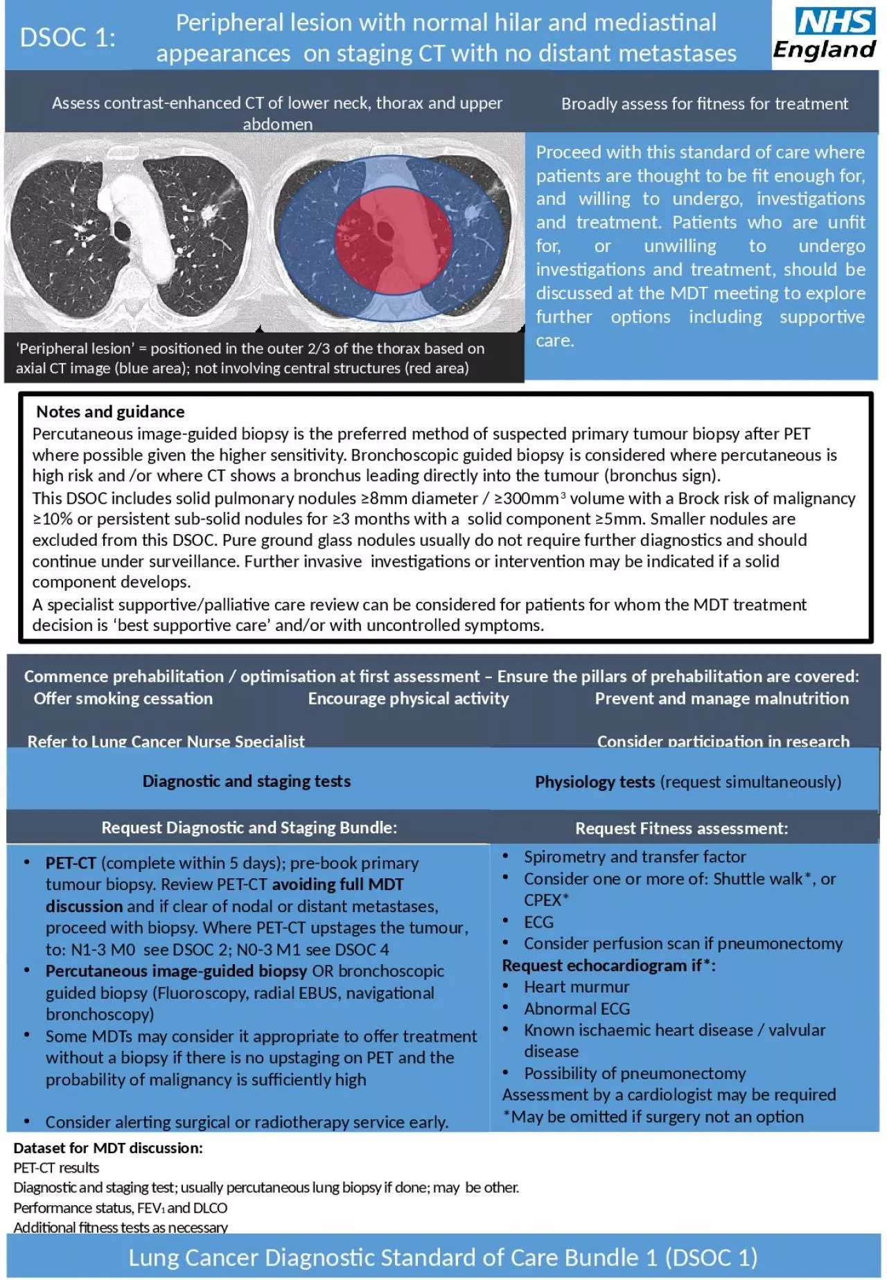
PPT-Peripheral lesion with normal hilar and mediastinal appearances on

Author : lily | Published Date : 2024-01-13

staging CT with no distant metastases Commence prehabilitation optimisation at first assessment Ensure the pillars of prehabilitation are covered Offer smoking

Presentation Embed Code

Download Presentation

Download Presentation The PPT/PDF document "Peripheral lesion with normal hilar and ..." is the property of its rightful owner. Permission is granted to download and print the materials on this website for personal, non-commercial use only, and to display it on your personal computer provided you do not modify the materials and that you retain all copyright notices contained in the materials. By downloading content from our website, you accept the terms of this agreement.

Peripheral lesion with normal hilar and mediastinal appearances on: Transcript


staging CT with no distant metastases Commence prehabilitation optimisation at first assessment Ensure the pillars of prehabilitation are covered Offer smoking cessation Encourage physical activity Prevent and manage malnutrition. Dr. Ackerman’s dedication to teaching.   This site is presented in recognition of Dr. Norman Ackerman and his contributions to the field of veterinary diagnostic imaging.. Sponsorship of the display supports the Dr. Norman Ackerman Memorial Fund, dedicated to the teaching of diagnostic imaging residents at the University of Florida College of Veterinary Medicine.. Know what your EMG might miss!. Introduction. Speakers. Tony . Chiodo. , MD. Timothy Dillingham, MD. W. David Arnold, MD. Shawn Jorgensen, MD. Introduction. Blind spots. Introduction. “. Electrodiagnostic impression. Alistair Ross MB FRCS. Consultant Orthopaedic Surgeon, Bath, UK. Associate Editor. The Bone & Joint Journal. Cambridge Annual Medico-Legal Conference . Peterhouse,Cambridge,2016. Peripheral nerve: organisation. VI medical unit. PROF.DR.C DHARMARAJ MD.DCH. DR M RAJKUMAR MD. DR SENTHUR RAJA PANDIAN MD.DM. DR ARUN GOVIND PG. 42 year old male patient . Presented with H/o. abnormal involuntary movements of Lt UL - 1yr. R. Campbell Garland, PGY-2. Overview. Review of normal platelet life cycle. Definition of thrombocytopenia. Mechanisms of thrombocytopenia. Production, destruction, sequestration. Outpatient a. pproach . Yueh. Z. Lee, MD/PhD. October 26, 2011. Case #1. 46 y/o female with history of NSCLC, who . presents with confusion. . . History . of . paraspinal. mass in right back/chest region, for which she received . . tumors (MT). Dr. . F.Hajmanoochehri. 93/10/25 . introduction. Appropriate therapy for various . mediastinal. tumors differs considerably according to the histological type and it may significantly impact on survival. Assistant Professor/School of Medicine. The Hashemite University. Anatomy. The . mediastinum. is anatomically defined as the space between the two lungs; it is demarcated by the thoracic inlet superiorly, the diaphragm inferiorly, the sternum anteriorly, and by the spine and paravertebral thoracic sulci posteriorly. . . Central nervous system. The CNS (upper motor neuron) includes the brain and spinal cord. . which includes :. Cerebral hemispheres. White matter tracts. Cerebellum. Brain stem with cranial nerves. (. lecture . №. 5). Inna Krupnik. Radiological department of Odessa State Medical University. 2021. . APPROACH TO THE CHEST X-RAY (CXR). An approach to reviewing a chest x-ray will create a foundation that will facilitate the detection of abnormalities. You should create your own strategy. There is no correct way to analyze the images. Consistency and thoroughness are good general strategies. With time, and repetition, the process will become subconscious. Repetitive viewing of images will help establish a baseline of normality and normal variation that will represent an internal yard stick for the detection of variation from normal.. Balance assessment . a Three Dimensional picture. Three pillars . Sensory . Part. Motor part. Functional part. Sensory Part. Vision. Somatosensory. Vestibular system. Central part/integration . Vestibular . 2020 Histomorphological Case Report Priyanka A. Kurdukar 1 , Abhishek A. Kurdukar 2 , Vishakha V. Chaudhari 3 CASE REPORT Introduction: Peripheral cemento-ossifying �broma is a hard f You are performing a routine lower limb peripheral neuropathy screening on a patient with weakness in the right lower limb and lower back pain.   . All sensory and motor studies are normal except for the right tibial motor study to abductor hallucis (AH) which you notice is low in amplitude compared to the other side (>50%) and lower in amplitude compared to your normal values. What are your thoughts about the reason for this finding and what would your next actions be?  . Lung. Capillary lumen. Type I pneumocyte. Type I pneumocyte. Type II. pneumocyte. Endothelium. Alveolar space. The respiratory acinus. Cartilage is present to level of proximal bronchioles. Beyond terminal bronchiole gas exchange occurs.

Download Document

Here is the link to download the presentation.
"Peripheral lesion with normal hilar and mediastinal appearances on"The content belongs to its owner. You may download and print it for personal use, without modification, and keep all copyright notices. By downloading, you agree to these terms.

Related Documents