/
Periodontal Plastic and Reconstructive Surgery Periodontal Plastic and Reconstructive Surgery

Periodontal Plastic and Reconstructive Surgery - PowerPoint Presentation

linda
linda . @linda
Follow
66 views
Uploaded On 2023-07-14

Periodontal Plastic and Reconstructive Surgery - PPT Presentation

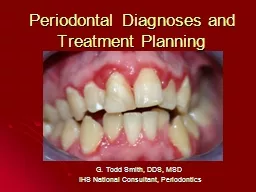
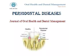
INTRODUCTION Surgical procedures for soft tissue management are designed to preserve gingiva remove aberrant frenulum or muscle attachments and increase the depth of the vestibule According ID: 1009078

soft tissue oral graft tissue soft graft oral surgery laser surgical gingiva vestibular electrosurgery mucosal depth procedures membrane extension

Share:

Link:

Embed:

Download Presentation from below link

Download Presentation The PPT/PDF document "Periodontal Plastic and Reconstructive S..." is the property of its rightful owner. Permission is granted to download and print the materials on this web site for personal, non-commercial use only, and to display it on your personal computer provided you do not modify the materials and that you retain all copyright notices contained in the materials. By downloading content from our website, you accept the terms of this agreement.


Presentation Transcript

1. Periodontal Plastic and Reconstructive Surgery

2. INTRODUCTIONSurgical procedures for soft tissue management are designed to preserve gingiva, remove aberrant frenulum or muscle attachments, and increase the depth of the vestibule.

3. According to the Glossary of Periodontal Terms (1992), mucogingival surgery is defined as "plastic surgical procedures designed to correct defects in the morphology, position and/or amount of gingivae surrounding the teeth".

4. Mucogingival therapy is defined as the correction of defects in morphology, position, or amount of soft tissue and underlying bone. This is the most comprehensive definition because it includes both nonsurgical and surgical mucogingival therapy of the gingiva, alveolar mucosa, and bone.

5. In 1988, Miller4 introduced the term periodontal plastic surgery because the term mucogingival surgery did not adequately describe all the periodontal procedures that were being performed under this classification. Such procedures include root coverage, functional crown lengthening, esthetic crown lengthening, ridgepreservation (after removal of periodontally involved teeth), ridge augmentation, maintenance of interdental papillae, reconstruction of papillae, esthetic soft tissue surgery around implants, and surgical exposure of teeth for orthodontic purposes.

6. FRENECTOMYThe word frenum is derived from the Latin word “fraenum”. Frena, are triangle-shaped folds found in the maxillary and mandibular alveolar mucosa, and are located between the central incisors and canine premolar area.

7. Frenum may be classified depending upon itsMorphology as:Long and thinShort and broadDepending upon the attachment level, frenumhas been classified as: (Placek et al. 1974)MucosalGingivalPapillaryPapillary penetrating

8. When the insertion point of the frena is at the gingival margin it may pose a problem (Corn 1964). This kind of abnormal insertion of the frenum may cause marginal recession of the gingiva.

9. Aberrant frenum can be treated by frenectomy or frenotomy procedures. The terms frenectomy and frenotomy signify operations that differ in degree of surgical approach. Frenectomy is a complete removal of the frenum, including its attachment to the underlying bone, and may be required for correction of abnormal diastema between maxillary central incisors (Friedman 1957). Frenotomy is the incision and relocation of the frenal attachment.

10. Dieffenbach V-plastyFrenectomy by V-plasty may result in scar formation that could prevent the mesial movement of the central incisors (West 1968).It is typically a safe surgical procedure with no notable complications.

11. Schuchardt Z-plasty -Useful when frenum is broad and shallow vestibule is present-Minimal scar tissue formation.-The method requires a skilled operator as it is tedious to perform.

12. Miller’s technique combined with lateral pedicled graft

13. Advantages :1. Healing takes place by primary intention.2. A zone of attached gingiva, matching with adjacent tissue, forms in midline which is pleasing to the individual.3. No unesthetic scar formation.4. No recession of interdental papilla occurs because the transseptal fibres are not severed out.5. The attached gingiva in midline may have a bracing effect which helps in prevention of orthodontic relapse.

14. VESTIBULOPLASTYA shallow vestibule is most of the times associated with an inadequate width of attached gingiva. Presence of attached gingiva & good vestibular depth can be of help to individuals whose oral hygiene practices are less than optimal.

15. DISCUSSIONA shallow vestibule is most of the times associated with an inadequate width of attached gingiva. Presence of attached gingiva & good vestibular depth can be of help to individuals whose oral hygiene practices are less than optimal.

16. depth of the vestibule hampers the proper placement of a tooth brush plaque accumulation

17. Most documented techniques for the creation of a good vestibular depth require a -high level of surgical dexterity on the clinician’s part -adequate motivation of the patient.

18. METHODSThree methods of cutting oral soft tissue are used commonly in dentistry: scalpel electrosurgery laserEach of these methods are different from the standpoints of hemostasis, healing time, cost of instruments, width of the cut, anesthetic required and disagreeable characteristics, such as smoke production, the odor of burning flesh and undesirable taste.

19. SURGICAL METHODSFenestration techniqueEdland and Mejcher sulcus extension procedure Mucosal advancement vestibuloplastySecondary epithelization vestibuloplastyKazanjian’s techniqueObwegeser’s technique Grafting vestibuloplastyMucosal graftSkin graft

20. Fenestration technique

21. Fenestration technique

22. Edlan and Mejcher sulcus extension procedureTrans-positional Flap Surgery It is a relatively difficult procedure requiring considerable amount of surgical skills. (Int Dent J 1963; 13 : 593)

23.

24. A retrospective study conducted on the progression of alveolar resorption and alteration of the soft tissue profile of the chin in 15 patients operated on by Edlan and Mejcher sulcus extension procedure reported -Accelerated bone resorption of the alveolar crest. Furthermore a significant reduction of lower lip and increase of mental soft tissue protrusion were recorded.(Keithley JL et al 1978)

25. Bohannan (1960) suggested that deepening of the vestibule by non-free graft procedures were not successful when evaluated years later because a nearly complete relapse could be proven during secondary healing with contraction and epithelialization of the vestibular periosteum.To date various autogenous soft tissue grafts from autogenous mucosal to allogeneic collagen membrane have been used for vestibular extension.

26. Numerous graft materials are available like Skin graft, mucosal grafts, palatal graft, Buccal graft.Biological membrane obtained from placenta opens new perspectives. The human amnion membrane is a biological graft which has unique properties like antiadhesive effects, bacteriostatic properties, wound protection, pain reduction and epithelisation effects. (Güler R et al,Br J Oral Maxillofac Surg 1997;35:280-3)

27. In this technique underlying submucosal tissue is either excised or repositioned to allow direct apposition of the labiovestibular mucosa to the periosteum.MUCOSAL ADVANCEMENT VESTIBULOPLASTY

28.

29. Indications-Adequate anterior mandibular length (at least 15mm) Inadequate facial vestibular depth from mucosal & muscular attachment Presence of adequate vestibular depth on the lingual aspect of mandibleKAZANJIAN’S TECHNIQUE

30.

31. ADVANTAGEIt is a well-tolerated surgical procedure, can be done under local anesthesia with satisfactory results.This eliminates the need for a free graft or splintAdequate amount of sulcus depth can be achieved with minimal relapse.Complications were minimal compared to other techniques

32. DISADVANTAGE Pain and oedemaTransient ParaesthesiaHypersensitivity of the surgical site Breakdown of the sutured margin of the mucosaDamage to the mucosal flapSagging chinunpredictability of the amount of relapse of vestibular depth (Kethley JL et al J Oral Surgery 1978; 36 :701–5.)

33. Its modification is Trans-positional Flap Surgery (Edlan and Mejcher sulcus extension procedure). Trans-positional Flap Surgery is a relatively difficult procedure requiring considerable amount of surgical skills. (Int Dent J 1963; 13 : 593)

34. AB

35. A retrospective study conducted on the progression of alveolar resorption and alteration of the soft tissue profile of the chin in 15 patients operated on by Edlan and Mejcher sulcus extension procedure reported -accelerated bone resorption of the alveolar crest. Furthermore a significant reduction of lower lip and increase of mental soft tissue protrusion were recorded.(Keithley JL et al 1978)

36. OBWEGESER’S TECHNIQUE

37. ADVANTAGEEarly covering of the exposed periosteal bed which improves patient’s comfortLong term results of vestibular extension are predictable

38. DISADVANTAGEModerate swellingDiscomfort experienced by patient

39. GRAFTING VESTIBULOPLASTYBohannan (1960) suggested that deepening of the vestibule by non-free graft procedures were not successful when evaluated years later because a nearly complete relapse could be proven during secondary healing with contraction and epithelialization of the vestibular periosteum.To date various autogenous soft tissue grafts from autogenous mucosal to allogeneic collagen membrane have been used for vestibular extension.

40. Numerous graft materials are available like Skin graft, mucosal grafts, palatal graft, Buccal graft.Autogenous Grafts may have disadvantages of increased morbiditypostoperative pain risk of surgical complications at the donor site.

41. Split skin graft is well-tolerated but can be subjected to postoperative shrinkageclinical difference in consistency and color. susceptibility to infectionPaindiscomfortdelayed healing

42. The problems with mucosal (buccal and palatal grafts) are-Limited amount of mucosa available for graftingPossibility of nerve damages Palatal mucosal wounds leave an open wound with a healing course of 4-6 and half weeks Possible complications of soreness and ulcers.

43. Biological membrane obtained from placenta opens new perspectives. The human amnion membrane is a biological graft which has unique properties like antiadhesive effects, bacteriostatic properties, wound protection, pain reduction and epithelisation effects. Its easy availability, low cost makes it the best material. (Güler R et al,Br J Oral Maxillofac Surg 1997;35:280-3)

44. The amniotic membrane is formed from the ectoderm of the fetus. It has a stromal matrix, a thick collagen layer, and an overlying basement membrane with a single layer of epithelium. It closely resembles the epidermis of the skin and has been used as a physiological wound dressing with great success. (Güler R et al,Br J Oral Maxillofac Surg 1997;35:280-3)

45. The amnion has the following advantages:It promotes secondary epithelialization, Vascularize healthy granulation tissue and stimulate the neovascularisation in neighbouring tissues Antibacterial.Lack of immunogenicity as it did not express antigens of histocompatibility, the allograft was never rejected.(Samandari MH et al Surg Oral Med Oral Pathol Oral Radiol Endod 2004;97:574-)

46. These are inexpensive, readily available, used fresh or lyophilised, and stored at room temperature after sterilisation by gamma irradiation.

47.

48. Conventional methods of surgery are often associated with pain, trauma, bleeding, post-operative edema and scarring which are problematic for both the patient and the clinician. To overcome these drawbacks, Diode laser was used to perform various surgical procedures including frenectomy, gingivectomy, vestibuloplasty, depigmentation and second stage implant surgery.

49. VESTIBULOPLASTY BY LASER The first laser was introduced by Maiman in 1960 , who used ruby to make laser. After a while,(Carbon Dioxide Laser) CO2 and Neodymium- Doped Yttrium Aluminium Garnet(Nd: YAG) lasers were developed. In medical field, laser was first used for photocoagulation of retina in 1960 . Goldman used lasers in dentistry in 1964 for the first time . (Mahajan A. Lasers in periodontics-a review. Eur J Dent Med. 2011;3:1-11.)

50. In 1989, Myers published the first article about using laser in periodontal surgery . Food and Drug Administration (FDA) allowed CO2, Nd: YAG, and diode lasers only for soft tissues .

51. The diode basically does not interact with dental hard tissues, this makes it an excellent soft tissue surgical laser, indicated for cutting and coagulating gingiva, oral mucosa and for soft tissue curettage or sulcular debridement.It is a solid-state semiconductor laser that typically uses a combination of Gallium (Ga), Arsenide (Ar), and other elements such as Aluminium (Al) and Indium (In) to change electrical energy into light energy. It is usually operated in contact mode using a flexible fiber optic delivery system, and emits laser in continuous- wave or gated-pulsed modes.

52. The power output for dental use is generally around 2 to 10 watts.Tissues can respond to laser light in four different ways: scatter transmit,reflectabsorb

53. Absorption is the most desired laser-tissue interaction in dental use It depends on three factors WavelengthTissue composition Tissue's water content Laser-tissue interaction is the use of light energy that is absorbed by the tissue to produce a photobiological effect

54. The diode laser exhibits thermal effects using the "hot-tip" effect caused by heat accumulation at the end of the fiber, and produces a relatively thick coagulation layer on the treated surface.

55. The diode laser causes minimal damage to the periosteum and bone under the gingiva being treated as well as exhibits the unique property of being able to remove a thin layer of epithelium cleanly. It can be used for a variety of soft tissue procedures without impacting the neighboring tissues. (Bains VK, Gupta S, Bains R. Lasers in periodontics: An Overview. J Oral Health Community Dent. 2010;4:29-34)

56.

57.

58. Diode laser at a pulsed mode, 2 watt & 1.0 ms pulse interval was used. -810 nm

59.

60.

61.

62. ADVANTAGESminimal or no anestheticdo not harm dental hard tissues judicious use does not injure the dental pulp antimicrobial

63. remove endotoxinsfrom root surfacesbecause of low heatproduction they can be used around dental implantspatients are more accepting of its use in their treatment than of electrosurgery.

64. less operative and post-operative pain hemostasis little wound contractionReduced edema

65. DISADVANTAGES costrequires a learning period and strict precautionscan cause eye damage protectiveglasses are required during its use

66. cutting slowerburning flesh odorsome techniques are time consumingcombustible gases must be turned off during laser uselaser plume requires use of a high-filtration face mask,because of the possible presence of pathogens in the plume.

67. ELECTROCAUTERYElectrosurgery is a controlled, precise application of heat to the soft-tissue site to be cut, achieved by means of carefully designed electrodes resulting to controlled, irreversible thermal alteration of the soft tissue.

68. There are two basic types of electrosurgery. Monopolar electro surgery units - the current begins with the electrosurgery device and travels along a wire to the oral site, then to an in different plate placed behind the patient’s back. As the surgicalelectrode contacts the patient’s oral soft tissues, heat is produced and cutting is achieved. -Smoke and pain also are produced as the tissue is cut, necessitating the use of anesthetic.

69. Bipolar electrosurgery devices- have two electrodes on the cutting tip. The current flows from one electrode to the other, making a broader cut than does the monopolar unit, but eliminating the need for the indifferent plate. Both types of electrosurgery units achieve their intended purposes well, but monopolar electrosurgery is used more often than is bipolar electrosurgery (R. Goldman,vice president, Clinical Products, Parkell, Edgewood, N.Y.,oral communication, May 6, 2008).

70. Currents used in oral electro-surgery are –Fully rectified filtered-usually called "cut" or "filtered" on the devices.Fully rectified-usually called "coagulate," "coagulate/cut," "coagulate/hemostasis"or "unfiltered" on the devices. Partially rectified –usually called "coagulate" or "fulgerate" on the devices. most dentists use fully rectified current on a medium setting so the electrode tip cuts but does not drag.

71.

72.

73.

74.

75.

76. ADVANTAGEScost much lesselectrode cuts on its sides as well as on its tipelectrode may be bent to meet the clinical needcuts are made with easehemostasis is immediate

77. cutting is consistentthe wound is nearly painlessthe tip is self-disinfecting.the soft tissue has minimal trauma

78. DISADVANTAGESAnesthetic required low tactile sense of exactly what is being cutan unavoidable burning-flesh odorboth the name and the use of electrosurgery cause fear in some patients

79. heat developed by monopolar electrosurgery units does not allow for their use around implants bone can be damageddangerous in an explosive environment

80.