/
Vol 20 No 2 May 2011Continuous exposure EMF double minute Vol 20 No 2 May 2011Continuous exposure EMF double minute

Vol 20 No 2 May 2011Continuous exposure EMF double minute - PDF document

linda
linda . @linda
Follow
348 views
Uploaded On 2021-07-02

Vol 20 No 2 May 2011Continuous exposure EMF double minute - PPT Presentation

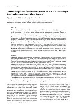
Continuous exposure of three successive generations of mice to electromagnetic elds implication on double minute frequency Med J IndonesSari et al 110 Epidemiological evidence was obtained in the U ID: 851222

minute double exposure elds double minute elds exposure emf chromosomes frequency leukemia cancer mice dna exposed generations chromosome offspring

Share:

Link:

Embed:

Download Presentation from below link

Download Pdf The PPT/PDF document "Vol 20 No 2 May 2011Continuous exposure ..." is the property of its rightful owner. Permission is granted to download and print the materials on this web site for personal, non-commercial use only, and to display it on your personal computer provided you do not modify the materials and that you retain all copyright notices contained in the materials. By downloading content from our website, you accept the terms of this agreement.


Presentation Transcript

1 Vol. 20, No. 2, May 2011Continuous expos
Vol. 20, No. 2, May 2011Continuous exposure EMF double minute Continuous exposure of three successive generations of mice to electromagnetic elds: implication on double minute frequency Med J IndonesSari et al. 110 Epidemiological evidence was obtained in the USA during 1950 until 1982 from mortality analysis in workers employed in occupations with intuitive exposure to high voltage EMF. These workers showed a signi cant increase in proportionate mortality ratios (PMR) associated with leukemia and non-Hodgkin lymphomas. Moreover, leukemia incidence was 2-3 times higher than among people who lived faster away from the Exposure to extremely low frequency elds of 5-500 T for 2 hours per day for two years was found to cause skin cancer. Two decades ago, Brain and Pool reported increasing risk of leukemia, lymphomas, and brain tumors among children who live close to power lines or among men whose jobs A Canadian study found that occupational exposure of elds may be related with leukemia of their children in two pooled retrospective studies. Increased risk of childhood leukemia was found elds above 0.3 or 0.4 T, respectively. These ndings support the hypothesis elds are carcinogenic. Epidemiological studies show links between exposure to EMF and cancer, especially leukemia’s, lymphomas, and brain tumors. According to the International Agency for elds probably play The biological mechanism by which human cancer could be induced by EMF is unknown. Nevertheless, experimental studies in Congenital anomalies in the offspring were found after elds of 6 and 7 kV 12 elds increased proliferation and chromosomal aberration of Continuous exposure of four successive mouse generations to EMF showed increased mortality rates and congenital anomalies in their offspring. Usually, life expectancy of offspring suffering anomalies or tumors was only 5 – 6 month as compared to 2 – 2.5 years of Further investigation has been done by Lai and Singh on the brain cells of rats exposed to EMF in which DNA stand breaks were signi cantly increased Double minute chromosomes are small a centric fragment cation, which become In connection with the continuous exposure to EMF will affect chromosomal alterations in mice. The purpose of the present study is to evaluat

2 e the effects of continuous exposure to
e the effects of continuous exposure to EMF of 3, 4 and 5 kV respectively, on chromosomal breakage L) of Swiss Webster strain, 3-4 months of age, and 7-40 gram of and fed a standard diet. Three couple were exposed to EMF at the doses of 3 kV, 4 kV, and 5 kV, and one couple served as control. Twelve plastic cages (26 x 20 x11 cm; length x width x height) with metal wire cm. Subsequently, the electrodes were connected to an alternating current power supply. All mice belonging and deliver the F1 generation. At maturity, the mice gestate, and bear their offspring of the F2 generation while being continuously exposed to EMF. Couples generation. A parallel procedure was followed for the offspring until weaning at about 4 weeks after birth.In all offspring, except one couple to be exposed to ced. Haemopoetic bone marrow cells C; mitosis xation with Carnoy xative solution and subsequent staining with Giemsa. Twenty metaphases of chromosomes were examined exposures. The frequency of double minutes was then analyzed using two-way ANOVA with Duncan test.RESULTS Data obtained in control group with score zero were Y + 0.5 to enable statistical The frequency of double minute formation in F1, F2, F3, after exposure of EMF is shown in Table 1. minute in mice exposed to EMF of 4 kV or 5 kV were Vol. 20, No. 2, May 2011Continuous exposure EMF double minutefound in F1 and F3, but not in F2. Whereas in group exposed to 3 kV, the signi cant different from control From our experimental setting we obtained two sets of results, one on the variation of strength of EMF and another one on the three successive generations. Table 2 represents the average double minute frequency in groups exposed to 3 kV, 4 kV, and 5 kV of EMF by grouping cant different were found between group of 4 kV and 5 kV compared to control group. Table 3 represents the average double minute frequency cant differences were found of double minute appears generally lower in F2 than in Table 1. M elds (values in control grouped were TreatmentF1F2F30.83 ± 0.11*Table 2. Average frequency of double minute in all generations of elds. TreatmentsControl 3 kV 4 kV 5 kVTable 3. Average frequency of double minute in three successiv

3 e generations of mice after continuous e
e generations of mice after continuous exposure to eld. Frequencies were taken from mean values of exposure to 3 kV, 4 kV, and 5 kV exposure. F1 F2 F3 Figure 1. Demonstrates double minute formation (Fig. 1A) compared to normal mouse chromosomes (Fig. 1B). A BFigure 1. Double minute is shown in A, and normal mouse chromosomes in B elds) from generations F1 to F3 normal cells without any treatment. This is different from the mice exposed to EMF at the doses of 3 kV, 4 kV, and 5 kV from generations F1 to F3 generation, in which double minute chromosomes can be formed, although not all exposure lead to large quantities of double-minute formation. Although not all double minute formation cant difference, it is clear that EMF exposure in Swiss Webster strain mice cantly higher double minute protect this generation from chromosomal alterations. Further exposure to EMF, especially at 4 or 5 kV Usually, double minute can be found in cancer cells cation of oncogenes. Gene cation is common in many tumors including neuroblastoma, squamous cell carcinoma of the head and Although double minute is rarely found in leukemia, several studies reported the frequency vary from 1% to 12.5% and double minute frequency found in myelodysplastic 19, In general, existence of double minute in leukemia is cation of oncogene c-MYC. The results of a study by Obe and Vijayalaksmi elds (ELF), 46% of the researchers Med J IndonesSari et al. 112 said there was no increase in DNA damage such as stated an increase in DNA damage. Further studies eld 2.45 GHz for 2 hours did not The result from Magnussen study, concluded from various studies on the effects of electromagnetic elds, that elds can trigger changes in membrane signal that involves changes in gene expression and in turn there is a change in DNA repair. Accumulation of DNA damage repair caused chromosome damage, thus increasing the risk of cell death such as diseases that are directly related to Alzheimer’s and cancer. If damage occurs on chromosome fragile sites will be promoting cation, which is then referred to as double Chromosome double minute chromosomes are structures that do not have a centro

4 mere and telomere. It is known that if t
mere and telomere. It is known that if there is a fault on the segment of chromosome arms (chromosome breakage), then the normal chromosome in the cell will make the settings again (rearrangement) which can be either stable or not. In the rearrangement process, centromere and telomere Telomere are nucleoprotein structures that contain DNA sequences, which among other functions to protect chromosome ends, protect chromosomes from fusion, as well as maintain the integrity of chromosomes Centromere is part of the DNA c proteins to form kinetokor. Kinetokor functions, among others, to attach chromosomes to microtubules and regulate In this case, the double minute chromosomes are structures that do not have a centromere and telomere. Thus, double minute can not maintain the integrity of chromosomes during mitosis. In the absence of the centromere, the double minute can kV, 4 kV and 5 kV may not always evoke the formation However, it suggests that continuous exposure may clinical relevance to carcinogenicity and mutagenicity. magnetic eld may cause chromosomal alterations, We thank University of Indonesia for the Research Lunquist, S. Biological effects of electromagnetic elds. Royal Swedish Academy of Engineering Sciences IV A-Meddelande 210. 1980, Stockholm. P.10www.elektroindonesia.com/Pengukura2. n Medan listrik dan medan magnit di bawah SUTET. (Cited 10 September 2006).Deventer, E.Environmental health criteria 238: Extremely elds World Health Organization. 2007Tynes T, Klaeboe L, Haldorsen T. Residental and occupational elds and malignant melanoma: a population based study. Br Med J. 2003;60:343-7.Brain JD, Kavet R, McCormick DL, Poole C, Silverman LB, Smith TJ, et al. Childhood leukemia: electric and elds as possible risk factors. Environ Health Perspect. 2003;111:962-70.genotoxic: The implications for the epidemiology of cancer and cardiac, neurological and reproductive effects. 2000, http://www.feb.se/EMFguru/EMF/genotoxi/ Genotoxic-EMR-paper.html. (Cited 27 November 2007).Infante-Rivard C, Deadman JE. Maternal occupational exposure elds during pregnancy and childhood leukemia. Epidemiology. 2003; 14: 437-41.Ahlbom A, Day N, Feychting M, Roman E, Skinner J, Dockerty J, et al. A pooled analysis of magnetic elds and childhood

5 leukemia. Br J Cancer. 2000;83(5):692-8.
leukemia. Br J Cancer. 2000;83(5):692-8. Greenland S, Sheppard AR, Kaune T, Poole C, Kelsh MA. A pooled analysis of magnetic elds, wire codes, and Group. Epidemiology. 2000;11(6):624-34.www.mindfully.org/Nucs/2002/Non-Ionizing-80- elds (Cited 8 January 2005).Lacy-Hulbert L, Metcalfe JC, Hesketh R. Biological responses 11. elds. FASEBSoeradi O, Tadjudin MK. Congenital anomalies in the offspring rats after exposure of the testis to an electrostatic eld. Int J Androl. 1986;9:152-60.Sari P. Pemajanan medan elektrostatik pada mencit strain Swiss Webster dan pengaruhnya terhadap kromosom serta proliferasi limfosit. Magister Thesis at the Postgraduate Soeradi O, Yurnadi, Sari P, Pujianto DA. The effect of eld on four successive generations of mice. Med J Indones. 2002;11:3-10.Lai H, Singh N. Magnetic- eld-induced DNA strand breaks in brain cells of the rat. Environ Health Perspect. 2004;112:1-12.Snustad DP, Simmons MJ. Principles of Genetics. 4edition. John Wiley & Sons, Inc. 2010. p. 659. Miller OJ, Therman E. Chromosomes and cancer: activation of oncogenes. In. Miller OJ, Therman E: Human edition, 2001. Springer, Berlin. P.405. Stell RGD, Torrie JH. Prinsip dan prosedur statistika. Suatu Vol. 20, No. 2, May 2011Continuous exposure EMF double minute113Mathew, S. Double minute chromosomes and c-MYC cation in a child with secondary myelodysplastic Von Hoff, D.D., Forseth, B, Clare, C.N., Haneen, K.L., and Van Deventer, D. Double minute arise from circular extra chromosomal DNA intermediates which integrate into Vijayalaxmi, Obe G. Controversial cytogenetic obser vations in mammalian somatic cells exposed to extremely low frequency elektromagnetic radiation: A review and Komatsubara Y, Hirose H, Sakurai T, Koyama S, Suzuki Y, Taki M, and Miyakoshi J. Effect of high-frequency elds with a wide range of SARs on 2005; 587: 114-9.23. Magnussen T. Key to understanding the EMF issue: Piecing 24. Nussabaum RL, McInnes RR, Willard HF. Principle of Willard HF, editors. Thompson & Thompson. Genetics in ed. 2001. WB. Saunders. Philadelphia.Bailey SM, and Murnane JP. Telomeres, chromosomes instability, and cancer. Nucleic Acid Res. 2006; 34:2408-17.26. Lo AWI, Sprung CN, Fouladi B, Pedram M, Sabatier L, Ricoul M, Reynolds GE, Murnane JP. Chromos