PDF-Hidradenitis suppurativa
24
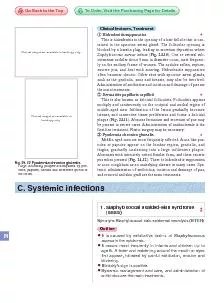
This is hidradenitis in the opening of a hair follicle that is con
Staphylococcus aureus
infects Fig 2410
Staphylococcus
in the epidermisIt occurs most frequently
Download Presentation
"Hidradenitis suppurativa" is the property of its rightful owner. Permission is granted to download and print materials on this website for personal, non-commercial use only, provided you retain all copyright notices. By downloading content from our website, you accept the terms of this agreement.
Presentation Transcript
Transcript not available.