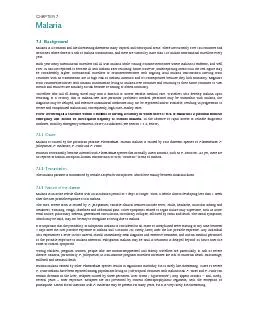
/
Laboratory diagnosis of malaria Laboratory diagnosis of malaria

Laboratory diagnosis of malaria - PDF document

lois-ondreau
lois-ondreau . @lois-ondreau
Follow
453 views
Uploaded On 2016-08-04

Laboratory diagnosis of malaria - PPT Presentation

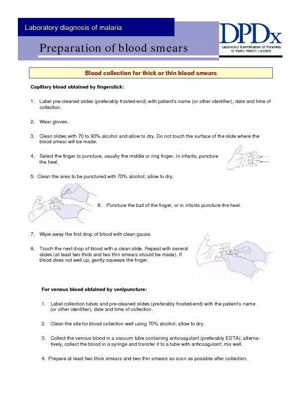
Capillary blood obtained by fingerstick Label precleaned slides preferably frostedend with patient146s name or other identifier date and time of collection 2 Wear gloves 3 Clean slide ID: 431650

Capillary blood obtained fingerstick:

Share:

Link:

Embed:

Download Presentation from below link

Download Pdf The PPT/PDF document "Laboratory diagnosis of malaria" is the property of its rightful owner. Permission is granted to download and print the materials on this web site for personal, non-commercial use only, and to display it on your personal computer provided you do not modify the materials and that you retain all copyright notices contained in the materials. By downloading content from our website, you accept the terms of this agreement.


Presentation Transcript

Laboratory diagnosis of malaria Capillary blood obtained by fingerstick: Label pre-cleaned slides (preferably frosted-end) with patient’s name (or other identifier), date and time of collection. 2. Wear gloves. 3. Clean slides with 70 to 90% alcohol and allow to dry. Do not touch the surface of the slide where the blood smear will be made. 4. Select the finger to puncture, usually the middle or ring finger. In infants, puncture 5. Clean the area to be punctured with 70% alcohol; allow to dry. 6. Puncture the ball of the finger, or in infants puncture the heel. 7. Wipe away the first drop of blood with clean gauze. 8. Touch the next drop of blood with a clean slide. Repeat with several slides (at least two thick and two thin smears should be made). If blood does not well up, gently squeeze the finger. For venous blood obtained by venipuncture: 1. Label collection tubes and pre-cleaned slides (preferably frosted-end) with the patient’s name (or other identifier), date and time of collection. 2. Clean the site for blood collection well using 70% alcohol; allow to dry. 3. Collect the venous blood in a vacuum tube containing anticoagulant (preferably EDTA); alterna-tively, collect the blood in a syringe and transfer it to a tube with anticoagulant; mix well. 4. Prepare at least two thick smears and two thin smears as soon as possible after collection. 1. Whenever possible, use separate slides for thick and thin smears. 2. Thin film (a): Bring a clean spreader slide, held at a 45° angle, toward the drop of blood on the specimen slide. 3. Thin film (b): Wait until the blood spreads alg the entire width of the spreader slide. 4. Thin film (c): While holding the spreader slide at the same angle, push it forward rapidlyand smoothly. 5. Thick film: Using the corner of a clean slide, spread the drop of blood in a circle the size of a dime (diameter 1-2 cm). Do not slide. (You should be able to read newsprint through it.) 6. Wait until the thin and thick films are completely dry before staining. Fix the thin film with methanol (100% or absolute) and let it dry completely before staining. The thick film should 7. If both thin and thick films need to be made on the same slide, fix only the thin film with methanol. The thick film should not be fixed. For a video of how to make a thick smear, please visit the DPDx website at: http://www.dpd.cdc.gov/dpdx/HTML/Frames/DiagnosticProcedures/body_dp_bloodthickavi.htm For a video of how to make a thin smear, please visit the DPDx website at: http://www.dpd.cdc.gov/dpdx/HTML/Frames/DiagnosticProcedures/body_dp_bloodthinavi.htm