/
Clinical Artefacts While the incidence of artefact on Clinical Artefacts While the incidence of artefact on

Clinical Artefacts While the incidence of artefact on - PDF document

luanne-stotts
luanne-stotts . @luanne-stotts
Follow
401 views
Uploaded On 2015-04-30

Clinical Artefacts While the incidence of artefact on - PPT Presentation

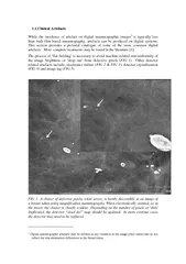
1Clinical Artefacts While the incidence of artefact on digital mammographic images is typically less than with film based mammography artefact s can be produced on digital systems This section provides a pictorial ID: 57900

1Clinical Artefacts While the incidence

Share:

Link:

Embed:

Download Presentation from below link

Download Pdf The PPT/PDF document "Clinical Artefacts While the incidence o..." is the property of its rightful owner. Permission is granted to download and print the materials on this web site for personal, non-commercial use only, and to display it on your personal computer provided you do not modify the materials and that you retain all copyright notices contained in the materials. By downloading content from our website, you accept the terms of this agreement.


Presentation Transcript

While the incidence of artefact on digital mammographic images is typically less than with film based mammography, artefacts can be produced on digital systems. This section provides a pictorial catalogue of some of the more common digital artefacts. More complete treatments may be found in the literature [1]. The process of ‘flat fielding’ is necessary to avoid machine-related non-uniformity of the image brightness or ‘drop out’ from defective pixels ( 1). Other detector related artefacts include; electronics failure (FIG 2 & ), detector crystallization (FIG 4) and image lag ( . A cluster of defective pixels, white arrow, is barely discernible in an image of a breast taken using magnification mammography. When electronically zoomed, as in the insert, the cluster is clearly evident. Depending on the number of pixels or ‘dels’ implicated, the detector “dead del” map should be updated. In more extreme cases the detector may need to be replaced Digital mammography artefacts may be defined as any variation in the image pixel values that do not reflect the true attenuation differences in the breast tissue. FIG 2. The image on the left demonstrates an odd looking well defined artefact which, when electronically zoomed, looks like a step wedge embedded in the breast. The direct cause of the artefact is a failure of an Application Specific Integrated Circuit (ASIC). In this case the fundamental cause was a failure of the room air conditioning, which allowed the temperature at the detector to exceed allowed tolerance FIG 3. An obvious example of an ASIC failure in an a-Se detector. Fortunately, in this instance it occurred near the nipple edge and not the chest wall so that the images of require repeating FIG 4. Detector crystallization. (a) The arrows top left indicate a subtle artefact in the MLO image which appears to mimic calcification. (b) The subsequent MLO view of the other breast indicates that the artefact is in fact caused by the a-Se detector beginning to crystallize. The window and level in this image have been adjusted to highlight the problem. (c) A more obvious and serious example of a-Se detector crystallization is apparent in this image (see arrows). FIG 5. The left image is a standard CC view of a breast and the right image is the MLO view acquired immediately afterwards. As indicated by the arrows, the CC view is still evident in this latter image. This is a totally unacceptable example of a-Se detector image lag and is caused, in this instance, by the room temperature not being temperature at the required levelExtreme examples of motion blurring may still occur as exemplified by . If the technologists do not view the images closely or fail to use the zooming tool, more subtle motion blurring may be missed, especially if the monitors on the digital acquisition workstation are of lower spatial resolution than those used by radiologists ). FIG 6. An extreme example of motion artefact. In this instance caused by a CR cassette not being firmly locked in the cassette holder when the MLO view was acquired FIG 7. A more subtle example of a motion artefact is shown in the left hand images. The artefact was observed on the radiologist’s reporting workstation but only became apparent when electronic zoom was utilised on the acquisition workstation. The repeated image shown on the right demonstrates the calcification more sharplyTalcum powder may mimic calcification as illustrated in and calcifications on the skin may be misinterpreted as being in the body of the breast in some circumstances ( FIG 8. Artefact mimicking calcification caused by talcum powder is clearly evident in left hand image (arrows). The subsequent image on right, after removal of powder, is devoid of artefact. Similar artefacts may also arise from zinc powder on the skin FIG 9. Right CC image, illustrated on left, appears to demonstrate calcification (arrows) in body of breast Although not unique to digital mammography and not strictly an artefact, poor collimation can result in large amounts of tissue being missed as illustrated in FIG 10. An example of poor collimator adjustment. In this magnification view the collimator has not been adjusted by the service organisation to allow the entire detector to be irradiated leaving a marked white border on the bottom and right margins of the image. Apart from being disconcerting for the radiologist interpreting the study, this allows an unacceptable amount of breast tissue to be missed, most seriously on the chest wallFinally, images must be checked before a case is closed in order to avoid mislabelling of images that cannot be corrected later. If the image processing for peripheral equalization is not performed well, then a “breast within a breast” appearance can be [1] VAN ONGEVAL, C., JACOBS, J., BOSMANS, H., ZANCA, F., "Classification of artifacts in clinical digital mammography", Digital Mammography, (BICK, U.DIEKMANN, F., Eds), Springer-Verlag, Berlin-