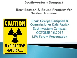
/
Protection against sealed sources Protection against sealed sources

Protection against sealed sources - PowerPoint Presentation

luanne-stotts
luanne-stotts . @luanne-stotts
Follow
391 views
Uploaded On 2016-07-09

Protection against sealed sources - PPT Presentation

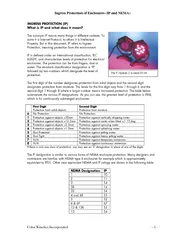
Sealed Source Radioactive Material Sealed sources are radioactive materials encased or sealed inside metal or plastic and can take many different forms sizes and shapes All forms share some type of encapsulation that prevents their radioactive contents from leaking or dispersing barring tampe ID: 397391

sealed sources radioactive radiation sources sealed radiation radioactive material radiography source detectors metal rays ray smoke medical gamma lightning equipment ionizing particles

Share:

Link:

Embed:

Download Presentation from below link

Download Presentation The PPT/PDF document "Protection against sealed sources" is the property of its rightful owner. Permission is granted to download and print the materials on this web site for personal, non-commercial use only, and to display it on your personal computer provided you do not modify the materials and that you retain all copyright notices contained in the materials. By downloading content from our website, you accept the terms of this agreement.


Presentation Transcript

Slide1

Protection against sealed sourcesSlide2

Sealed Source Radioactive Material

Sealed sources are radioactive materials encased or "sealed" inside metal or plastic and can take many different forms, sizes and shapes. All forms share some type of encapsulation that prevents their radioactive contents from leaking or dispersing, barring tampering or a severe accident. In some forms, the radioactive material is an inherent part of the source and cannot be separated. Slide3

Almost all "sealed sources" can be handled without concern that the radioactive material will rub-off or be dispersed onto hands or clothing. There is, however, reason to be concerned about exposure to the radiation emitted from the sealed source. Sealed sources are not a significant contamination hazard under normal conditions; however, they may present an external exposure hazard.Slide4

Sealed source forms include:

Plated sources

In this form, the radioactive material coats a disk or

planchette

. This coating may be covered, depending upon the type of radiation, by

mylar

, aluminum, steel, or plastic.Slide5

Capsules

In this form, a capsule usually made of metal surrounds the radioactive material. These sources are often placed onto the end of metal or plastic handling rods. Another example of a capsule is when a mixture of radioactive compounds is placed into a container and welded or sealed closed.Slide6

Activated metal

In this form, a metal wire or foil has been exposed to a neutron flux to irradiate the metal and create a radioactive isotope from the original material. This form of sealed source may have a plastic or epoxy coating to protect the activated metal. In some instances, however, the metal is not protected.

Many commonly used laboratory devices also contain sealed sources, such as gas chromatographs with electron capture detectors, liquid scintillation detectors, and static eliminators.Slide7
Slide8

Safe Use of Sealed Sources

Sealed sources present an external radiation hazard as opposed to a contamination hazard. Sealed sources can emit any type of ionizing radiation, including alpha particles, beta particles, gamma rays, x-rays, or neutrons.

1. Do not touch electroplated sources, as this may result in the removal of the active material.

2. Wear gloves when working with a plated or deposited source.

3. Monitor hands and fingers after handling a plated or deposited source.

4. Do not use handling tools in such a way as to penetrate the surface of the source.

5. Storage containers should not have material that abrades the surface of the electroplated sources.Slide9

6. Sealed Sources shall not be opened under any circumstances!

7. Only authorized individuals shall perform the repair and cleaning of sources. The safety and handling precautions furnished by the manufacturer shall be maintained in a location that is readily available to all workers and followed.

8. Storage containers must be properly labeled.Slide10

EQUIPMENT CONTAINING SEALED RADIOACTIVE SOURCES

The equipment which may contain sources of interest in this publication varies widely in construction and application. Descriptions of some of the main types of equipment are provided belowSlide11

1. Medical applications

1.1. Brachytherapy

(therapy at a short distance) is a term that is used to describe the interstitial application of radioactive sources by placing them directly in the tumor (e.g. breast, prostate), in moulds (e.g. skin, rectum) or in special applicators (e.g. vagina, cervix). Originally, brachytherapy techniques involved the use of individual needles or manual after loading. Relatively low activity sources were used in these applications. Historically,

226

Ra encapsulated in platinum in either needles or tubes of a few mm in diameter and up to 5 cm in length was used (Fig). Slide12

كبسولات نشطة إشعاعياً تُغرس في موضع سرطان البروستات

زراعة المواد النشطة في الصدر

زراعة المواد النشطة داخل الرحمSlide13

226

Ra applicator.

Slide14

FIG. 3. Typical brachytherapy machineSlide15

Emission of alpha particles (helium nucleus) leads to pressure buildup in a sealed capsule. A buildup of pressure may eventually damage the encapsulation, resulting in a release of radioactivity. Remote afterloading techniques were developed in the 1970s.

226

Ra was replaced mainly by

137

Cs and

60

Co and more recently by

192

Ir and

252

Cf. These techniques involve the use of machines which can contain a large number of relatively low activity sources, but which taken together represent a significant inventory stored in a single, relatively transportable container. An example of brachytherapy equipment is shown in Fig. 3. This type of equipment typically contains activities of up to 185 GBq (usually of

137

Cs). Remote afterloading equipment is used to arrange sources into an appropriate configuration and to transfer them either pneumatically or on the end of a cable into the patient applicator.Slide16

1.2. Teletherapy

The other principal medical application of sealed sources is

teletherapy

, where a large source (typically

60

Co but possibly

137

Cs) of several hundred

TBq

is used, external to the body, to irradiate a tumor.

60

Co Teletherapy heads can contain up to 500

TBq

of activity. An example of a

60

Co unit is shown in Fig. 4.Slide17

FIG. 4. Typical Teletherapy machineSlide18

1.3. Other medical applications

Sealed radioactive sources are also used in medicine for bone densitometry (

241

Am,

153

Gd and

125

I), for whole blood irradiation (

137

Co,

60

Co) and as gamma radiosurgery knives (

60

Co).Slide19
Slide20

2. Industrial applications

In heavy industries such as steel foundries or fabrication, portable, mobile or fixed radiographic equipment incorporating various radionuclides may be installed in purpose built enclosures. Mobile or fixed installations incorporate heavier shielding than portable source housingSlide21

Typical industrial applications with their main isotopes are shown below:

(a) Industrial radiography: 6oCo, 192Ir, 75Se, 170Tm, 169Yb, 137Cs (historical); 241Am/Be, 252Cf (neutron radio-

graphy

);

 (b) Moisture detectors: 241Am/Be, 137Cs, 226Ra/Be, 252Cf;

(c) Well logging: 241Am/Be, 137Cs;

(d) Gauges: 137Cs, 6oCo, 241Am, 85Kr, 90Sr(+90Y), 32P, 147Pm; Slide22

(e) Static eliminators:

241

Am,

210

Po,

226

Ra;

(f) Lightning rods

: 241

Am,

85

Kr, 226Ra (historical); (g) Dredgers: 60Co

(h) X ray fluorescence analysis:

55

Fe,

109

Cd,

238

Pu,

241

Am,

57

Co;

(

i

) Calibration: 60Co, 137Cs; (j) Smoke detectors: 241Am, 239

Pu.Slide23

(a) Industrial radiography

is the use of ionizing 

radiation

 to view objects in a way that cannot be seen otherwise. It is not to be confused with the use of ionizing radiation to change or modify objects; radiography's purpose is strictly viewing. Industrial radiography has grown out of engineering, and is a major element of nondestructive testing. It is a method of inspecting materials for hidden flaws by using the ability of short X-rays and gamma rays to penetrate various materials. One way to inspect materials for flaws is to utilize X-ray computed tomography or Industrial computed tomography scanning.Slide24

Gamma-ray image of intermodal 

cargo container

with stowaways

A portable wireless controlled battery powered X-ray generator for use in non-destructive

testing

and

security.Slide25

b) Moisture detectors

For example

Soil moisture sensors

 measure the water content in soil. A soil moisture probe is made up of multiple soil moisture sensors.

A simple soil moisture sensor for gardeners.Slide26

(c) Well logging

Nuclear logging was initially developed to measure the natural gamma radiation emitted by underground formations. However, the industry quickly moved to logs that actively bombard rocks with nuclear particles. The gamma ray log, measuring the natural

radioactivity.

These logs were important because they can be used in cased wells (wells with production casing). WSI quickly became part of Lane-Wells. During World War II, the US Government gave a near wartime monopoly on open-hole logging to Schlumberger, and a monopoly on cased-hole logging to 

Lane-Wells

 Nuclear logs continued to evolve after the war.Slide27

(d) Gauges

nuclear density gauge

 is a tool used in  petroleum

industry,

as well as for mining and archaeology purposes. It consists of a 

radiation

source

that emits a directed beam of particles and a 

sensor

that counts the received particles that are either reflected by the test material or pass through it. By calculating the percentage of particles that return to the sensor, the gauge can be calibrated to measure the density and inner structure of the test material.

Different variants are used for different purposes. For density analysis of very shallow objects such as roads or walls,

a

gama

 source emitter such as 

137

Cesium

is

used to produce gamma radiation. These isotopes are effective in analyzing the top 10 inches (25 centimeters) with high accuracy. 

226

Radium

is

used for depths of 328 yards (300 meters). Such instruments can help find underground caves or identify locations with lower density that would make tunnel construction hazardous.Slide28

(e) Static eliminators

Static eliminators may be used in research laboratories as part

of analytical

balances, or to neutralize static charges on membrane

filters

. These devices may contain Po

210

, a radioactive

material which

emits alpha radiation

.

These devices often contain 500

microcuries

of Po

210

. They are

often only useful for 1-2 years due to the decay of the Po

210

,

which

has a half live of only 138 days.Slide29
Slide30

(F) Lightning rods

is a metal rod or metallic object mounted on top of an elevated structure, such as a building, a ship, or even

a tree

, electrically bonded using a wire or electrical conductor to interface with ground or "

earth“ through

an electrode, engineered to protect the structure in the event

of lightning strike

. If lightning hits the structure, it will preferentially strike the rod and be conducted to ground through the wire, instead of passing through the structure, where it could start a fire or cause electrocution.

In

a lightning protection system, a lightning rod is a single component of the system. The lightning rod requires a connection to earth to perform its protective function.

The

main attribute common to all lightning rods is that they are all made of conductive

materialsSlide31
Slide32

From article :

the

lightening preventers are “highly dangerous” and should not be touched. They said that if someone comes across the material they should not approach it.

The Radiological Protection Institute of Ireland said ”if handled they will cause radioactive contamination to the person’s hands and clothing with the possibly of internal radioactive contamination. Close proximity to these sources will result in a person exceeding the annual radiation dose limit in a matter of hours”.

If the radioactive sources are cut up or dismantled they could cause local environmental contamination as well as contamination of the person.

Speaking to 

TheJournal.ie

 David Dawson from the Radiological Protection Institute of Ireland said the metal rods “may just look like scrap metal, but on that metal are tiny black squares which are highly radioactive”.

He added:

This material has a very high dose of radiation and is extremely hazardous to the health of anyone who comes into contact with them.Slide33

(h) X ray fluorescence analysis

X-ray fluorescence (XRF)

 is the emission of characteristic "secondary" (or fluorescent) X-rays from a material that has been excited by bombarding with high-energy X-rays or gamma rays. The phenomenon is widely used for elemental

analysis

and

 chemical analysis, particularly in the

investigation of

 metals, 

glass , ceramics

 and building materials, and for research in geochemistry, 

forensic science and archaeology.Slide34

(i) Calibration

It is a statutory requirement that all instruments used for the designation and  monitoring of controlled or supervised areas are tested on a regular basis.

A calibration will determine the instruments response to a range of well-defined radiations under reference conditions. This provides the user with evidence that the instrument’s response is satisfactory. It will usually confirm whether the response curve provided in the manual or type test data is satisfactory for use

.Slide35

(j) Smoke detectors

smoke detector

 is a device that senses smoke, typically as an indicator of fire. Commercial and residential security devices issue a signal to a fire alarm control panel as part of a fire alarm system, while household detectors, known as 

smoke alarms

, generally issue a local audible or visual 

alarm

from

the detector itself.

Smoke detectors are typically housed in a disk-shaped plastic enclosure about 150

millimetres (6 in) in diameter and 25 millimetres

(1 in) thick, but the shape can vary by manufacturer or product line. Most smoke detectors work either by optical detection (photoelectric) or by physical process (ionization), while others use both detection methods to increase sensitivity to smoke. Slide36

An ionization smoke detector uses a radioisotope such as americium-241 to produce ionization in air; a difference due to smoke is detected and an alarm is generated. Ionization detectors are more sensitive to the flaming stage of fires than optical detectors, while optical detectors are more sensitive to fires in the early smouldering stage.[2]Slide37

Radiography

Radiography

is the use of ionizing electromagnetic radiation such as X-rays to view objects. Although not technically radiographic techniques, imaging modalities such as PET and MRI are sometimes grouped in radiography because the radiology department of hospitals handle all forms of imaging. Slide38

history

Radiography started in 1895 with the discovery of X-rays , a type of electromagnetic radiation. Soon these found various applications, from helping to find shoes that fit, to the more lasting medical uses. Slide39

X-rays were put to diagnostic use very early, before the dangers of ionizing radiation were discovered. The medical specialty of radiology grew up around the new technology, and new diagnostic tests involving X-rays were developed, it was natural for the radiographers to be trained and adopt this new technology. This happened first

with fluoroscopy, computed tomography (1960s), and mammography

Ultrasound (1970s) and magnetic resonance imaging (1980s) was added to the list of skills used by radiographers because they are also medical imaging, but these disciplines do not use ionizing radiation or X-rays. Slide40

Diagnostic radiography

Diagnostic radiography involves the use of both ionizing radiation and non-ionizing radiation to create images for medical diagnoses. The predominant test is still the X-ray. X-rays are the second most commonly used medical tests, after laboratory tests. This application is known as diagnostic radiography. Slide41

Since the body is made up of various substances with differing densities, X-rays can be used to reveal the internal structure of the body on film by highlighting these differences using attenuation, or the

absorption

of X-ray photons by the denser substances (like calcium-rich bones). Medical diagnostic radiography is undertaken by a specially trained professional called a diagnostic radiographer in the UK, or a radiologic technologist in the USA.Slide42

Mammography is an X-ray examination of breasts and other soft tissues. This has been used mostly on women to screen for breast cancer, but is also used to view male breasts, and used in conjunction with a radiologist or a surgeon to localise suspicious tissues before a biopsy or a lumpectomy. Slide43

MRISlide44

(

Positron

Emission Tomography)

PETSlide45

Radiography of knee in modern x-ray machineSlide46

X-ray radiographsSlide47

fluoroscopySlide48
Slide49
Slide50
Slide51
Slide52
Slide53
Slide54
Slide55
Slide56
Slide57
Slide58
Slide59
Slide60
Slide61
Slide62
Slide63