/
Raghu  Maddela  MD; MPH, Paul Raghu  Maddela  MD; MPH, Paul

Raghu Maddela MD; MPH, Paul - PowerPoint Presentation

luanne-stotts
luanne-stotts . @luanne-stotts
Follow
344 views
Uploaded On 2019-11-19

Raghu Maddela MD; MPH, Paul - PPT Presentation

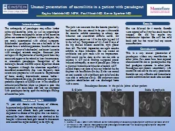
Raghu Maddela MD MPH Paul Nkadi MD Kevin Sperber MD The arthropathy of pseudogout may affect lumbar spine and sacroiliac joints as well as appendicular joints Whereas radiographic lesions of the sacroiliac joints are common in patients with ID: 765397

pain pseudogout disease joints pseudogout pain joints disease joint sacroiliitis severe steroids sacroiliac crystal cppd intraarticular presentation leg deposition

Share:

Link:

Embed:

Download Presentation from below link

Download Presentation The PPT/PDF document "Raghu Maddela MD; MPH, Paul" is the property of its rightful owner. Permission is granted to download and print the materials on this web site for personal, non-commercial use only, and to display it on your personal computer provided you do not modify the materials and that you retain all copyright notices contained in the materials. By downloading content from our website, you accept the terms of this agreement.


Presentation Transcript

Raghu Maddela MD; MPH, Paul Nkadi MD, Kevin Sperber MD The arthropathy of pseudogout may affect lumbar spine and sacroiliac joints, as well as appendicular joints. Whereas radiographic lesions of the sacroiliac joints are common in patients with pseudogout, they are rarely accompanied with clinical symptoms. Subchondral pseudocysts that are a hallmark of the disease have a variable appearance, but often occur as a typical cluster of subchondral, coalescent lucencies with smudged, sclerotic margins.1 Structural joint collapse with fragmentation of cartilage and bone may occur and appear to be related, at least in some cases, to antecedent pseudocysts. Recognition of the radiologic features in CPPD crystal deposition disease is important. Although the alterations superficially resembled osteoarthritis, they were frequently more severe and progressive with extensive fragmentation of bone, causing intra-articular osseous bodies.1,2 These characteristics allow the radiologist to suggest a probable diagnosis of CPPD even in the absence of articular calcification. Here we present a case who presented with acute back pain and was diagnosed with pseudogout basing upon the radiologic findings of sacroiliac joint. She was followed for 4 months. Steroids were tapered off to 5 mg but could never be stopped. She did not require any intraarticular injection. She still has mild back pain but has never had any severe exacerbations. This is a very unusual presentation of pseudogout without any manifesttions in other joints. Few cases have been reported about sacroiliitis due to pseudogout but they had psudogout symtoms in other joints. Pseudogout can present uncommonly with sacroiliitis and can be the only presentation. Steroids are very effective and intraarticular steroid administration needs to be evaluated. Results 73 year old female with history of diabetes, hypertension,dyslipidemia,myelodysplastic syndrome and chronic back pain secondary to degenerative disease of the spine who was recently treated for latent tuberculosis was admitted to the hospital with severe back pain located in the sacrum and sacroiliac region radiating to the right leg. The pain was so severe that she became practically bed bound. She was treated in the past with steroids for reactive arthritis secondary to urinary tract infection and clostridium difficile colitis. On examination power was 1/5 in the right leg and 3/5 in the left leg. Temperature of 101.5 and X rays of the hip showed bilateral sacroiliitis, right greater than left. The initial impression was again reactive arthritis to these infections. She was started on antibiotics and pain meds with no improvement. MRI could not be done as she was not able to tolerate it. CT pelvis findings suggested crystal induced arthropathy, as seen in pseudo gout. Prior to a planned intraarticular injection of steroid, a CTguided biopsy was done to rule out infection but the aspirate was insufficient. On day 20 she was started on oral steroids with significant pain relief and she was able to ambulate slowly. She underwent acute inpatient rehabilitation and was discharged home with a cane. Unusual presentation of sacroiliitis in a patient with pseudogout Introduction Case description Conclusion R SI joint L SI joint Pubic Symphysis Pseudogout features in the pelvic joints of our patient References: 1. Martel W etal . Further observations on the arthropathy of calcium pyrophosphate crystal deposition disease 2. Resnick D etal . Clinical, radiographic and pathologic abnormalities in calcium pyrophosphate dihydrate deposition disease (CPPD): pseudogout .