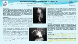
/
Disseminated Takayasu arteritis with neurovascular Disseminated Takayasu arteritis with neurovascular

Disseminated Takayasu arteritis with neurovascular - PDF document

mackenzie
mackenzie . @mackenzie
Follow
342 views
Uploaded On 2022-08-16

Disseminated Takayasu arteritis with neurovascular - PPT Presentation

53 small and medium vessel involvement 1 Peng Loon Cheah 1 Kartini Rahmat 1 Khairul Azmi Abdul Kadir 2 KhengSeang Lim 3 Fariz Yahya 2 MeiLing Sharon Tai 1 Department of Biomedical Imaging ID: 936487

arteritis vessel takayasu involvement vessel arteritis involvement takayasu artery imaging left mca small large vessels wall cerebral medium ica

Share:

Link:

Embed:

Download Presentation from below link

Download Pdf The PPT/PDF document "Disseminated Takayasu arteritis with neu..." is the property of its rightful owner. Permission is granted to download and print the materials on this web site for personal, non-commercial use only, and to display it on your personal computer provided you do not modify the materials and that you retain all copyright notices contained in the materials. By downloading content from our website, you accept the terms of this agreement.


Presentation Transcript

53 Disseminated Takayasu arteritis with neurovascular small and medium vessel involvement 1 Peng Loon Cheah, 1 Kartini Rahmat, 1 Khairul Azmi Abdul Kadir, 2 Kheng-Seang Lim, 3 Fariz Yahya, 2 Mei-Ling Sharon Tai 1 Department of Biomedical Imaging, University Malaya Research Imaging Centre; 2 Division of 3 Division of Rheumatology, Department of Medicine, Faculty of Medicine, University of Malaya, Kuala Lumpur, Malaysia Abstract Takayasu arteritis is a rare granulomatous vasculitis that is commonly classi�ed as a large vessel disease. Small and medium vessel involvement are extremely rare, with only a few case reports woman with pre-existing Takayasu arteritis con�ned to the large vessels, presenting one year later to our centre with status epilepticus. Extensive radiologic studies revealed diffuse encephalopathic changes and multifocal neurovascular involvement, with the vasculitis progressing to encompass discharged well with long term steroid and immunosuppressive therapy. Follow up MRI with vessel wall imaging sequence (VW-MR) and arterial spin labelling (ASL) perfusion imaging demonstrated intra and extracranial vessel wall in�ammation, with generalized reduction in left sided cerebral blood that small and medium vessel involvement does not preclude its diagnosis. Keywords: Takayasu arteritis, neurovascular, small and medium vessel, MRI, vessel wall imaging Neurology Asia 2019; 24(1) : 53 – 59 Address correspondence to: Dr Kartini Rahmat, Department of Biomedical Imaging, Faculty of Medicine, University of Malaya.Email: katt_xr2000@yahoo.com regaining consciousness. She was treated as status epilepticus with intravenous (IV) diazepam, phenytoin and phenobarbitone. Her seizures persisted and IV levetiracetam was subsequently added. The blood pressure was 200/100mmHg, and IV glyceryl trinitrate (GTN) infusion was started. This was preceded by headache and nausea �ve days earlier, for which the patient had presented to another hospital. She had recurrent episodes of syncope in 2016 and was subsequently found to have an ascending aorta aneurysm. Bentall and aortic valve replacement was done in April 2016. Histopathological examination (HPE) showed large vessel arteritis with degenerative aortic valve. The large vessel wall, media and adventitia showed granuloma formation, and a diagnosis of Takayasu arteritis was made. She wa

s treated with oral steroids and warfarin. On examination, her Glasgow coma scale (GCS) score was 12/15 (E3V4M5). The left radial pulse was weaker. She had left horizontal CASE REPORTS INTRODUCTION Takayasu arteritis is a rare disease manifesting as a granulomatous vasculitis that commonly affects the large vessels; predominantly the aorta and its major branches. 1 Cases of Takayasu arteritis have been reported to be more common in Asian countries, with approximately 100-200 new cases reported each year in Japan. 2 Annual incidence in Europe 3 and 2.6 per million population in North America. 4 As a primarily large vessel vasculitis, it is very rare for Takayasu arteritis to present with medium or small vessel involvement. We present a rare case of Takayasu arteritis in a young female with large, medium and small vessel involvement. CASE REPORT A 21-year-old female presented with a cluster of seizures in June 2017. She had a total of 5 seizures which consisted of up-rolling of eyeballs and drooling of saliva lasting for 15 minutes, without 54 nystagmus. There was no facial asymmetry, and neurological examination of the upper and lower limbs were normal. Cardiovascular examination did not reveal any heart murmurs. Computed tomography (CT) of the brain showed an early left frontal lobe infarct (Figure 1 a). Full blood count showed raised total white cell count of 18.8 x 10^9/L. The electroencephalography (EEG) showed intermittent frontal predominant diffuse rhythmic delta activities (IRDAs) indicating moderate diffuse cortical dysfunction. The magnetic resonance imaging (MRI) and angiography (MRA) of the brain showed multifocal T2/FLAIR hyperintensities involving the grey and white matter (Figure 1 b to d) with aneurysmal dilatation of the right common carotid artery (CCA) as well as beading of the basilar artery, both anterior cerebral arteries (ACA) and both middle cerebral arteries (MCA) (Figure 2 a to d). The previous MRI brain performed two years ago was normal. Echocardiogram performed on the third day of hospitalization showed aortic valve prosthesis with aortic area of 1.7cm 2 and trivial aortic regurgitation. Rheumatoid factor, p-ANCA, c-ANCA and ANA results were negative. Cerebral angiography showed aneurysmal dilatation of the right CCA with multifocal Figure 1 (a to d).(a) Non-enhanced CT showing ill-de�ned hypodensities in the left fronta

l lobe [arrow]. (b-d) MRI in coronal T2 FLAIR sequence showing multifocal hyperintensities in the right frontal lobe, both parietal lobes, both occipital lobes and left superior cerebellum. 55 Figure 2 (a to d).Magnetic Resonance Angiography (MRA) showing: (a) Aneurysmal dilatation of the right CCA [arrow]. (b) Beading of the basilar artery [arrow]. (c) Beading of bilateral ACA [arrows]. (d) Beading of bilateral MCA (arrows) stenoses involving the arch of aorta, proximal left subclavian artery, left CCA bulb and left external carotid artery (ECA) origin. There was also beading of the distal ECA branches (Figure 3 a to d). The descending aorta and the renal arteries were normal. Carotid Doppler study demonstrated vessel wall thickening in both CCAs. In both cerebral angiography and Doppler studies, antegrade �ow was noted in both vertebral arteries, with no evidence of left sided subclavian steal. She was treated with IV methylprednisolone 1g daily for three days followed by oral prednisolone. She was discharged well after 12 days of admission with oral steroids, levetiracetam and immunosuppresants (cyclophosphamide for 6 months followed by maintenance methotrexate). During the follow up period, she experienced a few episodes of headaches as well as chronic neck and left upper limb pain, but remained seizure free. A follow up MRI with vessel wall MR (VW- MR) and arterial spin labelling (ASL) perfusion imaging was done 17 months later. VW-MR imaging demonstrated vessel wall enhancement in bilateral CCA, proximal right internal carotid artery (ICA) and petrous segments of both ICA. MRA demonstrated unchanged vasculopathy involving the aorta, left subclavian artery, bilateral CCA and left ECA. However, there was resolution of the beaded appearance affecting both ACA, MCA and the basilar artery, with no vessel wall enhancement seen (Figure 4 a to e). ASL perfusion imaging with cerebral blood �ow (CBF) colour mapping demonstrated reduction in left sided cerebral blood �ow, predominantly in the ACA and MCA territories (Figure 5). DISCUSSION Our patient represents a rare case of Takayasu arteritis with initial isolated large vessel disease, now presenting with subsequent involvement of the small and medium sized vessels in the neurovascular circulation. The diagnosis of Takayasu arteritis was confirmed via 56 Figure 4 (a to e).MRA with VW-MR im

aging showing: (a) Vessel wall enhancement of both CCA (arrows), with wall enhancement and thickening of the proximal right ICA (arrow head). (b) Vessel wall enhancement of both petrous ICA (arrows). (c) Non-enhancement of the MCA walls (arrows). (d and e) Resolved vasculopathy in bilateral MCA, ACA and basilar artery. Figure 3 (a to d).Conventional digital subtraction cerebral angiography showing: (a) Focal stenosis at the arch of aorta, proximal to the branching of the brachiocephalic trunk [arrow]. (b) Short segment stenosis involving the proximal left subclavian artery before the branching of the vertebral artery [arrow]. This explains the weaker left radial pulse elicited on clinical examination. (c) Focal stenosis at the left CCA bulb [arrow] and at the origin of left ECA [arrow head]. (d) Beaded appearance of the distal ECA branches [arrows]. 57 histopathology in 2016, and the initial MRI brain was normal. However, in her second presentation one year later, extensive radiologic studies revealed diffuse bilateral encephalopathic changes with multi-focal neurovascular involvement of the large, medium and small-sized vessels, suggesting an ischaemic nature. In view of the persistent symptoms, follow up MRI was performed 17 months later with ASL perfusion and VW-MR imaging to further characterize and delineate the extent of disease. The ischaemic nature of the brain parenchymal changes were further supported by ASL perfusion imaging, which demonstrated reduction in left sided cerebral blood �ow likely due to the persistent stenosis at the left CCA bulb. ASL perfusion imaging utilizes the magnetic labelling of water protons in blood upstream to the imaged segment, with subsequent in�ow of labelled blood into the imaged segment causing alteration of tissue magnetization. The acquired signals are subtracted from pre-labelling control signals, with the resulting signal differences functioning as indicators of cerebral blood �ow. 5 While MRA demonstrated unchanged vasculopathy involving the extracranial vessels, contrast enhanced VW-MR imaging with fat saturation pulse demonstrated vessel wall enhancement involving both intra and extracranial vessels. No wall enhancement were seen in the previously involved smaller intracranial vessels (bilateral ACA, MCA and basilar artery), with MRA of these vessels also showing resolution of vasculopathy.

These �ndings are likely due to improving disease activity secondary to immunotherapy, with Hauenstein et al. having reported reduced sensitivity of VW-MR imaging in patients post corticosteroid therapy. 6 VW-MR Figure 5.ASL perfusion imaging with CBF colour mapping showing reduction in left sided cerebral blood �ow, predominantly in the ACA and MCA territories. imaging employs suppression pulses for fat and intraluminal blood, which after administration of contrast, enables the visualization of vessel wall enhancement in in�amed vessels. This technique has been shown to better demonstrate the disease extent in vasculitides such as Takayasu arteritis. 7,8 Takayasu arteritis with small and medium sized vessel involvement is extremely rare. Table 1 summarizes the previously reported cases that presented with small and medium sized vessel involvement. The majority of reported cases had cutaneous involvement, with a smaller proportion presenting with neurovascular and ocular manifestations. Amongst the cases with neurovascular involvement, Table 2 demonstrates that vertebral artery and MCA involvement were commonest, with 10 of the 17 reviewed cases exhibiting vertebral artery involvement and 8 cases exhibiting MCA involvement. While there was involvement of a single medium sized vessel in nearly half the cases (8 of 17), all of the reported cases had co-existing large vessel involvement. Though comparable, our patient exhibited more extensive and mixed involvement of the ECAs, basilar artery, ACAs and MCAs. This demonstrates that although Takayasu arteritis is primarily regarded as a large vessel vasculitis, small or medium sized vessel involvement do occur, with previous reports describing cutaneous, neurovascular and ocular involvement. Classi�cation of Takayasu arteritis as a large vessel vasculitis may cause diagnostic delays in patients presenting with small and medium sized vessel involvement. While brain biopsy is considered the gold standard in diagnosing cerebral vasculitis, it was not performed for this patient as histopathologic 58 con�rmation had already been obtained from the aortic sample in 2016, which when coupled with the suggestive clinical and radiological features, ensured that there was no diagnostic confusion. 9 Unfortunately, the Bentall procedure in 2016 was performed at a different hospi

tal, and attempts to obtain the histopathology slides were unsuccessful. In conclusion, despite Takayasu arteritis being regarded as a large vessel vasculitis, it is important to consider the diagnosis in patients presenting with small and medium sized vessel involvement. A thorough radiologic evaluation of the aorta and its main branches with histopathologic correlation is essential to ensure the diagnosis is not missed or delayed. VW-MR and perfusion imaging sequences should also be considered as part of the evaluation of disease extent and activity. ACKNOWLEDGEMENT This work was supported by University Malaya Research Grant RP 052B-17HTM DISCLOSURE Con�ict of interest: None Table 1:Summary of previously reported Takayasu arteritis with small or medium sized vessel involvement (in addition to large vessels) AuthorsInvolvement Pattern (in addition to large vessels)No. of Patients Kono M et al . 10 , 2017 Cutaneous vessels, scleritis, left posterior tibial 1 artery and right peroneal artery Rocha LK et al . 11 , 2013 Cutaneous vessels (skin biopsy proven vasculitis)3 Noel N et al . 12 , 2013 Small retinal vessels9 Ringleb PA et al . 13 , 2005 13 Pascual-Lopez et al . 14 , 2004 Cutaneous vessels (skin biopsy proven vasculitis)23 Klos K et al . 15 , 2003 Molnar P et al . 16 , 1984 Neurovascular (post mortem study) Table 2:Summary of previously reported Takayasu arteritis with small or medium sized neurovascular involvement (in addition to large vessels). ACA: anterior cerebral artery, MCA: middle cerebral artery, PCA: posterior cerebral artery, ICA: internal carotid artery, ECA: External carotid artery, VA: vertebral artery, BA: basilar artery, PICA: posterior inferior cerebellar artery, SCA: superior cerebellar artery Neurovascular Involvement PatternNo. of Patients (in addition to large vessels) Ringleb PA et al . 13 , 2005 1 1 2 ICA, MCA 1 4 ICA, VA, BA ICA, MCA, VA ICA, ECA, MCA 1 ICA, ECA, MCA, VA ICA, ECA, MCA, VA, BA Klos K et al . 15 , 2003 ICA, VA, BA, PICA, ACA, MCA PCA, SCA 1 Molnar P et al . 16 , 1984 All major intracranial vessels1 (post mortem study) 59 REFERENCES 1.Nicholson T, Patel J. Granulomatous vasculitis (Takayasu’s disease). In: Adam A, Dixon AK, Grainger RG, Allison DJ, eds: Grainger & Allison’s diagnostic radiology: A textbook of medical imaging. 1. Philadelphia: Churchill Livingstone/Elsevier, 2008:561-2. Numano

F, Kobayashi Y. Takayasu arteritis - beyond pulselessness. Intern Med 1999;38(3):226-32. 3.Onen F, Akkoc N. Epidemiology of Takayasu arteritis. Presse Med 2017;46(7-8 Pt2):192-203. Hall S, Barr W, Lie JT, Stanson AW, Kazmier FJ, Hunder GG. Takayasu arteritis. A study of 32 North American patients. Medicine 1985;64(2):89-99. 5.Haller S, Zaharchuk G, Thomas DL, Lovblad KO, Barkhof F, Golay X. Arterial spin labelling perfusion of the brain: Emerging clinical applications. Radiology 2016;281(2):337-56. 6.Hauenstein C, Reinhard M, Geiger J, et al. Effects of early corticosteroid treatment on magnetic resonance imaging and ultrasonography �ndings in giant cell arteritis. Rhematology 2012;51:1999-2003. Kato Y, Terashima M, Ohigashi H, et al . Vessel wall in�ammation of Takayasu arteritis detected by contrast-enhanced magnetic resonance imaging: Association with disease distribution and activity. PLoS ONE 2015;10(12):e0145855. 8.Mandell DM, Mossa-Basha M, Qiao Y, et al . Intracranial vessel wall MRI: Principles and expert consensus recommendations of the American Society of Neuroradiology. Am J Neuroradiol 2017;38:218-29. 9.Berlit P, Kraemer M. Cerebral vasculitis in adults: what are the steps in order to establish the diagnosis? Red �ags and pitfalls. Clin Exp Immunol 2014;175(3):419-24. Kono M, Yasuda S, Atsumi T. Takayasu arteritis with varied-size vessel involvement. J Rheumatol 2017;44(1):125-6. 11.Rocha LK, Romitti R, Shinjo S, Neto ML, Carvalho J, Criado P. Cutaneous manifestations and comorbidities in 60 cases of Takayasu arteritis. J Rheumatol 2013;40(5):734-8. 12.Noel N, Butel N, Hoang PL, et al . Small vessel involvement in Takayasu’s arteritis. Autoimmun Rev 2012;12(3):355-62. 13.Ringleb PA, Strittmatter EI, Loewer M, et al. Cerebrovascular manifestations of Takayasu arteritis in Europe. Rheumatology 2005;44(8):1012-5. Pascual-Lopez M, Hernandez-Nunez A, Aragues- Montanes M, Dauden E, Fraga J, Garcia-Diez A. Takayasu’s disease with cutaneous involvement. Dermatology 2004;208(1):10-5. 15.Klos K, Flemming KD, Petty GW, Luthra HS. Takayasu’s arteritis with arteriographic evidence of intracranial vessel involvement. Neurology 2003;60(9):1550-1. 16.Molnar P, Hegedus K. Direct involvement of intracerebral arteries in Takayasu’s arteritis. Acta Neuropathol 1984;63(1):83-6. Neurology Asia March 20