/
EPITHELIA = Epi (outside) EPITHELIA = Epi (outside)

EPITHELIA = Epi (outside) - PowerPoint Presentation

mackenzie
mackenzie . @mackenzie
Follow
342 views
Uploaded On 2022-06-11

EPITHELIA = Epi (outside) - PPT Presentation

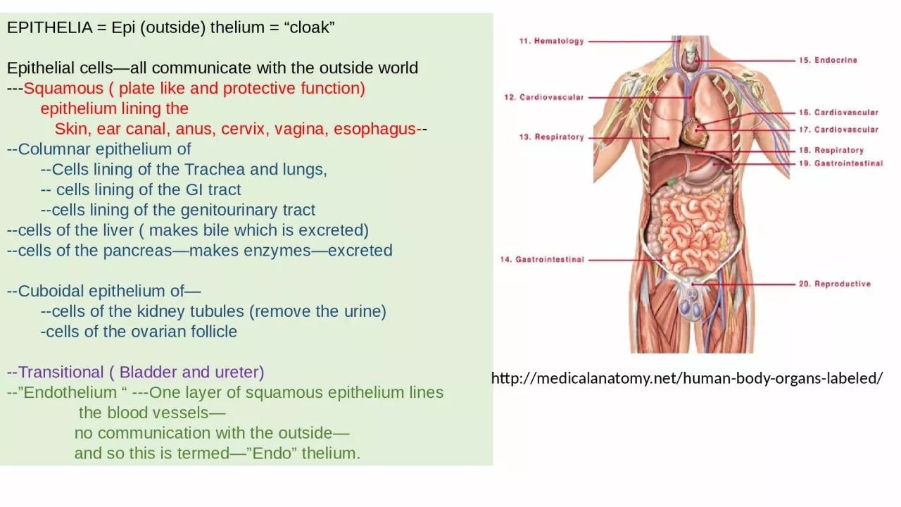
thelium cloak Epithelial cellsall communicate with the outside world Squamous plate like and protective function epithelium lining the Skin ear canal anus cervix vagina esophagus ID: 917352

epithelium cells lining blood cells epithelium blood lining squamous human epithelial columnar trachea layer keratin neoplastic body stomach mouse

Share:

Link:

Embed:

Download Presentation from below link

Download Presentation The PPT/PDF document "EPITHELIA = Epi (outside)" is the property of its rightful owner. Permission is granted to download and print the materials on this web site for personal, non-commercial use only, and to display it on your personal computer provided you do not modify the materials and that you retain all copyright notices contained in the materials. By downloading content from our website, you accept the terms of this agreement.


Presentation Transcript

Slide1

EPITHELIA = Epi (outside) thelium = “cloak”Epithelial cells—all communicate with the outside world---Squamous ( plate like and protective function) epithelium lining the Skin, ear canal, anus, cervix, vagina, esophagus----Columnar epithelium of --Cells lining of the Trachea and lungs, -- cells lining of the GI tract --cells lining of the genitourinary tract--cells of the liver ( makes bile which is excreted)--cells of the pancreas—makes enzymes—excreted--Cuboidal epithelium of— --cells of the kidney tubules (remove the urine) -cells of the ovarian follicle--Transitional ( Bladder and ureter)--”Endothelium “ ---One layer of squamous epithelium lines the blood vessels— no communication with the outside— and so this is termed—”Endo” thelium.

http://

medicalanatomy.net

/human-body-organs-labeled/

Slide2

EPITHELIUM is the term given to the cells that -cover the exterior surface of the body, -lines both the internal closed cavities of the body, -lines body tubes that communicate with the exterior (alimentary, respiratory, genitourinary) -comprise the various organs (liver) Epithelium can be -impervious (epidermis or bladder) , -secretory (stomach), -absorptive (intestines), -be a transport system(trachea), -receive sensory stimuli (taste buds of the tongue)

Slide3

Diagrammatic representation of epithelial types

Slide4

Epithelium can be impervious (epidermis or bladder) Stratified Squamous epithelium—stacked up like plates

Squamous epithelium function helps with shear forces that are encountered as in

-Skin, with

anuclear

keratin layer

-Esophagus(No keratin layer)

-Cervix (no keratin layer)

-External ear canal

-Anus

Slide5

Epithelium can be secretory (stomach), absorptive (intestines), Columnar epithelium—is so termed because the cells are arranged like columns

Epithelial lining of intestine

The height of the cell is greater than the width

Thus secreted material can be stored or secreted

Slide6

Cuboidal epitheliumOvary with developing folliclesPrimordial follicles lined by flat squamous epitheliumDeveloping Primary follicles lined by cuboidal epithelium

Stromal fibroblasts

Slide7

Pseudo-stratified columnar(lining of trachea and ..)Transitional:liningBladder and ureter

Slide8

Pseudo-stratified Columnar epitheliumHuman trachea

Mouse trachea

Slide9

Junctional zone epithelium: Where epithelium of one kind changes naturally to anotherSquamo-Columnar junction between esophagus and stomach in Human ( Left) and in the mouse stomach (right)

Slide10

Junctional zone epithelium: Where epithelium of one kind changes naturally to another

Slide11

Different cell types with different functions in the GI tract

Slide12

Colon is a good sample for use in a variety of histologic stains

Slide13

Slide14

Once the neoplastic cells (Neo=New growth) invade the basement membrane, they can access underlying blood vessels to metastasize to distant sitesBenign = “Normal”Malignant = neoplastic cells have invaded through basement membraneCarcinoma = malignancy of epithelial cellsSarcoma = malignancy of supporting cells

Slide15

Supporting cells or “Stromal cell” -Fibroblast -endothelial cells of blood vessels -Muscle—smooth, skeletal, cardiac -adipose -bone --cartilageCells circulating in the blood --red blood cells --platelets --white blood cells -Neutrophils, Eosinophils, basophils -Lymphocytes -Monocytes—when these settle in tissues they are called macrophages (Macro) --Macrophages in different organs have different functions --microglia in the brain` --Kupffer cells in the liver --alveolar macrophages in the lung

Slide16

Slide17

Slide18

Slide19

Human BreastHuman ProstateGreen= keratin expressing epithelial cellsRed = Smooth muscle actin expressing myo-epithelial cells

Slide20

Slide21

Slide22

Slide23

This is NOT a Neoplasm, but is necrotic( death due to pathology of infections or blood vessel compromise)Here, due to the invasive organisms ,which can be identified with a Gram stain

Slide24

Mouse Models of Human Ulcerative colitis

Slide25

Colon Polyp—projects into the lumen and is lined by layers of tubular Neoplastic (new growth) cells—but there is no invasion into stalkAdenocarcinoma—tubular structures with many layers of neoplastic cells—with lots of mitoses,and invasion of basement membrane

Slide26

Slide27

Slide28

Slide29

 A Panel of 66 Human Squamous Carcinomas on the left—show more expression of SiglecXII, compared to the panel with 47 Human Adenocarcinomas on the rightAn example of results using the 5 step with biotinyl tyramide enhancement with Mouse anti-SiglecXII on paraffin sections, Formalin Fixed Paraffin Embedded ( FFPE) Multi-tissue arrays ( MTA) from Novus Biological June 2019)