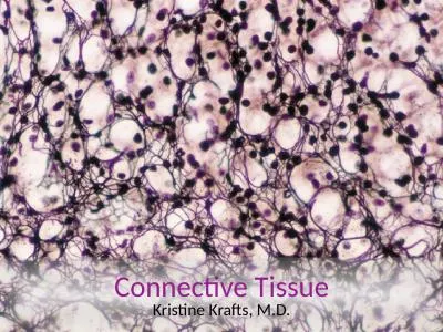
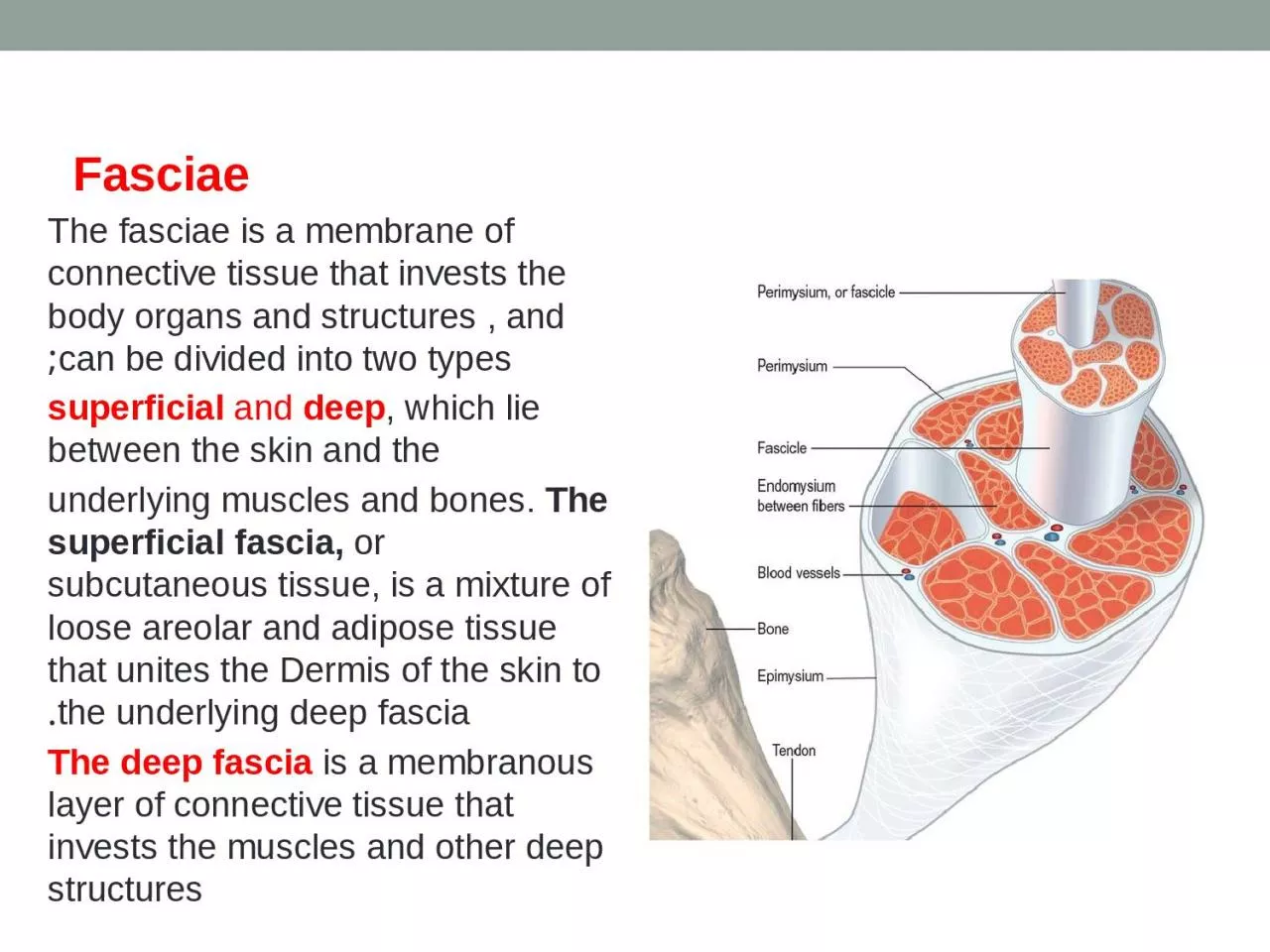
/
Fasciae   The fasciae is a membrane of connective tissue that invests the body organs Fasciae   The fasciae is a membrane of connective tissue that invests the body organs

Fasciae The fasciae is a membrane of connective tissue that invests the body organs - PowerPoint Presentation

madison
madison . @madison
Follow
0 views
Uploaded On 2024-03-15

Fasciae The fasciae is a membrane of connective tissue that invests the body organs - PPT Presentation

superficial and deep which lie between the skin and the underlying muscles and bones The superficial fascia or subcutaneous tissue is a mixture of loose areolar and adipose tissue that unites the Dermis of the skin to the underlying deep fascia ID: 1048388

system fibers nervous nerve fibers system nerve nervous muscle spinal vessels tissue cartilage consists muscles bones nerves called joint

Share:

Link:

Embed:

Download Presentation from below link

Download Presentation The PPT/PDF document "Fasciae The fasciae is a membrane of c..." is the property of its rightful owner. Permission is granted to download and print the materials on this web site for personal, non-commercial use only, and to display it on your personal computer provided you do not modify the materials and that you retain all copyright notices contained in the materials. By downloading content from our website, you accept the terms of this agreement.


Presentation Transcript

1. Fasciae The fasciae is a membrane of connective tissue that invests the body organs and structures , and can be divided into two types;superficial and deep, which lie between the skin and theunderlying muscles and bones. The superficial fascia, or subcutaneous tissue, is a mixture of loose areolar and adipose tissue that unites the Dermis of the skin to the underlying deep fascia. The deep fascia is a membranous layer of connective tissue that invests the muscles and other deep structures

2. MusclesThere are three types of muscle, skeletal, smooth, and cardiac muscles.Skeletal muscles are responsible for production of the movements of the skeleton; they are sometimes called voluntary muscles and are made up of striped muscle fibers. A skeletal muscle has two or more attachments. The attachment that moves the least is referred to as the origin, and the one that moves the most is referred to as the insertion.The fleshy part of the muscle is referred to as its belly, the ends of a muscle are attached to bones, cartilage, or ligaments by cords of fibrous tissue called tendons . Occasionally, flattened muscles are attached by a thin but strong sheet of fibrous tissue called an aponeurosis .

3. Nerve Supply of Skeletal Muscle (figure 2.10). The nerve supply to a muscle is a mixed nerve, about 60% is motor and 40% is sensory, and it also contains some sympathetic autonomic fibers. The nerve enters the muscle at about the midpoint on its deep surface, often near the margin; the place of entrance is known as the motor point. This arrangement allows the muscle to move with minimum interference with the nerve trunk.

4. Numenclature of muscleDeltoid TriangularTeres Round Brachii of the armProfundus DeepSuperficialis SuperficialExternus ExternalSternocleidomastoid From sternum and clavicle to mastoidProcess Coracobrachialis From coracoid Process to arm Extensor Extend Flexor Flex Constrictor Rectus Straight

5. Smooth muscles.Smooth muscle consists of long, spindle-shaped cells closely arranged in bundles or sheets. It forms the muscular wall of the digestive system, and consists of two layers; outer longitudinal and inner circular. A wave of contraction of the circularly arranged fibers passes along the tube, milking the contents onward. By their contraction, the longitudinal fibers pull the wall of the tube proximally over the contents. This method of propulsion is referred to as peristalsis. In the urinary bladder and the uterus, the smooth fibers are irregularly arranged and interlaced with one another. Their contraction is slow and sustained and brings about expulsion of the contents of the organs.In the walls of the blood vessels, the smooth muscle fibers are arranged circularly and serve to modify the caliber of the lumen. Depending on the organ, smooth muscle fibers may be made to contract by local stretching of the fibers, by nerve impulses from autonomic nerves, or by hormonal stimulation

6. Cardiac muscles figure.Cardiac muscle consists of striated muscle fibers that branch and unite with each other. It forms the myocardiu of the heart. Its fibers tend to be arranged in whorls and spirals, and they have the property of spontaneous and rhythmic contraction. Specialized cardiac muscle fibers form the conducting system of the heart. Cardiac muscle is supplied by autonomic nerve fibers that terminate in the nodes of the conducting system and in the myocardium.

7. Cartilaginous JointsCartilaginous joints can be divided into two types: primary and secondary. A primary cartilaginous joint is one in which the bones are united by a plate or a bar of hyaline cartilage. Thus, the union between the 1st rib and the manubrium sterni is an example of such a joint. No movement is possible.A secondary cartilaginous joint is one in which thebones are united by a plate of fibrocartilage and the articularsurfaces of the bones are covered by a thin layer of hyalinecartilage.

8. Synovial JointsThe articular surfaces of the bones are covered by a thin layer of hyaline cartilage separated by a joint cavity This arrangement permits a great degree of freedom of movement. The cavity of the joint is lined by synovial membrane, which extends from the margins of one articular surface to those of the other. The synovial membrane is protected on the outside by a tough fibrous membrane called the capsule.LigamentsA ligament is a cord or band of connective tissue uniting two structures. Commonly found in association with joints, ligaments are of two types. Most are composed of dense bundles of collagen fibers and are un stretchable under normal conditions.

9. Blood vessels are of three types: arteries, veins, and capillariesArteries transport blood from the heart and distribute it to the various tissues of the body by means of their branches . Arteries have a tick muscular wall consist of smooth muscles arranged circularly.The smallest arteries, <0.1 mm in diameter, are referred to as arterioles. The joining of branches of arteries is called an anastomosis. Arteries do not have valves. Anatomic end arteries are vessels whoseterminal branches do not anastomose with branches ofArteries.

10. Veins are vessels that transport blood back to the heart;many of them possess valves. The smallest veins are calledvenules. Medium-size deep arteries are often accompanied by two veins, one on each side, called venae comitantes. Veins have a thinner muscular wall than arteries.Veins leaving the gastrointestinal tract do not go directlyto the heart but converge on the portal vein; this vein enters the liver and breaks up again into veins of diminishing size, which ultimately join capillary-like vessels, termed sinusoids, in the liver. A portal system is thus a system of vessels interposed between two capillary beds.

11. Capillaries are microscopic vessels in the form of a network connecting the arterioles to the venules .Sinusoids resemble capillaries in that they are thin-walled blood vessels, but they have an irregular cross diameter and are wider than capillaries. They are found in thebone marrow, the spleen, the liver, and some endocrine glands.Lymphatic vessels are tubes that assist the cardiovascular system in the removal of tissue fluid from the tissue spaces of the body; the vessels then return thefluid to the blood. Lymphatic vessels are found in all tissues and organs of the body except the central nervous system, the eyeball, the internal ear, the epidermis of the skin, the cartilage, and the bone. The lymph vessels that carry lymph to a lymph node are referred to as afferent vessels, those that transport it away from a node are efferent vessels.

12. Basic structuresNervous system. The nervous system is divided into two main parts: the central nervous system, which consists of the brain and spinal cord, and the peripheral nervous system, which consists of 12 pairs of cranial nerves and 31 pairs of spinal nerves and their associated ganglia. Functionally, the nervous system can be further divided into the somatic nervous system, which controls voluntary activities, and the autonomic nervous system, which controlsinvoluntary activities.

13. Nervous system. The nervous system is divided into two main parts: the central nervous system, which consists of the brain and spinal cord, and the peripheral nervous system, which consists of 12 pairs of cranial nerves and 31 pairs of spinal nerves and their associated ganglia. Functionally, the nervous system can be further divided into the somatic nervous system, which controls voluntary activities, and the autonomic nervous system, which controlsinvoluntary activities.

14. Neuron is the term given to the nerve cell and all its processes. The nerve cell has two types of processes, called dendrites and an axon. Dendrites are the short processes of the cell body; the axon is the longest process of the cell body. The interior of the central nervous system is organizedinto gray and white matter. Gray matter consists of nerve cells embedded in neuroglia. White matter consists of nerve fibers (axons) embedded in neuroglia. Peripheral Nervous SystemThe peripheral nervous system consists of the cranial and spinal nerves and their associated ganglia. On dissection, the cranial and spinal nerves are seen as grayish white cords. They are made up of bundles of nerve fibers (axons)supported by delicate areolar tissue. Cranial NervesThere are 12 pairs of cranial nerves that leave the brain and pass through foramina in the skull. All the nerves are distributed in the head and neck except the Xth (vagus), which also supplies structures in the thorax and abdomen.

15. Spinal NervesA total of 31 pairs of spinal nerves leave the spinal cordand pass through intervertebral foramina in the vertebralcolumn . The spinal nerves are named according to the region of the vertebral column with whichthey are associated: 8 cervical, 12 thoracic, 5 lumbar,5 sacral, and 1 coccygeal.Each spinal nerve is connected to the spinal cord by tworoots: the anterior root and the posterior root.The anterior root consists of bundles of nerve fibers carrying nerve impulses away from the central nervous system. Such nerve fibers are called efferent fibers. Those efferent fibers that go to skeletal muscle and cause them to contract are called motor fibers. Their cells of origin lie in the anterior gray horn of the spinal cord.The posterior root consists of bundles of nerve fibersthat carry impulses to the central nervous system and arecalled afferent fibers. Because these fibers are concerned with conveying information about sensations of touch, pain, temperature, and vibrations, they are called sensory fibers. The cell bodies of these nerve fibers are situated in a swelling on the posterior root called the posterior root ganglion.

16. The anterior and posterior roots unite at intervertebral foramen to form a spinal nerve. Here, the motor and sensory fibers become mixed together, so that a spinal nerve is made up of a mixture of motor and sensory fibers. On emerging from the foramen, the spinal nerve divides into a large anterior ramus and a smaller posterior ramus. The posterior ramus passes posteriorly around the vertebral column to supply the muscles and skin of the back . The anterior ramus continues anteriorly to supply the muscles and skin over the anterolateral body wall and all the muscles and skin of the limbs .

17. BoneBone is a living tissue capable of supporting other body structures. bone is hard because of the calcification of its extracellular matrix and possesses a degree of elasticity because of the presence of organic fibers.Bone exists in two forms: compact and cancellous.Compact bone appears as a solid mass; cancellous bone consists of a branching network of trabeculae. The trabeculae are arranged in such a manner as to resist the stresses and strains to which the bone is exposed.Classification of BonesBones may be classified regionally or according to their general shape. Bones are grouped as follows based on their general shape: long bones, short bones, flat bones, irregular bones, and sesamoid bones

18. Figure 2.21

19. Cartilage. cartilage is a form of connective tissue in which the cells and fibers are embedded in a gel-like matrix. There are three types of cartilage:■■ Hyaline cartilage has a high proportion of amorphous hyaline matrix. it plays an important role in the growth in length of long bones. It covers the articular surfaces of nearly all synovial joints. Hyaline cartilage is incapable of repair. Hyaline cartilage covers the articular surfaces of synovial. Fibrocartilage has many collagen fibers embedded in a small amount of matrix and is found in the discs joints (e.g., the temporomandibular joint, sternoclavicular joint, and knee joint) and on the articular surfaces of the clavicle and mandible. Fibrocartilage, if damaged, repairs itself slowly in a manner similar to fibrous tissue elsewhere..■■ Elastic cartilage possesses large numbers of elastic fibersembedded in matrix. It is flexible and is found in the auricle of the ear,. Elastic cartilage, if damaged, repairs itself with fibrous tissue. Hyaline cartilage and fibrocartilage tend to calcify or even ossify in later life.