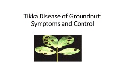
/
Tikka Disease of Groundnut: Symptoms and Control Tikka Disease of Groundnut: Symptoms and Control

Tikka Disease of Groundnut: Symptoms and Control - PowerPoint Presentation

madison
madison . @madison
Follow
27 views
Uploaded On 2024-02-09

Tikka Disease of Groundnut: Symptoms and Control - PPT Presentation

Causal Organism Cercospora personata Berk amp Curt Elle and Eve now known as Cercosporidium personatum Berk amp Curt Deighton amp ID: 1045096

leaf disease surface brown disease leaf brown surface tikka septate host infection conidia hyphae fungicides septa spots mycelium conidiophores

Share:

Link:

Embed:

Download Presentation from below link

Download Presentation The PPT/PDF document "Tikka Disease of Groundnut: Symptoms and..." is the property of its rightful owner. Permission is granted to download and print the materials on this web site for personal, non-commercial use only, and to display it on your personal computer provided you do not modify the materials and that you retain all copyright notices contained in the materials. By downloading content from our website, you accept the terms of this agreement.


Presentation Transcript

1. Tikka Disease of Groundnut: Symptoms and Control

2.

3. Causal Organism: Cercospora personata (Berk. & Curt.) Elle and Eve., now known as Cercosporidium personatum (Berk. & Curt.) Deighton & Cercospora arachidicola Hori. The two form-species differ from each other with respect to the size, shape and colour of necrotic lesions they produce, conidia formation and the nature of the mycelium.

4. C. personata Symptoms of Tikka Disease:Leaf spots are small, more circular and about 1-6 mm in diameter 2. The necrotic lesions appear on both the leaf surfaces and change from dark brown to dark in colour.3. Young spots lack bright yellow halo which, however, develops around the older ones.

5. Mycelium of Tikka Disease: The mycelium consists of hyphae which are entirely internal. The septate hyphae ramify in the intercellular spaces and obtain nutrition by sending branched haustoria into the mesophyll (both spongy and palisade) cells. The unbranched conidiophores arise in tufts from a dense, globular, and brown to black stroma (23-30 µ in dia.) and emerge by rupturing the host epidermis . They are geniculate, olivaceous brown, 24-54 p long and 5-8 µ broad and are either aseptate or septate with 1-2 septa. The conidiophores are confined to the lower surface of the host leaf and are seen arranged in concentric circles in the tuft. The conidia are obclavate or cylindrical, light coloured, 18-60 µ in length and 6-11 µ in breadth. They are septate with 1-7 septa and have bluntly rounded ends.

6.

7. C. arachidicola:Symptoms:The leaf spots are comparatively larger in size, irregularly circular in outline and 4-10 mm in diameter.2. The leaf spots are often confluent and necrotic lesions occur on both the surfaces. Those on the upper surface are reddish brown to black and on the lower surface are light brown.3. There is a yellow halo around each spot but halos on the lower surface are less distinct.

8. Mycelium: It consists of both external and internal hyphae. The internal hyphae are both intercellular and intracellular. The haustoria are absent. The unbranched conidiophores arise scattered from a deep brown stroma (25-100 p in dia.).They are geniculate, yellowish brown and 15 to 45 p long and 3-5 p broad and are either aseptate or septate with 1 to 2 septa. The conidiophores usually occur on the upper surface of the host leaf but occasionally are found on the lower surface as well and thus described as amphigenous. They are sparse and do not occur. The conidia are obclavate hyaline or pale yellow to slightly olivaceous, 38-108 µ long and 8-6 µ broad. They are septate with 4-12 septa and have rounded to distinctly truncate base and sub-acute apex.

9. Disease Cycle:Primary infection takes place by soil borne conidia, which in the soil and on seeds in the shells. The spread of the disease during the season is by means of wind disseminated conidia.Penetration is direct through the epidermis and also by way of stomata. Chiefly it occurs through the upper epidermis. However, there is possibility of infection through both the leaf surfaces.

10.

11. Predisposing Factors of Tikka Disease:Maximum predisposing factors for the development of disease prevail in the month of September. Prolonged low temperature and heavy dew favour severe infection.relative humidity plays an important role in infection.The application of nitrogen and phosphatic fertilizers increase disease incidence whereas potash decreases it slightly.Ascorbic acid and riboflavin content of host leaves appear to play some part in susceptibility and resistance to the disease.Increase in riboflavin content of the leaves is correlated with resistance whereas increase in ascorbic acid is directly related to susceptibility.

12. Control Measures of Tikka Disease:Rotation of crops, seed treatment and disposal of infected host debris by burning or burying in deep pits eliminate chances of primary infection from the soil borne inoculum.The seeds within the shells are disinfected with sulphuric acid.Without shells, they are soaked for half an hour in 0.5% copper sulphate solution. Agrosan GN dressing of naked seeds is equally effective.To check secondary spread of disease in the field, spraying with suitable fungicides is the only remedy. Among the fungicides the use of Bordeaux mixture has given good results.red oxide of copper plus sulphur as highly effective. Daconil and Benlate are the two more effective fungicides than Dithane M45 for pea nut leaf spot disease control.The sprays of these fungicides increased yield as much as 100%. Early maturing varieties are reported to possess resistance to the disease.