/
Dr.  Essam  AL- Fahadwi Measles Dr.  Essam  AL- Fahadwi Measles

Dr. Essam AL- Fahadwi Measles - PowerPoint Presentation

maisie
maisie . @maisie
Follow
27 views
Uploaded On 2024-02-03

Dr. Essam AL- Fahadwi Measles - PPT Presentation

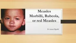
Measles Rubeola DEFINITION Measles is an acute highly contagious viral disease caused by RNA virus Paramyxovirus family genus  Morbillivirus   Morbillivirus structure ID: 1044743

rash measles virus days measles rash days virus infection body viral cell rubella cells respiratory years immune day brain

Share:

Link:

Embed:

Download Presentation from below link

Download Presentation The PPT/PDF document "Dr. Essam AL- Fahadwi Measles" is the property of its rightful owner. Permission is granted to download and print the materials on this web site for personal, non-commercial use only, and to display it on your personal computer provided you do not modify the materials and that you retain all copyright notices contained in the materials. By downloading content from our website, you accept the terms of this agreement.


Presentation Transcript

1. Dr. Essam AL-FahadwiMeasles

2. Measles (Rubeola)

3. DEFINITIONMeasles is an acute highly contagious viral disease caused by RNA virus ( Paramyxovirus family, genus Morbillivirus ).Morbillivirus structurenon-segmented., linear., ssRNA-, helical capsid, enveloped.

4. Agent Source of infection-cases of measles, but not carriers.No animal reservoirInfective material- Nasal secretion ,Respiratory tract &ThroatCommunicability- Highly infectious during prodromal period and at the time of eruption.Secondary attack rate- > 80%

5. Host factorsAge- 6 months to 3 years even up to 10 years Incidence equal in both sexesImmunity – life long immunityMalnourished children are susceptible

6. Environmental factorWinter season, over crowdingTransmission – Droplet infection 4 days before and 4 days after rashIncubation period 7 days

7. Virulence factorsPortal of entry:Respiratory mucus membrane.It first infects the respiratory mucosa, spreads through the lymphatics and bloodstream, and can then infect the conjunctiva, respiratory tract, urinary tract, GI tract, endothelial cells, and the central nervous system.

8. Virulence factors2. Attachment:HemagglutininHemagglutinin in an integral membrane protein found on the surface of the measles virus. Hemagglutinin binds to CD46 (cluster of differentiation), a glycoprotein found on the surface of most cells.(CD46 protects host cells from autoimmune destruction by binding to C3b and C4b and cleaving them).

9. Virulence factors3. Evade the immune system:Immunosuppression: The measles virus blocks TH proliferation response to IL-2.The measle’s Hemagglutinin protein and fusion proteins bind to lymphocytes and interrupt IL-2 cell signaling.

10. Virulence Factors4. Destruction of tissue: a serious febrile illness. The maculopapular rash, which starts at the hairline and spreads over the whole body, is caused by immune T-cells targeted to the infected endothelial cells of the small blood vessels. T-cell deficient individuals do not have the rash, but do have uncontrolled disease which usually results in death.The damage, as well as the control of the disease, is most probably caused by the immune system.

11. TransmissionMeasles transmission is primarily person to person via large respiratory droplets. Airborne transmission via aerosolized droplet has been documented in closed areas for up to 2 hours after a person with measles occupied the area.Measles is highly communicable, with >90% among susceptible persons. Measles may be transmitted from 4 days prior to 4 days after rash onset. Maximum communicability occurs from onset of symptoms through the first 3-4 days of rash.

12. Measles PathogenesisRespiratory transmission of virusReplication in nasopharynx and regional lymph nodesPrimary viremia 2-3 days after exposureSecondary viremia 5-7 days after exposure with spread to tissues

13.

14. Clinical featuresProdromal stageEruptive stagePost-measles stageClinical features3 Cs (Cough, Coryza & Conjunctivitis)Koplik spotsFour days fever (400c)Generalized, maculopapular,erythematous  rash.

15. Measles Clinical Features2-4 days after prodrome, 14 days after exposureMaculopapular, becomes confluentBegins on face and headPersists 5-6 daysFades in order of appearanceRash

16. Stage of Maculopapular Rash (day 4-5) -Temp. rises abruptly as the rash appears (40 C0) -Starts on upper lateral part of neck, -Behind the ear, posterior part of cheek -24hr upper chest -Next 24hr Back, Abdomen -On 3rd day reaches legs/feet and starts fading from face- Hemorrhagic, black Measles-confluent rash, ecchymosis. -Brawny desquamation and brownish discoloration – post measles staining-disappears 7-10 days

17. Koplik's spotsFound in the mouth, these spots look like tiny grains of white sand, each surrounded by a red ring. They are found especially on the inside of the cheek (the buccal mucosa) opposite the 1st and 2nd upper molars.

18. KOPLIK SPOT

19. Clinical signs of measles ( Rubeola virus ) belong to MorbilivirusI.p 9-10 dayErupting phase last for 2-7 day Characterized byRash occur due to1. Ag – Ab reaction on the endothelium of smallVasodilatation2. cytotoxic T cells attack viral infected vascular endothelia cellProdromal phase last for 2-4 day characterized byFlu – like illnessMalaiseFeverPhotophobiaRunning nosesneezing and coughConjunctivitis)redness of eye(Koplik spotsmacula popular rash Charact.by Rash spread over all the body ( from the face and peripherally within 2-4 day(Rash become brownish in 5-10 daysFound in bucchalMucosa ContainWhich are small red macula's or ulcer with centerGiant cellesViral nucleocupsid protein ( NC) Viral Ag complicationVascuilties

20. Complication Diarrhea,PneumoniaOtitis mediaConvulsions,SSPE (sub acute sclerosing panencephalitis)

21. complicationEyeGutEntritis with diarrheaEarOtitis media due to secondary bact infectionCorneal ulcerRareConjunctivitisCNSSymptomatic encephalitis 1/1000 cases Progressive measles inclusion body encephalitisSubacute sclerosing pan encephalitis ( S.S.P.E )Infect teenager and young adult Characterized byFailure of immune response to prevent viral infection Resp. infection Bronchitis Croup Giant cell pneumonia Amount large of measles Ag ( M – Ag ) within inclusion body in infected brain cell Occure after 5-15 years after initial measles Slow progressive demyelination in CNS Lead to Death

22. Failure of immune response to prevent viral infection2virus become defective ( lack of measles protein in brain tissue(1Expression of viral Ag on the surface of Host cell is modulated measles AbVirus become defective in brain cell without coat or lamenting brain cellStart to MultiplyS.S.P.ESo because 1/2S.S.P.E occurThat meanFailure of immune response to complete removal of virus from bodyDue to Agic modulation of viral Ag on surface of host cell

23. Feature of S.S.P.EProgressive mental deteriorationInvoluntary movementPatient with S.S.P.EexhibitMuscular rigidityComaHigh titer of measles Ab in C.S.f serumDefective measles virus in Brain cell* so defective virusEitherSever persistent infectionNo infection

24. Rubella German MeaslesTypically mildOften unrecognizedDifficult to diagnoseSignificant infection in pregnant womenSymptomsSlight fever with mild cold symptomsEnlarged lymph nodes behind ears and back of neckFaint rash on faceRash consists of light pink spotsAdults commonly complain of joint painSymptoms last only a few daysJoint pain may last up to 3 weeksCongenital rubella syndromeFirst trimester susceptibility highestCan lead to fetal death, or neurological disease in survivors (deafness, mental retardation)

25. RubellaCausative AgentRubella virusMember of Togaviridae familySmall, envelopedSingle-stranded RNA genomePathogenesisEnters body via respiratory routeVirus multiplies in nasopharynx, then enters bloodstreamCauses sustained viremiaBlood transports virus to body tissuesImmunity develops against viral antigens resulting antigen-antibody complexes most likely responsible for rash and joint pain

26. RubellaEpidemiologyHumans are only natural hostDisease is highly contagiousLess so than measles 40% of infected people fail to develop symptomsThese individuals can spread virusInfectious 7 days before appearance of rash to 7 days afterPrevention and TreatmentVaccination with attenuated rubella virus vaccineAdministered at 12 months and boosted at 4 to 6 years of ageProduces long-lasting immunity in 95% of recipientsVaccine not given to pregnant women due to potential complicationsWomen are advised not to become pregnant for 28 days post vaccination

27. Clinical signs of rubella1postnatal rubellaI.P 17 – 25 dayIn adolescent and adultIn young childrenMild prodromal symptom with adenopahyRash) morbill form (Then l – 5 dayProdromal symptomRashSome cases enlargement of L – N without skin rash)B) Permanent manifestation appear at birth and became recognize during firstyear(A) transient effect in infantHepatospleenomegallyGrowth retardationFailure to thriveMeningitis2congenital rubella syndromeThe present of rubella virus in uterus lead toFetal birth Birth defect live birthSo the effectTotal or partial deafnessTotal or partial blindnessCongenital Heart diseaseThrombocytopenia puerperal Otitis

28. )C) Developmental abnormalitiesPsychiatric disorder and behavioral manifestationAppear and continue during childhood and adolescenceOccur inOccur in preschool and age school children3 complication of RubellaTransient arthralgea and arthritisInfection of mother with Rubella virus during 1st trimesterThrombocytopenia puerperalEncephalitis) post – infection (Notice 1Notice 2Infection of the mother( month ) during pregnancy First month of pregnancy 2nd month of pregnancy 3rd month of pregnancyApparent and in apparent infection in the motherBirth defect in uncommon at 18 week of pregnancy5-10 year of infection in living born which are apparent the first years of livePercentage of abnormalities in infant50 %20 %4 %-----------------------------------------------------------------------------------------------------------------------

29. Rubella Single serotypeSo Long live immunityRout of entry through resp. tractAfter multiplication in Resp. tractCervical L.N Further multiplicationViraemia after 7 day last until Ab appear on 15 dayRash appearance* virus remain detectable in nasopharynx for several weeks* 25% of cases in subclinicalMeaslesHave 2 spike ( HA , F ) Virus enter the body via resp. tractMultiply in epithet. Of Resp. TractSpread to Reginald L.N ( Further multiplication)Viraemia ( primary )Dissemination Reticulo endothelial systemSecondary viraemiaDissemination toConjunctivaSkinRespiratory tractEpithelial surface of the body

30. ProphylaxisMeasles vaccine is a live, attenuated measles virus grown in chick embryo tissue culture. It is given as part of the MMR vaccine.Vaccine is about 95% effectiveCases continue to occur among those who do not develop or retain good immunity from vaccinations.Most of these infections are caused by contact with infected people who come from outside.

31. ProphylaxisThe first dose of MMR should be given on or after the first birthday; the recommended range is from 12-15 months. The second dose is usually given when the child is 4-6 years old, or before he or she enters kindergarten or first grade. The second dose can be given anytime as long as it is at least four weeks after the first dose.

32. Thank You