/
JOP J Pancreas Online 2010 May 5 113234236 JOP Journal of th JOP J Pancreas Online 2010 May 5 113234236 JOP Journal of th

JOP J Pancreas Online 2010 May 5 113234236 JOP Journal of th - PDF document

martin
martin . @martin
Follow
342 views
Uploaded On 2022-09-06

JOP J Pancreas Online 2010 May 5 113234236 JOP Journal of th - PPT Presentation

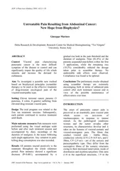
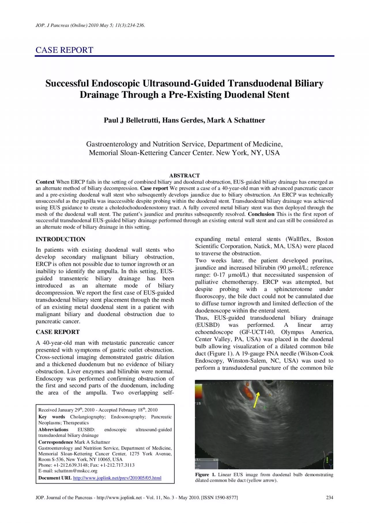
ice Department of Medicine Memorial SloanKettering Cancer Center New York NY USA ABSTRACT When ERCP fails in the setting of combined biliary and duode Received January 29 2010 Accepted Febru ID: 951621

stent biliary bile duodenal biliary stent duodenal bile stents metal figure duct obstruction 2010 pmid pancreas ercp jop drainage

Share:

Link:

Embed:

Download Presentation from below link

Download Pdf The PPT/PDF document "JOP J Pancreas Online 2010 May 5 1132342..." is the property of its rightful owner. Permission is granted to download and print the materials on this web site for personal, non-commercial use only, and to display it on your personal computer provided you do not modify the materials and that you retain all copyright notices contained in the materials. By downloading content from our website, you accept the terms of this agreement.


Presentation Transcript

JOP. J Pancreas (Online) 2010 May 5; 11(3):234-236. JOP. Journal of the Pancreas - http://www.joplink.net - Vol. 11, No. 3 - May 2010. [ISSN 1590-8577] ice, Department of Medicine, Memorial Sloan-Kettering Cancer Center. New York, NY, USA ABSTRACT When ERCP fails in the setting of combined biliary and duode Received January 29, 2010 - Accepted February 18Key words Cholangiography; Endosonography; Pancreatic Neoplasms; Therapeutics EUSBD: endoscopic ultrasound-guided transduodenal biliary drainage Figure 1. Linear EUS image from duodenal bulb demonstratingdilated common bile duct (yellow arrow). JOP. J Pancreas (Online) 2010 May 5; 11(3):234-236. JOP. Journal of the Pancreas - http://www.joplink.net - Vol. 11, No. 3 - May 2010. [ISSN 1590-8577] duct through the proximal end of the duodenal stent. Bile was aspirated and a cholangiogram demonstrated good filling of the right and left duct systems with no evidence of a stricture proximal to the puncture site (Figure 2). Under fluoroscopy, a 0.035-inch straight guidewire (Jagwire, Boston Scientific Corporation, Natick, MA, USA) was passed into the biliary tree and directed toward the hilum. The FNA needle was exchanged for a biliary dilating balloon (Hurricane RX, Boston Scientific Corporation, Natick, MA, USA). The choledochoduodenostomy tract was dilated to 6 mm. A 10x60 mm fully covered self-expanding metal biliary stent (Wallfex, Boston Scientific Corporation, Natick, MA, USA) was placed over the guidewire and deployed under fluoroscopy. One cm of the stent was left protruding into the duodenal bulb through the mesh at the proximal end of the duodenal stent (Figure 3). Good bile and contrast efflux was seen. The final fluoroscopic image did not demonstrate a bile leak (Figure 4). Post-procedure CT imaging demonstrated good positioning of the biliary stent and no evidence of a bile leak (Figure 5). Post-procedure pain and nausea resolved within 48 hours. The patient was discharged from hospital 4 days later. At the time of discharge, the patient was tolerating a full diet. One week post-procedure, the bilirubin had dropped to 56 µmol/L (pre-procedure: 90 µmol/L) and normalized within two weeks. Three months later, the bilirubin remains normal (7 µmol/L). The patient continues to show no signs or symptoms of gastric outlet obstruction or stent dysfunction. He is currently free of pruritus and undergoing palliative chemotherapy. DISCUSSION Patients with advanced pancreatic cancer often develop malignant obstruction of the common bile duct and duodenum. In the setting of simultaneous biliary and duodenal obstruct

ion, combined placement of self-expanding metal biliary and duodenal stents can be performed safely with a high technical success rate in experienced centers [1]. If biliary obstruction is not apparent at the time of clinically significant gastric outlet obstruction, enteral stenting is usually performed in isolation. If possible, the enteral stent is placed proximally or distally to the ampulla to preserve future biliary access. Even if the ampulla is covered by an enteral stent, ERCP with placement of a biliary stent can be subsequently performed by cannulating through the mesh of the duodenal stent [2]. Argon plasma coagulation can also be used to create a larger fenestration at the site of the ampulla to improve biliary access [3]. Thus ERCP is still the endoscopic procedure of choice. Figure 3. Deployed metal biliary stent protruding through mesh o f duodenal wall stent. Figure 2. Cholangiogram obtained through 19-gauge FNA needle in distal common bile duct. Figure 4. Final fluoroscopic image of biliary and duodenal stents. JOP. J Pancreas (Online) 2010 May 5; 11(3):234-236. JOP. Journal of the Pancreas - http://www.joplink.net - Vol. 11, No. 3 - May 2010. [ISSN 1590-8577] If ERCP fails in this scenario, EUSBD has emerged as an alternate intervention. High technical and functional success has been reported with two drainage approaches: 1) transduodenal via the common bile duct and 2) transgastric via a left hepatic duct. The overall technical success rate is 80-90% [4, 5, 6, 7]. These series all report resolution of jaundice and pruritus following stent placement. Complications occur at a rate of 1-20% and include bile peritonitis, pneumoperitoneum, bleeding and cholangitis. The optimal choice of stent is unclear. The use of plastic stents was first described, but re-intervention was often required due to dysfunction over time. With uncovered metal stents, bile leak/peritonitis is a concern, thus fully covered metal stents have been employed more In the case presented here, when ERCP failed, EUSBD was successfully performed through the mesh of the existing duodenal stent. This is the first such report to our knowledge. The bile duct was readily seen through the duodenal bulb and manipulation of the 19-gauge FNA needle, dilating balloon and biliary stent were not hampered by the presence of the duodenal stent. Although the biliary stent protruded into the lumen of the duodenal stent, this did not interfere with passage of gastric contents as the patient was able to maintain adequate oral nutrition through his subsequent period of chemotherapy. We have not

observed duodenal injury secondary to the bare metal end of the biliary stent (Wallfex, Boston Scientific Corporation, Natick, MA, USA). In general, metal biliary stents are less prone to dysfunction over time and placing a fully covered stent across the choledochoduodenostomy tract may reduce the risk of bile leak. Others have reported similar success and safety with the initial use of metal biliary stents for EUSBD [4, 7]. If ERCP fails, even in the presence of a pre-existing duodenal wall stent, EUSBD can provide effective biliary decompression and should be considered as a viable alternative to percutaneous or surgical techniques. Conflict of interest The authors have no potential conflict of interest References Maire F, Hammel P, Ponsot P, Aubert A, O'Toole D, Hentic O, Levy P, Ruszniewski P. Long-term outcome of biliary and duodenal stents in palliative treatment of patients with unresectable adenocarcinoma of the head of the pancreas. Am J Gastroenterol 2006; 101:735-42. [PMID 16635221] Vanbiervliet G, Demarquay JF, Dumas R, Caroli-Bosc FX, Piche T, Tran A. Endoscopic insertion of biliary stents in 18 patients with metallic duodenal stents who developed secondary malignant obstructive jaundice. Gastroenterol Clin Biol 2004; 28:1209-13. [PMID 15671930] Topazian M, Baron TH. Endoscopic fenestration of duodenal stents using argon plasma to facilitate ERCP. Gastrointest Endosc 2009; 69:166-9. [PMID 19111700] Park DH, Koo JE, Oh J, Lee YH, Moon SH, Lee SS, et al. EUS-guided biliary drainage with one-step placement of a fully covered metal stent for malignant biliary obstruction: a prospective feasibility study. Am J Gastroenterol 2009; 104:2168-74. [PMID 19513026] Yamao K, Bhatia V, Mizuno N, Sawaki A, Ishikawa H, Tajika M, et al. EUS-guided choledochoduodenostomy for palliative biliary drainage in patients with malignant biliary obstruction: results of long-term follow-up. Endoscopy 2008; 40:340-2. [PMID 18389451] Taratino I, Barresi L, Repici A, Traina M. EUS-guided biliary drainage: a case series. Endoscopy 2008; 40:336-9. [PMID 18264890] Will U, Thieme A, Fueldner F, Gerlach R, Waznar I, Meyer F. Treatment of biliary obstruction in selected patients by endoscopic ultrasonography (EUS)-guided transluminal biliary drainage. Endoscopy 2007; 39:292-5. [PMID 17357950] Figure 5. Post-procedure CT demonstrating: juxtaposition o f metal biliary (yellow arrow) and duodenal (green arrow) stents withno evidence of a bile leak; fully covered metal biliary stent(yellow arrow) terminating in the common bile duct; no evidenceof pelvic ascites to suggest a bile leak