/
Vascular Disease Thrombi, Emboli Vascular Disease Thrombi, Emboli

Vascular Disease Thrombi, Emboli - PowerPoint Presentation

mila-milly
mila-milly . @mila-milly
Follow
0 views
Uploaded On 2024-03-15

Vascular Disease Thrombi, Emboli - PPT Presentation

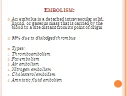
Venous Disease Arterial Disease Thrombus A thrombus is a clot that forms in a blood vessel and remains attached Locations include veins systemic and coronary arteries Cause conditions that encourage ID: 1048386

heart blood left disease blood heart disease left vessel venous pulmonary ductus veins cardiac artery ventricular shock vascular septal

Share:

Link:

Embed:

Download Presentation from below link

Download Presentation The PPT/PDF document "Vascular Disease Thrombi, Emboli" is the property of its rightful owner. Permission is granted to download and print the materials on this web site for personal, non-commercial use only, and to display it on your personal computer provided you do not modify the materials and that you retain all copyright notices contained in the materials. By downloading content from our website, you accept the terms of this agreement.


Presentation Transcript

1. Vascular DiseaseThrombi, Emboli Venous DiseaseArterial Disease

2. ThrombusA thrombus is a clot that forms in a blood vessel and remains attachedLocations include veins, systemic and coronary arteriesCause: conditions that encourage activation of the clotting cascadeRoughing of vessel wallStasis or pooling of bloodInfectious agents

3. ThrombusThrombophlebitisThrombi form in veins (with associated inflamation)Thrombi of heart valvesDue to endocardial inflammation or rheumatic heart diseaseThrombi can occlude vessels and cause ischemia or infarction of organs

4. Embolus TypesEmbolism (emboli): obstruction of a vessel by a moving chunk of materialThromboemboliThrombi will break loose and travel to another siteAir emboliAir bubble from injection, punctured lung or open vesselFat emboliHip replacement or broken bonePlaqueatherosclerosis

5. Embolus SourcesPulmonary emboliUsually arise from thrombophlebitis of veins in lower extremitySystemic (arterial) emboliOrigination in the left circulationLeft heart (thrombi after MI, endocarditis, or dysrhythmiaRenal emboliMesenteric emboliCerebral emboli

6. Thromboembolic disease: TreatmentAdministration of anticoagulants to prevent clot formationAspirinHeparinWarfarin (Coumadin)“Clot busters”Slow or stop thrombus growthAggressive reversal of risk factors

7. Diseases of the VeinsVaricose Veins are distended, tortuous, and palpableVaricose veins in the legs are caused by damage to one or more venous valves due to standing for extended periods of time and the contributing force of gravityA damaged valve permits back flow of blood (venous incompetence) and distended veins

8. PHYSICAL DIFFERENCES BETWEENARTERIES AND VEINSKnow the main differences in the structure ofan artery and vein

9. Varicose VeinsBecause the vein becomes swollen and engorged, the surrounding tissues become edematous.Varicose veins and valvular incompetence can progress to chronic venous insufficiencyChronic pooling of blood leading to hypercoagulability, severe edema, cell death, and necrosis (venous, stasis ulcers)

10. Venous ThrombiWhy would venous thrombi be more common than arterial thrombi?With age, deep veins in the legs become susceptible to thrombiDeep vein thrombosis (DVT)DVT is often asymptomatic, but can lead to pulmonary emboliTreatmentThrombolyticsAnti-embolism stockingAvoid prolonged sitting/immobility

11. Arterial DiseaseHypertension is caused by an increase in cardiac output, total peripheral resistance, or both.Cardiac output: increase in heart rate or stroke volumePeripheral resistance: increase in blood viscosity or reduction in vessel diameter (i.e. atherosclerosis, less elasticity)Types:Primary, secondary, and complicated

12. HypertensionNormal – 120/80Stage 1 – 140/90 (elevations maybe occasional or intermittent)Stage 2 – 140-160/80-100 (sustained)Stage 3 – greater than 160/100 (sustained with markers for cardiovascular disease)Often called the “silent killer”

13. Primary or “essential” HypertensionIn primary (aka: essential or ideopathic) hypertension, the cause is unknown (multifactorial)90-95% of hypertensive patients fall into this groupContributing factors:Family history, age, gender, race, diet, diabetes, obesity, cigarettes, heavy alcohol consumption

14. Secondary HypertensionRenalRenal vascular stenosis, renin producing tumors, renal failure, primary sodium retention (angiotensin)EndocrineThyroid or adrenal disordersVascularArterioscerosis, constriction of the aortaPregnancy induced hypertension (PIH)Pre-eclampsia, eclampsiaStressEpinephrine, norepinephrine, glucocorticoids (cortisol)

15. Complicated HypertensionProlonged hypertensionDemonstrates pathologic effects beyond hemodynamic alterationsVascular remodeling (smooth muscle hypertrophy)Hypo-perfusion: heart, kidneys, eyes, brainExamples of problems, aneurysm rupture, arterioscerosis, vessel occlusion, ventricular enlargement, heart failure, stroke

16. Treatments for HypertensionNon-pharmacologic c/o eliminating risk factorsPharmacologic:Diuretics – decrease blood volume and cardiac outputBlock the renin-angiotensin/alderosterone systemACE inhibitors and Angiotensin II blockersDrugs which relax blood vessel smooth muscleCalcium channel blockers (relax smooth muscles)Beta Blockers (block catecholamines)

17. Orthostatic (postural) HypotensionDecrease in blood pressure upon standing from a reclining positionThe physiologic response to this drop in blood flow is dizzinessCauses: age, drugs (antihypertensives, and antidepressants), prolonged immobility (chronic illness), starvation, physical exhaustion

18. AneurysmsAn aneurysm is a dilation or pouching of a vessel wall or cardiac chamberAs tension increases, the wall become thinnerTrue aneurysms involve all three layers

19. Locations and CausesCommon locations include:Abdominal AortaThoracic AortaCerebral, femoral, & popliteal arteriesRisk Factors include: congenital defect, atherosclerosis, hypertension, Marfans syndrome, diabetes, tobacco, age, infection, and trauma

20. ArteriosclerosisArteriosclerosis is a chronic disease of the arterial system that is characterized by abnormal hardening and thickening of the vessel wallsSmooth muscle cells and collagen fibers migrate in to the intima, causing it to stiffen and thickenThis restricts the artery’s ability to change lumen size (increased vascular resistance)Atherosclerosis is a form of arterioscerosis

21. AtherosclerosisAtherosclerosis is a form of arteriosclerosisThickening and hardening of the vessel walls are caused by deposits of intra-arterial fat (i.e. LDL cholesterol) and fibrin that harden over timeAtherosclerosis is not a single organ/area disease, it varies depending on location, age and geneticsLeads to:Coronary artery disease (CAD)Cardiovascular disease (CVD)Peripheral vascular disease (PVD)

22. Atherosclorotic Plaques

23. AtherosclerosisInflammatory diseasePossible causes: smoking, high cholesterol, hypertensionStep One – InjuryRelease of chemicals announcing the injuryStep Two – Formation of Fatty streakMacrophages engulf oxidized LDL (foam cells)Fatty streak is an accumulation of foam cells

24. Atherosclerosis

25. Atherosclerosis Cont.Step Three:Smooth muscle cells proliferate and migrate over the fatty streak forming a cap (fibrous plaque)Step Four:Fibrous plaques are not stableUlcerationsCalcificationsApoptosis causes rupturePlatelets now have an adherence site and become activatedThis is called a complicated lesion

26. Peripheral Vascular DiseasesThromboangitis Obliterans (Buerger Disease)An inflammatory autoimmune disease of the peripheral arteries of the hands and feet resulting in the formation of non-athersclerotic lesions that block the vesselsCauses pain and hair loss (not on the head) in young men who smoke or use smokeless tobaccoProgress to ulcers and gangrenous lesions of fingers and toes

27. Raynaud DiseaseRaynaud disease is a primary vasospastic disorder of unknown etiology causing episodic vasospasm in arteries of the fingers, and less commonly the toes (pallor/cyanosis)Raynaud phenomenon is the same thing, but it is secondary to other systemic diseasesCollagen vascular disease (scleroderma), smoking, pulmonary hypertension, and environmental factors (cold, vibrating machinery)The treatment is to simply remove the stimulus e.g. cold, drugs, or treat the primary disease

28.

29. MAJOR BLOOD VESSELS WHICH SUPPLY HEART

30.

31.

32.

33.

34.

35.

36. KNOW THE PATHWAYS

37.

38. DysrhythmiasClassified by abnormalities of rate or rhythmAtrial or ventricular Conduction – 1st, 2nd and 3rd degree block, bundle branch blockRate – increased rate give less time for each of the chambers to fill (only the rest time is shortened). Cardiac output may be inadequate despite the increase in rate.

39. Coronary Artery DiseaseNontraditional risk factorsMarkers of inflammation and thrombosisC-reactive protein, fibrinogen, protein C, Factor V Leiden, andplasminogen activator inhibitorElevated homocystine levelsInfectionRisk factorsDyslipidemiaHypertensionCigarette smokingDiabetes mellitusObesity/sedentary lifestyle

40. Ischemia, Injury, Infarction/NecrosisIschemia: Temporary decreased tissue perfusion/oxygenation to myocardial cells“Angina Pectoris”AtherosclerosisCoronary Artery DiseaseInjury: Can become necrotic but is reversiblewith oxygen/tissue perfusionInfarction: Area of tissue that undergoes necrosis following cessation of blood supply

41. ACUTE MYOCARDIALINFARCTIONDefinition: Permanently destroyed myocardial cellsRelated to an alteration in blood flowLeft coronary arteryLeft anterior descending arteryCircumflexRight coronary arterySpecific occluded arteries cause specific area damage

42. Areas Affected by InterruptedBlood SupplyLeft and Anterior Descending (Anterior MI)Along anterior section of left ventricleSeptum separating ventriclesCircumflex (Lateral MI)Upper lateral wall of left ventricleLeft AtriumRight CoronaryRight ventriclePosterior Descending BranchInferior section of left ventricle (Inferior MI)Posterior section of left ventricle (Posterior MI)

43.

44. Areas of Pain Due to MI

45.

46. Cardiac EnzymesResult of Release of Destruction of Lysosomes in CellsCK (CPK) - CKMBElevates 3-6 hours, peaks at 12-24 hoursNormal values:– CK Women 40-150 U/L Men 60-400 U/L– CKMB If less than 5 U/L, probably normal but varies.MyoglobinElevates 1.5 to 6.5 hours, peaks at 4-16 hoursNormal value: 50 to 120 mcg (.5 to 1.5 mg)Troponin I (Gold Standard for MI)Elevates 4-6 hours, peaks at 10-24, stays 10 daysNormal value: 0.6 to 1.5 ng/ml

47. Clinical Manifestations of MITypes of PainChest pain that occurs suddenly and continues despite rest and medicationCrushing/pressureEpigastric burningAnxiety and restlessnessDiaphoresisDyspneaCardiogenic shock

48. What is a “Rule Out MI”?Non-diagnostic ST changes on EKGChest pain relieved by vasodilators and oxygenNon-elevated enzymes (Troponin, CK MB)

49. Dysrhymia ComplicationsAfter AMILife ThreateningFrequent multifocal PVCs from irritable fociLeads to V Tach or V FibDecreases cardiac outputSeptal AMIs lead to heart blocksLeads to severe bradycardiaDecreases cardiac outputReperfusion causes frequent multifocal PVC’sAfter fibrinolytic therapyWait and watchCorrelate with decreased pain at same timeRepeat EKG to see if it improves

50. ShockDefinitionShock is not a withdrawn psychological state as in “the victim was in a state of shock”medically it is Circulatory Collapse – cells don’t get what they needGeneralized severe reduction in blood supply to the body tissues. Inadequate tissue perfusion leads to cellular hypoxia and tissue damage.

51.

52. ShockHypovolemic shockHemorrhageburnssurgery or traumaloss of fluid and electrolytes from the gutCardiogenic shockmyocardial infarctionheart failurearrhythmiasventricular septal rupturemitral valve papillary ruptureNeurogenic Lack of Sympathetic ToneOrthostasisemotional trauma (fear, horror, sight of blood)intense painLow resistance shockSepsisAnaphylaxis

53. Signs and SymptomsVarious causes of shock but many similarsigns and symptomsrapid pulse (tachycardia)rapid breathing (tachypnoea)decreased urine outputconfusion, restlessnesscold clammy skin (except in low resistance shock-warm shock)Decreased urine output

54.

55. Neurogenic shockSudden transient loss of consciousness that occurs when cerebral blood flow falls to less than half normal owing to an abrupt fall in arterial pressure.Caused by activation of Autonomic Nervous Systemincreased parasympathetic activityreduced heart rate (vagal nerve)decreased sympathetic activityVasodilationBradycardia (reduced heart rate)Color Pink

56. Loss of resistance shock

57. LOSS OF BODY FLUID IN HYPOVOLEMIC SHOCK

58. Cardiogenic shock

59.

60.

61.

62. Acquired Heart DiseaseRheumatic heart diseaseBacterial (infective) endocarditisMyocarditisPericarditisCardiomyopathiesValvular dysfunctionStenosisRegurgitationProlapse (can be congenital)Valve related problems resulting fromdiseases

63.

64. CardiomyopathiesDilated (Congestive) All four chambers are dilated, and there is also hypertrophy. The most common cause is chronic alcoholism, though some may be the end-stage of remote viral myocarditis.Hypertrophic The most common form, idiopathic hypertrophic subaortic stenosis (IHSS) results from asymmetricinterventricular septal hypertrophy, resultingin left ventricular outflow obstruction.Restrictive The myocardium is infiltrated with a material that results in impaired ventricular filling. The most common causes are amyloidosis and hemochromatosis.

65.

66. Fetal circulationEmbryologyCardiogenesis begins at approximately 3 weeks’gestationThe heart arises from the mesenchyme– Develops as an enlarged blood vessel with a largelumen and muscular wall– Midsection grows faster than the endsThe heart tube elongates and rotates to the right,creating a bulboventricular loopFetal heart contractions begin by approximatelythe 28th day

67. Developmental Anatomy of theCardiovascular System

68. Septum formation

69. Fetal circulationThe developing fetus has special circulatoryneeds because their lungs, kidneys, anddigestive tracts are non-functionalThe fetus derives it’s oxygen and nutrientsand eliminates its waste through the maternal blood supply by way of the placentaNormally there is no fetal/maternal blood mixing: the fetus is dependant of capillary exchange

70. Fetal CirculationPlacentaUmbilical VeinDuctus Venousus: between umbilical vein and inferior vena cava (bypasses the liver)Inferior Vena cava to heartForamen Ovale (bypasses the lungs)Ductus Arteriousus (bypassess the lungs)AortaUmbilical Arteries

71.

72. Circulation after BirthNon functional Structures:Placenta (delivered)Umbilical vein atrophy (round ligament)Ductus Venousus (atrophy about 1wk.Ligamentum venosum)Foramen ovalae (fossa ovale about 1 month)Ductus arteriousus (atrophy: 3wks ligamentumarteriousum)Umbilical arteries: (atrophy: become lateralumbilical ligaments

73. Congentital Heart DefectsUnderlying cause is known in only 10% of defectsPrenatal, environmental, and genetic risk factorsMaternal rubella, insulin-dependent diabetes, alcoholism, PKU and Hyper-calcemiaDrugsChromosome aberrations

74. Hypoxemic/Cyanotic DefectsAssessment/Clinical ManifestationsPolycythemiaClubbingFatigue with feedingPoor weight gainTachypneaDyspneaAbnormal positioningFlaccid with extremities extendedSide-lying with knees toward chestSquatting in children

75. Acyanotic DefectsWith Increased Pulmonary Blood FlowASD (Atrial Septal Defect)Due to failure of flap to close orHole to big for flap to closeVSD (Ventricular Septal Defect)Structural hole between ventriclesPDA (Patent Ductus Arteriosus)Duct between pulmonary artery and aorta fails toClose after birth (fetal circulation bypasses lungs) due to decreased oxygenation or atelectasis of the lungs (usually RDS)– Oxygen in lungs usually stimulates production ofprostaglandin which closes the ductResults in left to right shunt after birth

76. Atrial Septal Defect (ASD)Hole between the two atria Incidence 1/4000Symptoms– Right sided CHF without murmurEnlarged LiverAtrial conduction defectsPremature Atrial ContractionsAtrial FlutterParoxysmal Atrial TachycardiaQuestion:– Why is there no cyanosis?

77. Ventricular Septal Defect (VSD)Hole between the two ventriclesMost common type of congenital heart defect (3/1000 people)In large VSD’s, pressures become equal, blood flows into the Right ventricle due to systemic pressures, ad large aounts of blood flow into the pulmonary systemSymptomsRight sided CHFMurmur

78.

79. Patent Ductus ArteriosusPDADefined as failure of the ductus arteriosus to close within the first few weeks of lifeThis increases the pressure in the pulmonary system and overworks the ventriclesBlood flows from the aorta (higher pressure) to lower pressure pulmonary artery (left to right shunt)Patients present with a murmur and pulmonary vascular obstructive disease

80. Obstructive DefectsCoarctation of the Aorta Narrowing of the Thoracic AortaPulmonic Stenosis Pulmonary Artery is Narrowed

81. Coarctation of AortaSymptoms– High BP and boundingpulses in arms but weak in legs– CHF in infants (Left sided failure due to back up of blood and increased pressures)– Severe acidosis– Eventual hypotension

82. Pulmonic StenosisSymptomsCHFCyanosis withsevere narrowingIncreasedPressure opens foramenovale)Murmur

83. Cyanotic HeartTetralogy of FallotFour defectsTransposition of Great VesselsExchanged positions of arota and pulmonaryarteryTAVR (total anomalous venous return)Oxygenated blood returned to heart dumps intothe right heart instead of the left heartTruncus arteriosusNo division between the great vessels

84. Tetralogy of FallotFour defectsVentricular Septal DefectPulmonic StenosisBlocks normal blood flow to the lungsRight ventricular hypertrophyOverriding AortaEmerges from both ventriclesUsually right to left shunt

85. Transposition of Great VesselsNo real circulation unless fetal shunts remain open Foramon ovalePatent Ductus ArteriosusCalled “Ductus dependent”

86. Total Anomalous Venous ReturnPulmonaryveins returningoxygenatedblood returnto right side ofheart insteadof left atriumDuctus Dependent

87. Truncus ArteriosusOnly one great vessel leaving the heartMixed circulationOxygenated and un-oxygenated blood to arterial system