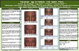
/
Perceived Age of Patients with Eyelid Ptosis Perceived Age of Patients with Eyelid Ptosis

Perceived Age of Patients with Eyelid Ptosis - PowerPoint Presentation

mitsue-stanley
mitsue-stanley . @mitsue-stanley
Follow
388 views
Uploaded On 2017-05-12

Perceived Age of Patients with Eyelid Ptosis - PPT Presentation

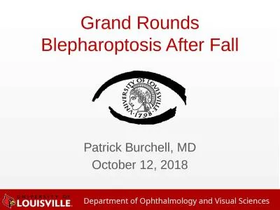
A drooping eyelid is associated with a tired aged and cosmetically unappealing facial appearance  Patients pursue upper eyelid ptosis repair for functional benefit however repair does change the ID: 547499

perceived age ptosis eyelid age perceived eyelid ptosis average ptotic patients group photograph photographs physician face facial study surgery

Share:

Link:

Embed:

Download Presentation from below link

Download Presentation The PPT/PDF document "Perceived Age of Patients with Eyelid Pt..." is the property of its rightful owner. Permission is granted to download and print the materials on this web site for personal, non-commercial use only, and to display it on your personal computer provided you do not modify the materials and that you retain all copyright notices contained in the materials. By downloading content from our website, you accept the terms of this agreement.


Presentation Transcript

Slide1

Perceived Age of Patients with Eyelid Ptosis

A drooping eyelid is associated with a tired, aged and cosmetically unappealing facial appearance.  Patients pursue upper eyelid ptosis repair for functional benefit, however repair does change the cosmesis of the face. Recent research from otolaryngology indicates that for patients undergoing aesthetic facial surgery there is a reduction of 3.1 years of perceived age1. However, no clinical research on the perceived age of patients based on eyelid ptosis has ever been performed. Our study examines the perceived age of a patient with a variable degree of upper eyelid ptosis.

Introduction

Srinivas Sai Kondapalli1, Craig Czyz2, 3, Jill Foster2, 4, Kenneth Cahill2, 4

AverageStandard DeviationLayperson 29.26.5Physician32.24.2Overall30.75.7

1Ophthalmology, Loyola University Medical Center, Chicago, IL, 2The Eye Center of Columbus, Columbus, OH, 3Oculofacial Plastic & Reconstructive Surgery, Doctors Hospital, Columbus, OH 4Ophthalmology, The Ohio State University, Columbus, OH

Methods

Methods

Results

There were 50 total patients enrolled in the study. There were ultimately 100 photographs that were created. The mean measured MRD1 was 1.3 (0.00-3.6) in the

ptotic

group. The mean measured MRD1 was 2.3 (0.40-5.5) in the non-ptotic group. The average perceived age for the ptotic group was 65 (29-90, +13). The average perceived age for the non-ptotic side was 63 (30-88 +14). The p-value was 0.40. Physician average perceived age for the ptotic group was 65 (29-90 +13). Lay person average perceived age for the ptotic group was 63 (30-88 +14). Physician average perceived age for the non-ptotic group was 63 (30-88 +14). Lay person average perceived age for the non-ptotic group was 61 (30-85 +13).

Conclusions

By creating mirror images of a patient with unilateral ptosis, we were able to create two photographs with comparable facial wrinkles, fat distribution and bony contour but with different eyelid positions. In doing so, this study is the first of its kind to determine the effect of eyelid ptosis on perceived age. Although our data does not demonstrate an effect on perceived age with an eyelid ptosis, there are some important limitations. The difference of eyelid position in our patient group was on average 1 mm, which may be too small for any changes in perceived age. Additionally, other clues including hairline and rhytides may have influenced our graders.Further work will include enrolling patients with larger degrees of eyelid ptosis, altering photographs to limit confounding facial clues, and prospectively obtaining pre and post-op photographs of patients with bilateral ptosis to eliminate a artificial and awkward appearing altered mirror image photograph.

Contact

Srinivas Sai A. Kondapalli, M.D.Loyola University Medical center2160 S. 1st AvenueMaywood, IL 601531Zimm et. al. “Objective Assessment of Perceived Age Reversal and Improvement in Attractiveness After Aging Face Surgery.” JAMA Facial Plastic Surgery. 2013 Aug 1. doi: 10.1001/jamafacial.2013.268.

This is a retrospective

study examining the affect of eyelid ptosis on perceived age. Patients from a single oculoplastic surgery group were enrolled. Using Current Procedural Terminology codes 67903 (levator recession), 67904 (levator advancement) and 67908 (conjunctivo-tarso-Muller’s muscle-levator resection), we obtained pre-procedure photographs of patients with unilateral ptosis. The standardized pre-procedure photographs were reviewed. The full face frontal image was selected. The photographs were modified using Photoshop CS2 Version 9.0 on a Windows 8 operating system. Each facial photograph was divided in the vertical midline (Fig 1). A mirror image of each resulting half image was produced. These images were merged to provide two resulting images (Fig 2 and 3).The photographs were cropped from hairline to upper philtrum of the lip. The marginal reflex distance 1 was measured in the Photoshop program. The final two resulting images were electronically compiled and randomized with Microsoft PowerPoint 2010. These resulting 100 images were then evaluated electronically for perceived age by 5 oculoplastic surgeons and 5 laymen. The evaluators were asked to provide the age of the patient in the photograph. The graders were not informed of the purpose of the study. Results were compiled. Ages were averaged between layperson, physician and overall. Appropriate statistical methods were employed and a p-value of 0.05 was used.

Average

Standard Deviation

Layperson

29.8

5.3

Physician

30.0

7.2

Overall

29.9

6.3

Average

Standard Deviation

Layperson

80.6

7.1

Physician

78.6

6.7

Overall

79.6

8.0

Average

Standard Deviation

Layperson

80.2

5.3

Physician

82.6

5.8

Overall

81.4

5.7

Figure 1. Full frontal face photograph, cropped from

philtrum

to hairline, divided on vertical midline

Figure 2. Full frontal face altered photograph derived from Figure 1. Non-

ptotic

photograph.

Figure 3. Full frontal face altered photograph derived from Figure 1.

Ptotic

photograph.