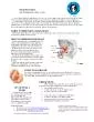
/
Testis   Must know 1-Classification of testicular Testis   Must know 1-Classification of testicular

Testis   Must know 1-Classification of testicular - PowerPoint Presentation

molly
molly . @molly
Follow
342 views
Uploaded On 2022-05-18

Testis   Must know 1-Classification of testicular - PPT Presentation

tumor 2Tumor markers in diagnosis 3Morphology of Seminoma Embryonal carcinoma Yolk sac tumor 4Cryptorchidism Testicular lesion Congenital Anomalies Regressive Changes Inflammation ID: 911916

tumours cell tumour germ cell tumours germ tumour cells testis tumor testicular seminoma carcinoma common sac features grossly cases

Share:

Link:

Embed:

Download Presentation from below link

Download Presentation The PPT/PDF document "Testis   Must know 1-Classification of..." is the property of its rightful owner. Permission is granted to download and print the materials on this web site for personal, non-commercial use only, and to display it on your personal computer provided you do not modify the materials and that you retain all copyright notices contained in the materials. By downloading content from our website, you accept the terms of this agreement.


Presentation Transcript

Slide1

Testis  

Slide2

Must know

1-Classification of testicular

tumor

2-Tumor markers in diagnosis

3-Morphology of

Seminoma

Embryonal carcinoma

Yolk sac

tumor

4-Cryptorchidism

Slide3

Testicular lesion

Congenital Anomalies

Regressive Changes

Inflammation (

Nonspecific ,

Specific

Inflammations,

Granulomatous (Autoimmune)

Orchitis

)

Vascular Disorders(torsion)

Spermatic Cord and 

Paratesticular

Tumors

Testicular 

Tumors

Slide4

Slide5

INFLAMMATIONS-

Inflammation of the testis is termed as

orchitis

and

of epididymis

is called as epididymitis; the latter being

more common

Slide6

1-Non-specific

Epididymitis and

Orchitis

-

may

be acute or

chronic

common

routes of

spread

are via the

vas deferens, or

lymphatic and haematogenous routes

caused

by urethritis, cystitis, prostatitis and

seminal

vesiculitis

common infecting organisms in

Neisseria

gonorrhoeae

and

Chlamydia trachomatis

Slide7

Grossly

,

acute

stage-

firm

, tense, swollen and

congested

may be

multiple abscesses, especially in gonorrhoeal

infection

chronic

stage-

variable

degree

of atrophy

and

fibrosis

Slide8

Histologically,

acute-

congestion, oedema and diffuse infiltration by neutrophils,

or

formation of neutrophilic abscesses

Chronic-

focal or diffuse

chronic inflammation, disappearance

of seminiferous

tubules, fibrous scarring and destruction

of interstitial

Leydig

cells

Slide9

2-Granulomatous

(Autoimmune)

Orchitis

-

Non-tuberculous granulomatous

orchitis

-

unilateral

, painless testicular enlargement

may

resemble a

testicular tumour

clinically

autoimmune

basis is

suspected

Slide10

Gross-

enlarged

Cut

section of

the testicle

is greyish-white to

tan-brown

Histologically,

granulomatous

reaction(non

caseating

)

is present

diffusely throughout

the testis and is confined to the

seminiferous tubules

Peritubular fibrosis

interstitial lymphocytes

and plasma

cells

Slide11

3-Tuberculous

Epididymo-orchitis

-

invariably

begins

in the epididymis

and spreads

to

the

testis

May spread via -tuberculous

seminal

vesiculitis

, prostatitis

and renal

tuberculosis

haematogenous

spread- from tuberculosis of

the

lungs

Slide12

Grossly,

discrete

, yellowish

,

caseous

necrotic

areas

Microscopically,

numerous tubercles which may

coalesce to

form large

caseous

mass

Characteristics of typical

tubercles such as epithelioid cells,

peripheral mantle

of lymphocytes, occasional multinucleate

giant cells

and central areas of

caseation necrosis

are

seen

AFB positive

Slide13

Slide14

4-Spermatic Granuloma(

epididymitis

nodosa

)

Spermatic granuloma is the term used for development

of inflammatory

lesions due to invasion of

spermatozoa

into the stroma

Slide15

MORPHOLOGIC

FEATURES

-

Grossly

,

a

small nodule, 3 mm to 3 cm in

diameter in

head of epididymis

firm, white

to

yellowish-brown

Slide16

Histologically,

it consists of a granuloma composed

of

histiocytes

, epithelioid cells, lymphocytes and

some neutrophils

Characteristically, the centre of

spermatic granuloma

contains spermatozoa and necrotic debris

Slide17

Vascular disorder

Torsion of

Testis

Twisting of the spermatic cord

sudden cessation of venous drainage and arterial supply

usually

followed by

sudden muscular effort or physical

trauma

Trauma may

occure

in either

in a

fully-descended testis

or in an

undescended

testis

Slide18

1-Neonatal torsion-

occurs either in utero or shortly

after birth

It

lacks any associated anatomic

defect in testis

2-“Adult

torsion-

is typically seen in

adolescence

and

presents with the sudden onset of

testicular pain

bell-clapper

abnormality

-

bilateral anatomic

defect that leads to increased mobility of

the testes

(

bell-clapper abnormality

)

Slide19

Viable- manually untwisted within

approximately 6 hours of the onset of

torsion

Slide20

MORPHOLOGIC

FEATURES-

duration

and severity

of vascular occlusion

may

be coagulative necrosis

of the

testis and

epididymis

may

be

haemorrhagic infarction

Slide21

Spermatic Cord and Paratesticular

Tumors

1-Lipomas

common lesions involving the

proximal

spermatic cord

, identified at the time of inguinal hernia

repair

represent

retroperitoneal adipose tissue that

has been

pulled into the inguinal canal along with the

hernia sac

, rather than a true

neoplasm

Slide22

2-A

denomatoid

tumor

-

most

common benign

paratesticular

tumor

typically occurring near the upper pole of the

epididymis

Slide23

grossly ,

well circumscribed small nodules

Microscopically- Proliferation of glandular

structures, irregularly lined by cuboidal

to flattened epithelial cell

Treatmet

-

local

excision

Slide24

Malignant tumor

rhabdomyosarcomas -children

liposarcomas

- adults

Slide25

CLASSIFICATION OF TESTICULAR TUMOR

most useful 

classification of

tumors

 is 

histogenetic

Named according to from which tissue

they arise and of which they consist

Slide26

Slide27

WHO histological classification of testis tumours

Germ cell tumours

Tumours of one histological type (pure forms)

Tumours of more than one histological type (mixed forms)

Sex cord/gonadal stromal tumours Pure forms

Miscellaneous tumours of the testis

Haematopoietic tumours

Tumours of collecting ducts and rete

Tumours of

paratesticular

structures

Mesenchymal tumours of the spermatic cord and testicular

adnexae

Secondary tumours of the testis

Slide28

Slide29

Testicular cancer is staged using the TNM system

created by the American Joint Committee on Cancer (AJCC

)

It’s

based on 4 key pieces of information:

refers to how much the main (primary) 

tumor

 has spread to tissues next to the

testicle

describes how much the cancer has spread to regional (nearby)

lymph 

nodes

indicates whether the cancer has 

metastasized

 (spread to distant lymph nodes or other organs of the body

)

indicates the serum (blood) levels of

tumor

markers that are made by some testicular

cancers

Slide30

Letters or numbers appear after

T, N, M, and S to provide more details about each piece of information.

The

numbers 0 through 4 indicate

increasing

severity

The

letters “IS” after the T stand

for in situ,

which means the

tumor

is contained in

one place

and has not yet penetrated to a deeper layer of tissue.

The

letter X after T, N, M, or S means “

cannot be assessed

” because the information is not

known

Slide31

TNM classification of germ cell tumours of the testis

pTNM

pathological classification

pTx

–Primary tumour

cannot be assessed

pT0

No evidence

of primary tumour (e.g. histologic scar in testis)

pTis

Intratubular

germ cell neoplasia (

carcinoma in situ)

pT1 Tumour

limited to testis and epididymis without

vascular/lymphatic invasion; tumour may invade tunica albuginea but not tunica

vaginalis

pT2 Tumour

pT1

+

involvement of tunica

vaginalis

pT3 Tumour invades

spermatic cord

with or without vascular/lymphatic invasion

pT4 Tumour invades

scrotum

with or without vascular/lymphatic invasion

Slide32

pN

– Regional lymph nodes

pNX

Regional lymph nodes cannot be assessed

N0 No regional lymph node metastasis

pN1 <

2 cm or less

in greatest dimension and

5 or fewer positive nodes

pN2

2 to 5 cm in greatest dimension

; or

more than 5 nodes positive

, none more than 5 cm; or evidence of

extranodal

extension of tumour

pN3 Metastasis with a lymph node mass

more than 5 cm

in greatest dimension

Slide33

S – Serum tumour markers

SX Serum marker studies not available or not performed

S0 Serum marker study levels within normal limits

LDH,

hCG

(

mIU

/ml) ,AFP (ng/ml)

Slide34

Serum tumor

markers (S)

For staging, serum (blood) levels of

tumor

markers are measured 

after

 the testicle containing the cancer has been removed with

surgery

LDH (U/liter)

HCG (

mIU

/ml)

AFP (ng/ml)

SX

Marker studies not available or not done.

S0

Normal

Normal

Normal

S1

*

<1.5 x Normal

<5,000

<1,000

S2

+

1.5 - 10 x Normal

5,000 - 50,000

1,000 - 10,000

S3

+

>10 x Normal

>50,000

>10,000

Slide35

Cryptorchidism

Cryptorchidism

is a complete or partial failure of the intra-abdominal testes to descend into the scrotal sac

associated with

testicular dysfunction

an increased risk of testicular

cancer

Slide36

In 70%

of cases

, the undescended testis lies in the inguinal

ring

in

25

% in

the abdomen

Slide37

ETIOLOGY.

exact

etiology

is

not known

in

majority

of cases

Mechanical factors-

short

spermatic

cord

narrow inguinal canal

adhesions

to the

peritoneum

problems

with development of the

gubernaculum

a

patent

processus

vaginalis

, or impaired intra-abdominal pressure have also been hypothesized to contribute to

cryptorchidism

Slide38

2

.

Genetic

factors-

up to 23% of cases suggesting an underlying genetic mutation

 Mutations in insulin-like factor 3 and its receptor, LGR8, have been demonstrated in a small number of cases

trisomy 13

Slide39

3.

Hormonal factor-

cryptorchidism

is only

rarely associated

with a well-defined hormonal disorder

deficient androgenic secretions 

mullerian

inhibiting

substance

insulin-like

3

hormone

4.

Neuromuscular

- abnormalities of the

genitofemoral

nerve's calcitonin gene-related peptide

or the

cremasteric

nucleus have been postulated to cause cryptorchidism

Slide40

Miscellaneous-

Maternal

alcohol consumption, 

analgesic

consumption,  and smoking 

have

also been associated with an increased

risk

Gestational diabetes has been shown to be related to the development of

cryptorchidism

Slide41

MORPHOLOGIC FEATURES.

Cryptorchidism is

unilateral in

80%

cases

Grossly

,

the

cryptorchid

testis is small in size, firm

and fibrotic

Slide42

Histology-

1-Seminiferous tubules

tubular

basement membrane is

thickened

hyalinised

tubules

with

a few

Sertoli

cells

foci

of

spermatogenesis

are discernible in 10% of cases

2

.

Interstitial stroma

:

usually

increase in the interstitial

fibrovascular

stroma

and conspicuous presence of

Leydig

cells

, seen singly or in small

clusters

Slide43

Slide44

Slide45

CLINICAL FEATURES.

asymptomatic

and is discovered only on

physical examination

1. Sterility-infertility.

Bilateral cryptorchidism is

associated with

sterility while unilateral disease may result

in infertility

2. Inguinal hernia.

A concomitant inguinal hernia

is

frequently

present along with

cryptorchidism

Slide46

3-Malignancy

.

Cryptorchid

testis is at 30-50 times

increased risk

of developing testicular

malignancy

most commonly seminoma

and embryonal carcinoma, than a

normally descended testis

risk of malignancy is greater in

intraabdominal

testis

than in testis in the inguinal

canal

Slide47

current

recommendations are for correction at 6 to 12 months of

age

carcinoma

arises from foci of

intratubular

germ

cell neoplasia within the atrophic

tubules

Orchiopexy

reduces

the risk of sterility and

cancer

Slide48

Tumour marker

Tumour

markers-

Germ cell tumours of the

testis secrete

polypeptide hormones and certain enzymes

which can

be detected in the

blood

There

are two principal serum

tumour markers

alpha

fetoprotein (AFP)

and

beta

subunit of human chorionic

gonadotropin (

shCG

)

In addition

, carcinoembryonic antigen (CEA), human

placental

lactogen

(HPL), placental alkaline phosphatase,

testosterone, oestrogen

and luteinising hormone may also be

elevated

Slide49

AFP-

synthesized

by

fetal

yolk sac

and also the

liver and

intestine

elevated

in

50-70% of testicular

germ cell

tumours

Markedly elevated in yolk

sac

tumor

a

serum half life

of 4.5

days

However,

elevated serum

AFP levels are

also found in liver cell carcinoma

hCG

-

secreted by

placental

trophoblastic cells

elevated in

non-

seminomatous

germ cell tumours

of the

testis

(e.g. in

choriocarcinoma

, yolk sac tumour

and embryonal

carcinoma

)

elevated in 50

% of patients with germ cell

tumours

elevation in

seminoma

in

10-25%

of cases

Slide50

Lactate dehydrogenase (LDH

)-

may also be elevated

direct relationship between

LDH and tumour

burden

However

, this test is

nonspecific although

its degree of elevation

correlates with

bulk of

disease

Slide51

Applications-

In

the evaluation of testicular

masses

In the staging of testicular germ cell

tumors

.

For example, after

orchiectomy, persistent elevation

of HCG

or AFP concentrations indicates stage II

disease even

if the lymph nodes appear of normal size

by imaging studies

In assessing

tumor

burden

In monitoring the

respons

to

therapy

. After

eradication of

tumors

there is a rapid fall in serum AFP

and HCG

. With serial measurements it is often

possible to

predict recurrence before the patients

become symptomatic

or develop any other clinical signs

of relapse

Slide52

TESTICULAR TUMOR

Slide53

Testicular tumor

Most

germ cell tumours

occur between

the

ages of 20 and 50

years

Before puberty, seminoma is

extremely uncommon

usual

germ cell tumours

,

yolk sac tumour and the better differentiated types of

teratoma

Spermatocytic

seminoma and

malignant lymphoma

usually occur in

older patients

incidence increases

shortly after the onset

of puberty

and reaches a

maximum

in

men in

the late

twenties and

thirties

Slide54

ETIOLOGIC

FACTORS

Cryptorchidism

Other developmental

disorders-

Dysgenetic

gonads associated

with endocrine abnormalities such as

androgen insensitivity syndrome

Genetic

factors-

high incidence in first-degree family members, twins

Other

factors.

A few less common

factors

Orchitis

Trauma

Carcinogens.

LSD

, hormonal therapy for sterility

, copper

, zinc

etc

Slide55

Prenatal risk

factors

consistent associations

of testicular cancer

with

low

birth

weight

Exposures in

adulthood

Possible

etiological clues, however

, include

a

low level of physical activity

and high

socioeconomic class

Slide56

PATHOGENESIS-

vast

majority of these tumours originate

from germ cells

1-Developmental disorders-

contribute to the pathogenesis

2-Molecular

genetic

features-

common molecular pathogenesis of all

germ cell

tumours

:

Hyperdiploidy

is almost a constant

feature

isochromosome

of short arm of

chromosome 12

Telomerase activity

is present in all germ cell tumours

of the testis

Other mutations include

p53

,

cyclin E

and

FAS

gene

Slide57

3-Intratubular

germ cell neoplasia (

ITGCN) or carcinoma

insitu

-

Most

testicular germ cell

tumors

originate from

a precursor lesion called

intratubular

germ cell

neoplasia (ITGCN)

exceptions

to this rule are

pediatric

yolk

sac

tumors

Teratomas

adult

spermatocytic

seminoma

Slide58

4-Three

hit’ process.

Germ cells in seminiferous tubules undergo

first hit-

activate the cell

second

hit-

occure

in CIS cell and further activate

third hit-

via some epigenetic

phenomena

cell become invasive

this sequential tumorigenesis

explains the development of

seminomatous

tumours

Slide59

Slide60

CLINICAL FEATURES AND DIAGNOSIS

gradual gonadal enlargement and a dragging

sensation in

the

testis

secondary symptoms

such as pain, lymphadenopathy,

haemoptsis

and urinary

obstruction (

Metastatic

involvement

)

SPREAD-

Lymphatic spread-

retroperitoneal para-aortic lymph

nodes, mediastinal lymph nodes and

supraclavicular lymph nodes

Haematogenous

spread

-

primarily

occurs to the lungs, liver

, brain

and

bones

Slide61

1-Intratubular

germ cell neoplasia

, unclassified

type (IGCNU

)

Also called

carcinoma

in situ

(CIS) stage of germ cell tumours

preinvasive

stage of germ cell

tumours

intratubular

seminoma

and

intratubular

embryonal carcinoma

are common

2-4

% of cryptorchidism

pt

show

Clinical

features -

atrophic testis

, infertility,

maldescended

testis

, and

intersex features

Slide62

gross-

no

grossly visible lesion

Histopathology

- Germ cells with abundant vacuolated cytoplasm, large, irregular nuclei and prominent nucleoli located within the seminiferous tubules

restricted to the seminiferous tubules without evident invasion into the

interstitium

Immunoprofile

-

PLAP

can be demonstrated in 83-99%

of

intratubular

germ cell neoplasia of

the unclassified type (IGCNU) cases and is widely

used for diagnosis

Slide63

Comparison of morphological features of normal seminiferous tubules (left part) and

intratubular

germ cell neoplasia (IGCNU) in seminiferous tubules (right part).

Slide64

Seminoma

Seminomas are the most common type of germ

cell

tumor

, making up about

(50%)

peak incidence

is the

third decade

and they almost never

occur in infants

An identical

tumor

arises in the ovary, where

it is called

dysgerminoma

Slide65

.

Slide66

MORPHOLOGY

-

cut

surface has a homogeneous,

graywhite

, lobulated,

usually devoid of

hemorrhage

or necrosis

Generally

the tunica albuginea is

not penetrated

but occasionally extension to the epididymis,

spermatic cord

, or scrotal sac

occurs

Slide67

Microscopy-

typical seminoma is composed of

sheets of

uniform cells divided into poorly demarcated lobules by

delicate fibrous

septa containing a lymphocytic infiltrate

Tumor

cell-

cell

is large and round

to polyhedral

and has a distinct cell membrane; clear

or watery-appearing

cytoplasm; and a large, central

nucleus with

one or two prominent

nucleoli

Slide68

Stroma

-

delicate fibrous tissue

which divides the tumour into

lobules

characteristic

lymphocytic infiltration, indicative of immunologic response of the host to the

tumour

Variable

features-

tumor

giant cells and greater mitotic

activity

15% of seminomas contain

syncytiotrophoblasts

ill-defined granulomatous reaction (20%)

Slide69

Special stain

-Cytoplasm contains

variable amount of glycogen that

stains positively

with

PAS reaction

IHC-

seminoma cells stain positively for KIT

,

(regardless of KIT mutational status), OCT4, and placental alkaline phosphatase (PLAP

)

Slide70

Slide71

Prognosis-

better

than

other germ

cell

tumours

tumour

is highly

radiosensitive

Slide72

Spermatocytic

Seminoma-

Spermatocytic

seminoma is both clinically

and morphologically

a distinctive tumour from classic

seminoma

Incidence of

about 5% of all germ cell

tumours

older patients

generally

in

6

th

decade

of

life

bilateral

in 10% of

patients

Slide73

Grossly,

spermatocytic

seminoma

is homogeneous, larger, softer and

more yellowish

and gelatinous than the classic

seminoma

Slide74

Histologically,

the distinctive features are as under:

1. Tumour cells.

lymphocyte-like

to huge

mononucleate

or multinucleate

giant cells.

Majority of

cells

are

, however

, of

intermediate size.

Mitoses

are

often frequent

.

2. Stroma

.

stroma lacks lymphocytic and

granulomatous reaction

seen in classic seminoma.

Slide75

prognosis

of

spermatocytic

seminoma is excellent

slow-growing

and rarely

metastasises

tumour

is radiosensitive

Slide76

Embryonal Carcinoma-

30

% of germ

cell tumours more

common

2nd

to 3rd decades of

life

90% cases are

associated with

elevation of AFP or

hCG

or both

Slide77

Grossly

,-

a

small tumour in the

testis

distorts the

contour of the testis

as it frequently invades the

tunica and

the

epididymis

Cut surface-

greywhite

, soft

with areas of haemorrhages and

necrosis

Slide78

Slide79

Microscopy-

1

.

tumour

cells are arranged in a variety of

patterns

— glandular

, tubular, papillary and

solid

2.

tumour

cells

are

highly anaplastic

carcinomatous

cells

having large size, indistinct cell borders,

amphophilic

cytoplasm

and prominent hyperchromatic

nuclei

Slide80

Slide81

Yolk Sac

Tumour

(

Synonyms: Endodermal Sinus Tumour

,

Orchioblastoma

, Infantile Embryonal Carcinoma

)

most

common

testicular tumour

of infants and young children

upto

the age of 4

year

may be present as the major component in 40% of germ

cell tumours

AFP levels are elevated in 100% cases of yolk

sac tumours

Slide82

Grossly,

the tumour

is generally

soft, yellow-white, mucoid with areas of

necrosis and haemorrhages

Microscopically

,

yolk sac tumour has the

following features

patterns—

loose

reticular

network, papillary, tubular and solid

arrangement

2

.

flattened

to cuboid epithelial

cells with

clear vacuolated

cytoplasm

3.A pathognomonic feature is

Schiller -

Duvel

body 

a

central vessel surrounded by

tumor


cells

 in a cystic space often lined by flattened

tumor

cells

4.

presence of both intracellular

and extracellular

PAS-positive

hyaline globules

Slide83

Slide84

Choriocarcinoma

highly

malignant tumour

composed of

syncytiotrophoblast

and

cytotrophoblast

2nd

decade of

life

serum

and urinary

levels of

hCG

are greatly elevated in 100%

cases

Slide85

Grossly,

the tumour

is usually

small and may appear as a

soft, haemorrhagic

and necrotic mass

Microscopically,

the characteristic feature is

syncytiotrophoblast

and

cytotrophoblast

without formation of

definite placental-type villi

Slide86

Slide87

Teratoma

-

Teratomas

are complex tumours composed of tissues

derived from

more than one of the three germ cell layers—endoderm

, mesoderm

and

ectoderm

more common

in infants and children and constitute

(

40

%)

in adults they comprise

5% of all germ cell tumours

Slide88

MORPHOLOGIC FEATURES.

Testicular

teratomas

are classified

into 3 types:

1. Mature (differentiated)

teratoma

2. Immature

teratoma

3.

Teratoma

with malignant

transformation

Slide89

Gross-

large

, grey-white

masses enlarging testis

Cut

surface

shows characteristic

variegated appearance—grey-white

solid areas

, cystic and honey-combed areas, and foci of

cartilage and

bone

Slide90

Germ cell layer derivatives

Slide91

Microscopicy

-

three

categories of

teratomas

show different

appearances

:

1-Mature

(differentiated)

teratoma

.

Well differentiated structures

such as cartilage, smooth muscle

, intestinal

and respiratory epithelium, mucus glands,

cysts lined

by squamous and transitional epithelium,

neural tissue

, fat and

bone

Slide92

2-Immature

teratoma

.

incompletely

differentiated and

primitive or

embryonic tissues

along with some mature elements

present are poorly-formed

cartilage, mesenchyme, neural tissues

, abortive

eye, intestinal and respiratory tissue elements

etc

Mitoses

are usually

frequent

Slide93

Slide94

Immature

stroma

Slide95

3-Teratoma

with malignant

transformation

-

extremely

rare form of

teratoma

one

or more

of the

tissue elements show malignant

transformation

Transformation

may take the form of

a squamous

cell carcinoma, mucin-secreting adenocarcinoma

, sarcoma

, or other cancers.

importance of recognizing

a non–germ

cell malignancy arising in a

teratoma

is that

these secondary

tumors

are

chemoresistant

Slide96

Mixed Germ Cell Tumours

About 60% of germ cell tumours have more than one of

the above

histologic types (except

spermatocytic

seminoma)

and are

called mixed germ cell

tumours

most common combinations of mixed germ

cell tumours

are as under:

1.

Teratoma

, embryonal carcinoma, yolk sac tumour

and

syncytiotrophoblast

2. Embryonal carcinoma and teratoma (teratocarcinoma

)

3. Seminoma and embryonal

carcinoma

Slide97

SEX CORD-STROMAL

TUMOURS

-

Tumours arising from specialised gonadal

stroma

primitive

mesenchyme>>>

specialised

stroma of gonads in either

sex gives

rise to theca, granulosa and lutein cells in the

female

Sertoli

and

interstitial

Leydig

cells

in the male

Slide98

Leydig

(Interstitial) Cell

Tumour-

20

to

50

yr

,

secrete

androgen, or

both androgen

and

oestrogen

Grossly,

as

a small, well-demarcated and

lobulated nodule

. Cut surface is homogeneously yellowish

or brown

Histologically,

the tumour is composed of sheets

and cords

of normal-looking

Leydig

cells

These

cells

contain abundant

eosinophilic cytoplasm and

Reinke’s

crystals and

a small central nucleus

Slide99

Sertoli

Cell Tumours (

Androblastoma

)-

infants

and

children

Oestrogen or

androgen

gynaecomastia

in an

adult

precocious sexual development in a

child

Slide100

Grossly,

the tumour

is

large, firm, round, and well

circumscribed

.

Cut surface

is

yellowish or

yellow-grey

Microscopically,

Sertoli

cell tumour is composed

of benign

Sertoli

cells arranged in well-defined

tubules

Slide101

MIXED GERM CELL-SEX CORD STROMAL

TUMOURS-

Gonadoblastoma

-

secrete androgen

Grossly,

the tumour is

of variable

size, yellowish-white and

soft

Microscopically,

gonadoblastoma

is composed of

2 principal

cell types—large germ cells

resembling seminoma

cells, and small cells resembling

immature

Sertoli

,

Leydig

and granulosa

cells

Slide102

OTHER TUMOURS

Malignant

Lymphoma-

comprises

5% of

testicular

malignancies

most common testicular tumour in the elderly

Bilaterality

is seen in half the

cases

Most common are

large cell

non-Hodgkin’s lymphoma of B cell

type

Slide103

Must know

1-classification

2-Tumor markers in diagnosis

3-Morphology of

Seminoma

Embryonal carcinoma

Yolk sac

tumor

4-Cryporchidism