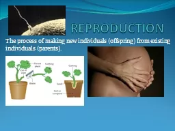
Embryonic development into malefemale A Embryos identical until about 8 weeks The same embryonic structures give rise to ovaries and testes clitoris and penis B Females genes on 2 X chromosomes interact to produce high estrogen and progesterone levels gt female reproducti ID: 744918
Download Presentation The PPT/PDF document "6.6 Part II and 11.4- Reproduction" is the property of its rightful owner. Permission is granted to download and print the materials on this web site for personal, non-commercial use only, and to display it on your personal computer provided you do not modify the materials and that you retain all copyright notices contained in the materials. By downloading content from our website, you accept the terms of this agreement.
Slide1
6.6 Part II and 11.4-
ReproductionSlide2
Embryonic development into male/female
A. Embryos identical until about 8 weeks - The same embryonic structures give rise to ovaries and testes, clitoris and penis
B. Females – genes on 2 X chromosomes interact to produce high estrogen and progesterone levels -> female reproductive structures
C. Males – genes on Y chromosome responsible for testosterone production and testes developmentSlide3
II. Male Reproductive SystemSlide4Slide5
Spermatogenesis – starts at puberty and continues for life
Seminiferous tubules of testesSlide6
Arise from
spermatogonia
(germinal epithelial cells) near outer edge of seminiferous tubules
a. Undergo mitosis to replenish supply of
spermatogonia
b. Undergo meiosis to produce spermatozoa (millions/day) – stimulated by follicle stimulating hormone (produced by pituitary)Slide7
Daughter cells must mature into fully motile spermatozoa
a.
Sertoli
cells – provide nutrients for developing sperm
b. Leydig cells – leutinizing hormone from pituitary stimulates production of testosterone c
. Move closer to
the center of the
seminiferous tubule as they develop d. Move to epididymis to finish maturationSlide8
Mature sperm
a. Acrosome – contains hydrolytic enzymes to help get into egg
b. Mitochondria – produce ATP for swimming
c. Flagellum – tail for swimming
Slide9
Production of semen
a. Mature sperm moves from epididymis to vas deferens
b. As it moves through vas deferens, fluid is added
i. Seminal vesicle adds 70% of fluid - high in fructose (sugar) to provide energy ii. Prostate adds alkaline fluid – to help neutralize low pH in vaginaSlide10
Hormones involved
Leutinizing
hormone (LH) - stimulates
Leydig
cells in testes to produce testosterone
Follicle Stimulating Hormone (FSH) - along with testosterone, stimulates meiosis of
spermatogoniaSlide11
III. Female Reproductive SystemSlide12Slide13Slide14
Oogenesis
1. Events before birth - in ovaries of female fetus
a.
O
ogonia in the ovaries undergo mitosis to increase in number b. Oogonia grow into larger cells called primary oocytes c. Primary oocytes start meiosis, but pause in prophase I
d. Follicle cells undergo mitosis forming a layer around the primary oocyte (together this is called a primary follicle)
e. At birth, ovaries contain about ½ million primary follicles – remain unchanged until pubertySlide15
Events during the menstrual cycle
a. Each month, a few primary follicles finish meiosis I
b. Results in 1 polar body and 1 secondary oocyte (both haploid)
c. Ring of follicle cells divides and forms fluid, resulting in a double ring of cells with fluid in between called Graafian follicle Slide16
Increase of fluid causes
Graafian
follicle to push on wall of ovary until the secondary oocyte is released from the ovary
Left over follicle cells become corpus luteum – secretes progesteroneSlide17
The Mature Egg
A. Secondary Oocyte – largest cell in most species – contains all nutrients for early developmentSlide18
IV.
Comparison of spermatogenesis and oogenesis
Spermatogenesis
Oogenesis
Millions of sperm/day from puberty throughout life
1
secondary oocyte/month between puberty and menopause
4 gametes from each germinal
cell
1 gamete from each germinal cell (+ 3 polar bodies)
Gametes are small with little cytoplasm and organellesGamete is large with substantial cytoplasm, organelles, and nutrients
Mitosis replaces germinal cells daily
Mitosis only replaces germinal cells in early development
Some cell
growth before meiosis I
Substantial
cell growth before meiosis I
Spermatids remain in seminiferous
tubules until differentiation into spermatozoa occurs
Differentiation begins in the ovary but finishes
after fertilization
Occurs in testes
Occurs in ovariesSlide19
The Menstrual Cycle
A. Purpose – release ovum for potential fertilization and implantation into uterus
B. Implantation must occur when endometrium is highly vascularized (increased number of blood vessels)
C. If implantation does not occur, excess blood vessels are broken down and discarded by menstruationSlide20
Hormones involved
1.
GnRH
– gonadotropin-releasing hormone – from hypothalamus, stimulates pituitary to release LH and FSH
2. Follicle Stimulating Hormone and
Leutinizing
Hormone a. Stimulate production of estrogen by the ovary b. Stimulate production of
Graafian
follicles
c. Lead to ovulation and creation of corpus luteum 3. Estrogen – increases vascularization of endometrium 4. Progesterone – maintains thickened endometrium - Slide21Slide22Slide23
VI. Fertilization
In animals, can be internal or external
1. External usually results in large numbers of offspringSlide24
Events of fertilization in humans
Sperm make their way along endometrium to Fallopian tubes
Many sperm start breaking down the
zona
pellucida
(glycoprotein layer surrounding the secondary oocyte) by releasing hydrolytic enzymes from acrosome (acrosome reaction) a. Also triggers oocyte to finish meiosis II3. One
spermatozoan
reaches plasma membrane, releases more hydrolytic enzymes to penetrate egg
4. Plasma membranes fuse
http://www.youtube.com/watch?v=BFrVmDgh4v4Slide25
5.
Cortical reaction – prevents more than one sperm from fertilizing egg – cortical granules fuse with plasma membrane and release enzymes
outside, chemically changing the
zona
pellucida
so that no other sperm can penetrate6. Nuclei of egg and sperm fuse to restore diploid chromosome numberSlide26
VII. Early development
A. Zygote starts undergoing mitosis within 24 hours of fertilization
B. Moves down Fallopian tube to uterus
C. Consists of about 100 cells when it reaches uterus = blastocyst Slide27
Implantation – embryo sinks down into endometrial lining
Around a week after fertilization
Has not increased in size, just number of cells (nutrients used for metabolism, not growth)
Embryo can start getting nutrients from endometrium rather than yolk
http://www.youtube.com/watch?v=UgT5rUQ9EmQSlide28
The Placenta
1. Forms from both mother and embryo
2. Umbilical cord forms on fetal side, consists of 3 blood vessels Slide29
http://www.youtube.com/watch?v=kZeZ62UXyzMSlide30
Hormones involved
1.
HCG (human chorionic gonadotropin) – secreted by embryo, enters mother’s bloodstream, goes to her ovaries to maintain the corpus luteum
2. Progesterone – first secreted by corpus luteum, later by placenta, maintains vascularization of the endometrium and suppresses contractions of the uterus
3. Estrogen – secreted by placenta, stimulates muscle growth of uterus, antagonizes progesterone’s suppression of contractions, stimulates mammary glands development, and induces production of oxytocin receptors in uterine muscles
*Both progesterone and estrogen inhibit production of further oocytes during pregnancySlide31Slide32
VIII. Childbirth
A. Oxytocin – secreted by pituitary to stimulate uterine contractions
1. Positive Feedback Slide33
In Vitro
Fertilization (IVF)
A. Reasons for infertility
1. low sperm count/motility in males
2. males with impotence 3. females who can’t ovulate normally 4. females with blocked Fallopian tubes
B. Harvest eggs from female
1. Injections of FSH to stimulate production of many
Graafian follicles (superovulation) 2. Eggs surgically removedSlide34
Eggs mixed with sperm in culture dishes
Use microscope to see which eggs are fertilized and healthy
1-3 eggs introduced into uterus
Extra embryos can be frozen