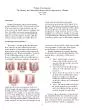
In general estrogen promotes amp progesterone inhibits squamous cell maturation Hormones may influence the morphology staining characteristics of the cervical endometrial vaginal urethral amp bladder mucosa cell ID: 717875
Download Presentation The PPT/PDF document "WELCOME INTRODUCTION The epithelia of ..." is the property of its rightful owner. Permission is granted to download and print the materials on this web site for personal, non-commercial use only, and to display it on your personal computer provided you do not modify the materials and that you retain all copyright notices contained in the materials. By downloading content from our website, you accept the terms of this agreement.
Slide1
WELCOMESlide2
INTRODUCTION
The epithelia of the female genital tract are sensitive to estrogen , progesterone , androgens & various additional steroid hormone derivatives & metabolites.
In general estrogen promotes & progesterone inhibits squamous cell maturation.
Hormones may influence the morphology staining characteristics of the cervical , endometrial , vaginal , urethral & bladder mucosa cell.Slide3
H
ormonal
evaluation is based on the degree of maturation & glycogen storage of the exfoliated squamous cells.
For useful cytologic evaluation , some information about the age , menstrual history & previous hormonal , surgical or treatment of the patient should accompany the smear. Slide4
INDICATIONS FOR CYTOLOGIC HORMONAL EVALUATION
1)Assessment of ovarian function
.
During puberty & reproductive , menopausal & post menopausal years after hysterectomy.During menstrual disorders.In premature menses (childhood).To determine ovulation time.Slide5
2)Assessment
of abnormal hormonal production (placenta, ovaries & other endocrine organs).
Before , during & after a pregnancy (fertility study , threatened abortion , retained placenta
).Existence of functioning (hormone producing) tumors.Various endocrine disorders
.
Assessment & guidance for hormonal therapy
.
Slide6
NORMAL HORMONAL PATTERNS
ESTROGEN
It increases cellular maturation & proliferation of all layers of squamous epithelium
mostly Superficial cells and Intermediate cells. DECREASING LEVELDeficiency of estrogen increases parabasal cells
.
The mucosa becomes thin & superficial layer is absent in deficient
estrogen stimulation
.
Slide7
INCREASING
LEVEL
Increase in estrogen level enlarges the parabasal cells to intermediate .this continue to enlarge & mature into keratinized superficial cells
.Increases the amount of intra cellular glycogen ( by protein & nucleic acid synthesis )High estrogenic effect in a vaginal smear is recognized by a clean background & increased eosinophilic & karyopyknotic indices. Slide8
PROGESTERONE
It produce proliferation of intermediate squamous epithelium at the time of exfoliation.
INCREASING LEVELClusters of intermediate cells dominate the smear.The amount of intracellular glycogen-increasesNo. of leukocytes –decreases Slide9
ANDROGEN
Male sex hormone
.
Small amount is present in female.INCREASING LEVEL It increases the no. of exfoliated large parabasal cells with central , almost pyknotic nuclei & dense , glycogen containing cytoplasm.Slide10
GUIDELINES FOR SPECIMEN COLLECTION & SUITABLE FOR HORMONAL ASSESSMENT
1
)SPECIMEN
COLLECTION GUIDELINES:Smear taken from the lateral vaginal wall Smear taken from the junction of the middle & upper 1/3rd of the vagina.
2)FACTORS THAT MAKE A SMEAR UNSUITABLE FOR HORMONAL ASSESSMENT:
Material not collected properly.
Adequate clinical information not provided (age , menstrual status, treatment etc)
Evidence of an infection.
Inflammation
.
Air-drying artifact
.Slide11
HORMONAL PATTERN AT DIFFERENT STAGES OF LIFE
LIFE STAGE
HORMONAL PATTERN
NewbornSterileNo inflammationIntermediate cells superficial cells
1
st
- 2
week of life
Acquires bacteria
Neutrophils appear
Atrophic cells
Childhood
Maturation
& cyclic alternations occur months (or)years before
menarche,
due to cycles that are initially
anovulatory
Cyclic women , proliferative phase
Increasing squamous cell maturation
Cyclic women , post
ovulatory
phase
Decreasing squamous cell maturationSlide12
Pregnancy
Navicular cells may be numerous
LactationAtrophy persists but some maturation may appear slowly .
Post partum
Atrophy
Menopause
Atrophy (or) intermediate cell predominateSlide13
CELLS
1) SUPERFICIAL
CELLS
Cell – flat , delicate & transparent.Size – 35-45 micro meter in diameter.Nuclei – small , dark & pyknotic (nuclear material become condensed & shrunken.Cytoplasm – delicate pink (eosinophilic / acidophilic)Slide14
2) INTERMEDIATE
SQAMOUS CELLS
Cell – flat
Size – 35-45 micro meterNucleus – spherical / oval -vesicularCytoplasm – basophilicA variant of intermediate cells –boat shaped navicular cells.
Navicular cells – common in early menopause & pregnancy.
Oval shaped cells store glycogen in the form of cytoplasmic deposits – stains yellow in PAP stain Slide15
PARABASAL CELLS
Size – 12 -30 micro
meter.
Nuclei – vesicular.Shape – round / oval.Cytoplasm – basophilic.Slide16
4) BASAL
CELLS
Shape – round / oval.
Cytoplasm – scanty , basophilic.Nuclei – vesicular -fine chromatic granules -occasionally tiny round nucleoliSlide17
CYTOLOGICAL INDICES
The degree of proliferation , maturation & desquamation of vaginal cells is influenced by various hormones
.
The cytological assessment of these hormones is given by different types of indices . All are based on recognition & exact typing of the epithelial cells exfoliated from the surface of the stratified squamous vaginal mucosa .Slide18
If the smear is to inflammatory & a repeat specimen is not possible after the proper treatment , the exfoliated urethral cells in the first portion of a voided urine specimen can be examined for this evaluation
.
The terminal part of the urethral mucosa is almost as sensitive to hormonal changes as in the vaginal mucosa but more difficult to evaluate.Slide19
TYPES OF INDICES
Maturation index (MI)
It is determined by the percentage of the basal , intermediate & superficial cells as presented as
3 part ratio with the basal cells started 1st , intermediate cells 2nd & superficial cells 3
rd
.
Example
:
MI =
80 /20 /
0
Indicates
:
80 % - Parabasal cells
20 % - Intermediate cells
0 %
- Superficial cells
Slide20
RESULTS READ AS:
Basal Intermediate superficial
0 0 100 Shift - left 0 100 0
Shift -middle
100 0
0
Shift – right.Slide21
RESULT READ
AS
Shift to left – atrophic
effect. Shift to right – increased estrogen like effect. Shift to midzone – progesterone like effectSlide22
2)KARYOPYKNOTIC (KPI) / CORNIFICATION
INDEX
The percentage of squamous cell with sharp squared cytoplasmic edges with pyknotic nuclei compared with other mature squamous cells with vesicular nucleus
. Parabasal cells are not counted.Slide23
3)MATURATION VALUE (MV)
This method is useful for providing hormonal evaluation data to a computer
.
Each parabasal cell is counted as – 0 Intermediate cell as – 0.5 superficial cell as - 1.0 The addition of all the values given to the first 100 epithelial cells is recorded. Slide24
Example : MI =
0 / 35 / 65
0 x 0 =0
35x 0.5 = 17.5 65x1.0 = 65.0 MV = 82.5 MV of 100 indicates – pure population of superficial
squamous cells
MV of 0
indicates – pure population of parabasal cells. Slide25
STAGES
MI
MV
VARIATIONNewborn 0 / 90 / 10
55
+/ - 10
Infancy
80
/ 20 / 0
10
+ / - 20
Pre
ovulatory
0 / 40 / 60
80
+
/ - 10
Post
ovulatory
0 / 70 / 30
65
+
/ - 15
Menopause
0 / 80 / 20
60
+
/ - 20
Post menopause
50 / 50 / 0
25
+
/ - 40
Estrogen therapy
0 / 10 / 90
95
+ / - 10
Progesterone therapy
0 / 90 / 10
55
+ / - 10
Androgen therapy
20 / 80 / 0
40
+ / - 10Slide26
4)EOSINOPHILIC INDEX (EI)
The no. of mature squamous cells with eosinophilic (pink) cytoplasm , nuclear appearance is compared with the no. of other mature squamous cells with basophilic (blue) cytoplasm.
The result is given in ratio.
Parabasal cells not counted. Example : 2:14 2 – eosinophilic sq. cell 14 – basophilic sq. cellsSlide27
5)FOLDED CELL INDEX (FCI)
No . of mature sq. cells with folded cytoplasmic rims are compared with no. of mature cells with flat non folded cytoplasm
.
The folded cells with cytoplasmic glycogen are considered as less mature than flat cells.Slide28
6)SUPERFICIAL CELLS INDEX (SCI
)
Superficial sq. cells with any type of nucleus is compared with any other sq. cell present in the smear.
7)CROWED CELL INDEX (CCI) The no. of mature sq. cells that are clustered together in groups of 5 or 6 cells is compared to the no. of similar cells found singly / less than 3 or 4. Slide29
INDICES USED FOR HORMONAL ASSESSMENT
TYPE OF INDEX
CELL TYPE RATIO COMPARED
Maturation indexParabasal : intermediate : superficialKaryopyknotic index
Superficial : intermediate
Eosinophilic index
Eosinophilic : basophilic
Folded cell index
Folded cytoplasm : non folded cytoplasm
Crowed cell index
Clusters of more than 4 cells : single cellSlide30
NORMAL CYTOHORMONAL AVERAGES
1)NEW BORN (UPTO 8 WEEKS
)
MI =0 / 90 / 10 Intermediate cell – increase with glycogen in their cytoplasm. - this is due to maternal hormones in infant’s blood. 1st 4 – 8 days -
Very little cellular debris , bacteria ,
leucocytes , lymphocytes (or)mucous can
be
found.
2 – 3 weeks
–
increased no. of leucocytes &
micro
organism occurs.
3 – 4 weeks
–
intermediate
cell decreases.
Slide31
2
) INFANCY & NEWBORN (3 WEEKS TO PUBERTY
)
MI = 80 / 20/ /0 Vaginal pool smears are scanty in cells & contain mainly parabasal cells. Leucocytes – few. These smears are very similar to the cells found in the late postmenopausal period except for their smaller size & the absence of inflammatory background
.Slide32
3)
MENSTRUAL AGE (REPRODUCTIVE PERIOD
)
MI = 0 / 70 / 30 The vaginal cellular changes start to appear several months before the beginning of menstruation. A nucleated eosinophilic sq. cells are present.
Basal cells & polymorphonuclear cells are the main cells found during the reminder of the cycle
.
Superficial & intermediate cells – always present.
Parabasal cells-few
. Slide33
4)DURING
PRE OVULATORY TIME
.
(Proliferative phase, 3-14 day of the cycle) MI=0/40/60 8th day of cycle -the cyanophilic intermediate cells
gradually increases in size with
progressive cytoplasmic
eosinophilia
& nuclear
pyknosis
.
- no. of leukocytes – decreases.
mucous – decreases. Slide34
5) DURING
OVULATION & POST OVULATORY TIME
(
Secretory phase 15 – 19th days) MI = 0 / 70 / 30 Super ficial
& eosinophilic cells –
decreases
Intermediate cells – increases & the cells show a
tendency
to cluster together.
Slide35
6
) DURING
MENSTRUAL FLOW
. MI = 0 / 60 / 40 Increase of fresh & old red blood cells , cyanophilic intermediate cells, histiocytes, mucous & leukocytes. Grandular
&
stromal
endometrial cells are seen singly in clusters /in
acinic
formation
.
The evaluation of smears taken at 3 day intervals will indicate if ovulation has
occurred
& the level of estrogenic effect. Slide36
7) MENOPAUSE
MI = 0 / 80 /
20
Exfoliated superficial & intermediate sq. cells become smaller & show some decrease in their staining capacity. Slide37
8) EARLY
POST
MENOPAUSE
2-6 years cessation of menstruation a decrease in estrogenic activity occurs. Parabasal & intermediate cells – increases Glycogen is found in some of the cells.Slide38
9
) LATE
POST
MENOPAUSE MI = 100 / 0 / 0 Some smear may become completely atrophic. Atrophic parabasal cells: size – varies
shape – oval / round with mild irregularities
cytoplasm –
cyanophilic
-show degenerative changes in
the form of vacuolization.
No glycogen is present.Slide39
THANK YOU