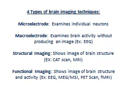
Microelectrode Examines individual neurons Macroelectrode Examines brain activity without producing an image Ex EEG Structural Imaging Shows image of brain structure EX CAT scan MRI ID: 593651
Download Presentation The PPT/PDF document "4 Types of brain imaging techniques:" is the property of its rightful owner. Permission is granted to download and print the materials on this web site for personal, non-commercial use only, and to display it on your personal computer provided you do not modify the materials and that you retain all copyright notices contained in the materials. By downloading content from our website, you accept the terms of this agreement.
Slide1
4 Types of brain imaging techniques:
Microelectrode
: Examines individual neurons
Macroelectrode
: Examines brain activity without producing an image (Ex: EEG)
Structural Imaging:
Shows image of brain structure (EX: CAT scan, MRI)
Functional Imaging:
Shows image of brain structure and activity (Ex: EEG, MEG/MSI, PET Scan, fMRI)Slide2
Microelectrode
Studies the function of a single neuron. The microelectrode is thinner than a human hair. Ending is charged and inserted into neuron. Changes in neurons documented. Mostly used to study effects of neurotransmitters Slide3
Macroelectrode
Used to obtain an overall picture of the activity in particular regions of the brain.EEG: (electroencephalograph)
Electrodes are taped to the scalp to detect brain waves (alpha waves when asleep/relaxed, beta waves when awake/alert). Can’t see through skull, but can show activity in brain.Slide4
Structural Imaging
CAT Scan (aka CT Scan)(computerized axial tomography)Creates 3D images of the brain by combining X-rays from many angles.Slide5
Structural Imaging (cont’d)
MRI (magnetic resonance imaging)Better images than CT scan. Head surrounded by magnetic field & brain exposed to radio waves, which causes hydrogen atoms to be released. Released energy mapped out and made into image.Slide6
Functional Imaging
To see brain’s activity, not just the structure.EEG Imaging: Same as EEG, but detected brain waves transformed into an image instead.Slide7
Functional Imaging (cont’d)
MEG/MSI: Like EEG Imaging, but more precise in where activity is taking place in the brain.(
magnetoencephalography
/magnetic source imaging)Slide8
Functional Imaging (cont’d)
PET scan (positron emission tomography)Uses radioactive energy to map brain activity. Person injected with radioactive substance. When substance decays due to activity of cells, positrons emitted. More activity, higher emissions. Mapped into image.Slide9
Functional Imaging (cont’d)
fMRI: (functional MRI)Same as PET scan, but instead measures movement of blood molecules, which is an index of neuron activity
Extremely precise images collected rapidly, many times per second. Doesn’t require injection of radioactive materials.Slide10
4 Types of brain imaging techniques:
Microelectrode: Examines individual neurons
Macroelectrode
: Examines brain activity without producing an image (Ex: EEG)
Structural Imaging: Shows image of brain structure (EX: CAT scan, MRI)Functional Imaging: Shows image of brain structure and activity (Ex: EEG, MEG/MSI, PET Scan, fMRI)