/
Gaolin Liang, Keming Xu, Lihua Li, Ling Wang, Yi Kuang, Zhimou Yang, a Gaolin Liang, Keming Xu, Lihua Li, Ling Wang, Yi Kuang, Zhimou Yang, a

Gaolin Liang, Keming Xu, Lihua Li, Ling Wang, Yi Kuang, Zhimou Yang, a - PDF document

natalia-silvester
natalia-silvester . @natalia-silvester
Follow
377 views
Uploaded On 2015-10-11

Gaolin Liang, Keming Xu, Lihua Li, Ling Wang, Yi Kuang, Zhimou Yang, a - PPT Presentation

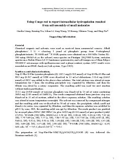
2 A CD spectra of Gel TEM images of Gel inset optical image formed by via enzymatic gelation in A TEM image of Congo red stained Gel 3 A Supramolecular hydrogel of Gel left and ID: 157539

(A) spectra

Share:

Link:

Embed:

Download Presentation from below link

Download Pdf The PPT/PDF document "Gaolin Liang, Keming Xu, Lihua Li, Ling ..." is the property of its rightful owner. Permission is granted to download and print the materials on this web site for personal, non-commercial use only, and to display it on your personal computer provided you do not modify the materials and that you retain all copyright notices contained in the materials. By downloading content from our website, you accept the terms of this agreement.


Presentation Transcript

Gaolin Liang, Keming Xu, Lihua Li, Ling Wang, Yi Kuang, Zhimou Yang, and Bing Xu* Chemical reagents and solvents were used as received from commercial sources. Alkali phosphatase: 1 U = cleaving 1 mol of phosphate group from 4-nitrophenyl phosphate/minute. P NMR spectra were obtained on a 300 MHz Varian XL- as the solvent, mass-spectra on Finnigan TSQ7000 System, emission spectra on a Perkin-Elmer LS-55 luminance spectrometer, and cell images on a Nikon Edipse TE2000-U microscope with epifluorescence and a phase contrast system. MTT results were recorded on an IEMS Analyzer (Lab-system, Type 1401). Syntheses and characterizations. Nap-D-Phe-D-Phe-tyrosine phosphate (): 203.5 mg (0.424 mmol) of Nap-D-Phe-D-Phe and 49.1 mg (0.427 mmol) of NHS were dissolved in 12 ml of chloroform. 128.6 mg (0.623 mmol) of DCC was added to the above clear solution. The total mixture was stirred at room temperature for 3 hour, the resulting precipitate was separated by filtration, and then the filtrate was dried by a rotary evaporator. The resulting solid was used for next reaction 111.1 mg (0.426 mmol) of tyrosine phosphate was dissolved in 15 ml of water containing 164.0 mg (1.95 mmol) of NaHCO. The crude compound obtained from previous step was dissolved in 25 ml of acetone, added to the above aqueous solution. The resulting solution was stirred at room temperature overnight. The solvent was removed by compressed nitrogen and the resulting solid was re-dissolved in 30 ml of water, the precipitate, which could not dissolve in water, was separated by filtration, and then the aqueous solution was acidified to pH~1 by conc. HCl, the resulting solid was got by filtration. The crude product was purified by reversed-phase HPLC (RP-HPLC) to give 164.9 mg (0.228 mmol) of pure product (yield: H-NMR (300 MHz, DMSO-): 8.45 (d, 1H), 8.40 (d, 1H), 8.20 (d, 1H), 7.82 (d, 1H), 7.74-7.69 (m, 2H), 7.57 (s, 1H), 7.46-7.43 (m, 2H), 7.28-7.05 (m, 13H), 6.93 (d, 2H), 4.60 (t, 1H), 4.51 (t, 1H), 4.42 (t, 1H), 3.50 (dd, 2H), 3.06 (dd, 1H), 2.98 (dd, 1H), 2.85 (dd, 1H), 2.69 (dd, 1H), 2.57 (dd, 1H), 2.40 (dd, 1H); P-NMR (300 MHz, DMSO-): -6.24; MS: calc. MThe preparation of Nap-Phe-Phe-NH(CHOCO(CHCOOH () was followed the literature methods [1]. 2 (A) CD spectra of Gel TEM images of Gel (inset: optical image) formed by via enzymatic gelation in . (A) TEM image of Congo red stained Gel 3 . (A) Supramolecular hydrogel of Gel (left) and polymer hydrogel polyacrylamide and polyacrylamide stained by Congo red; (C) and polyacrylamide after washing by PBS buffer.cells/well in 100 µL MEM medium with 10% FBS. Compounds at different concentrations were added when cells were plated. Then, the cell cultures were incubated for 24 hours at 37 and 5% CO. For cell survival assay, MTT assays were carried out to measure the proliferation of HeLa cells. The optical density (OD) of the dissolved formazan crystals was measured at wavelengths of 595 nm. The percentafollowing equation: viability (%) = ODtreatment groupcontrol group595 nm Cytotoxicity of 2 and 4. Extracellular Congo red staining. Before Congo red staining, were dissolved in PBS buffer to final concentrations of 8.3 mM (pH 7.4). After adding of alkaline phosphatase (500 u/mL), transparent hydrogel was obtained (Fig. 2A) in a few minut) was added to the hydrogel to a calculated concentration of 10 µM, the hydrogel was slowly stained by Congo red during the permeation of Intracellular Congo red stainingth cells for 24h, the culture medium with the precursors were removed, and the cells were washed with PBS for three times. Then the cells were incubated with 10 µM of in culture medium at 37 C, 5% CO and a humid environment for 30 min. was replaced with fresh PBS buffer and cell images were taken on a reverse fluorescence mias positive control, they were incubated with 1% Triton X-100 at 37 C, 5% CO and a humid environment for 1 min before Congo red staining. . (A, B, C) Merged microscope images (stained by Congo red: (A) Triton X-100 treated for 1 min before staining; (B) 250 µM and (C) 1 mM of treated for 24 h; (D, E, F) Merged microscope images (optical and ongo red: (D) Triton X-100 treated for 1 min microscope images (optical and flCongo red: (G) Control; (H) 1.73 µM and (I) 3.46 µM of [1] Z. M. Yang, K. M. Xu, Z. F. Guo, Z. H. Guo, and B. Xu, Adv. Mater., 2007, In press.