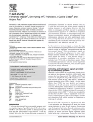
/
Induction of T Cell Anergy by Low Numbers of Agonist L Induction of T Cell Anergy by Low Numbers of Agonist L

Induction of T Cell Anergy by Low Numbers of Agonist L - PDF document

natalia-silvester
natalia-silvester . @natalia-silvester
Follow
392 views
Uploaded On 2015-04-28

Induction of T Cell Anergy by Low Numbers of Agonist L - PPT Presentation

Korb Saied Mirshahidi Kasra Ramyar Amir A Sadighi Akha and Scheherazade SadeghNasseri Engagement of TCR by its ligand the MHCpeptide complex causes T cell activation T cells respond positively to stimulation with agonists and are inhibited by antago ID: 56169

Korb Saied Mirshahidi Kasra

Share:

Link:

Embed:

Download Presentation from below link

Download Pdf The PPT/PDF document "Induction of T Cell Anergy by Low Number..." is the property of its rightful owner. Permission is granted to download and print the materials on this web site for personal, non-commercial use only, and to display it on your personal computer provided you do not modify the materials and that you retain all copyright notices contained in the materials. By downloading content from our website, you accept the terms of this agreement.


Presentation Transcript

InductionofTCellAnergybyLowNumbersofAgonistLigandsLauraC.Korb,SaiedMirshahidi,KasraRamyar,AmirA.SadighiAkha,andScheherazadeSadegh-NasseriEngagementofTCRbyitsligand,theMHC/peptidecomplex,causesTcellactivation.Tcellsrespondpositivelytostimulationwithagonists,andareinhibitedbyantagonistMHC/peptideligands.FailuretoinduceproperconformationalchangesintheTCRorfastTCR/MHCdissociationaretheleadingmodelsproposedtoexplainanergyinductionbyantagonistligands.Inthisstudy,wedemonstratethatpresentationofbetween1and10complexesofagonist/MHCIIbyun®xedAPCinducesTcellanergythatpersistsupto7daysandhascharacteristicssimilartoanergyinducedbyantagonistligandorTCRoccupancywithoutcostimu-lation.Furthermore,anergy-inducingdosesofhemagglutinin306±318peptideledtotheengagementoflessthan1000TCR/CD3complexes.Thus,engagementofasubthresholdnumberofTCRbyeitheralowdensityofagonist/MHCora2±3ordersofmagnitudehigherdensityofantagonist/MHCcausesanergy.Moreover,weshowthatanergyinducedbylowagonistconcentra-tionsisinhibitedinthepresenceofIL-2orcyclosporinA,suggestinginvolvementofthecalcineurinsignalingpathway.JournalofImmunology,1999,162:6401±6409.cellactivationrequirestheengagementofTCRwiththespeci®cMHC/peptideligand(signal1)andCD28onTcellswithB7onAPCs(signal2).Transmissionofsignal1intheabsenceofsignal2leadstoinductionofanergy(1,2).Inthepresenceofcostimulation,however,differencesinsignal1canalsodetermineanergyoractivation.Alteredpeptideligandshaveaidedourunderstandingofthemoleculareventsin-volvedintransmissionofsignal1attheinterfaceofTcellsandAPCsaswellasearlysignaltransductionprocesses(3,4).APLaresimilartotheagonistpeptide,exceptinoneormoreaminoacidresidues,generallythosethatcontacttheTCR,andcanfunctionasantagonists,orpartialagonistligands.AgonistpeptidesinducefullTcellactivation(bothproliferationandcytokineproduction).An-tagonistligandsarenotcapableofinducingTcellactivationandpreventTcellactivationinresponsetotheagonist,andpartialagonistpeptidescauseTcellstoexecutesome,butnotall,effectorfunctions.SeveralgroupshavedemonstratedthatTcellsstimu-latedwitheitherantagonistorpartialagonistpeptidesenterastateofanergyinwhichtheyareunresponsivetosubsequentstimula-tionwiththeagonistpeptide(5±9).Currently,theconformationalmodelandthekineticdiscrimi-nationmodelarethetwoleadingtheoriestoexplainhowAPLandagonistpeptidesgeneratetheirdifferenteffects.Theconforma-tionalmodelsuggeststhatTcellactivationdependsoninductionofacertainstructureintheindividualTCRthathasbeenengaged.Thus,APLinduceTcellunresponsivenessbytransmittinganeg-ativesignalduetotheirfailuretoinduceacorrectconformationintheTCR(10,11).Alternatively,thekineticdiscriminationmodelsuggeststhatagonistpeptidesandAPLengageTCRinasimilarconformation,butthatdifferencesinTcellresponsesresultfromtheshorterengagementtimebyAPL/MHCcomplexesascom-paredwiththeagonistligand(12,13).Speci®cally,therateofdissociationoftheTCRfromtheMHC/peptideliganddeterminesanergyoractivation.Aderivativeofthismodelistheaviditydis-criminationde®nedasthecollectiveaf®nitybetweenmultipleTCRinteractingwithMHC/peptidecomplexes.Therefore,antagonistpeptidesmaysendanegativesignaltotheTcellbecauseoffailuretoengagesimultaneouslyathresholdnumberofTCRsasaresultoffastdissociationoftheTCRfromMHCligand.Tcellactivationappearstobeacontinuoushierarchicalprocessinwhichdifferenteffectorfunctionsareinducedatdifferentden-sitiesofAg.Forexample,theligandconcentrationneededtobringaboutearlyactivationevents,suchasTCRdown-regulation,andin¯uxislowerthanthatneededforlaterresponses,suchasproliferationandcytokinesecretion(14±16).ActivationofThproliferationrequires100ormoreMHC/peptidecomplexes(17±19).ActivationofCD8cellsrequiresasimilarnumberofclassI/peptidecomplexes(20),althoughamuchlowerdensityofclassI-peptideligandforcytolytickillinghasalsobeenpre-dicted(21).IFN-productionseemstorequirealowerconcen-trationofMHC/peptideligandthanthatneededforIL-2pro-duction.Thus,selectiveactivationofTcelleffectorfunctionscanbeachievedbyeitherlowagonistdensityorpartialagonistpeptides.Itisunclear,however,whetherbothclassesofpep-tidesactthroughthesamemechanism.SinceAPLcouldpotentiallyalterboththeconformationandthekineticsofTCRinteractingwithligand,differencesinthesemod-elsarebestaddressedintheabsenceofthemultiplevariablesintroducedbytheuseofAPL.Inthisstudy,weinvestigatetheoutcomeofTcellstimulationwithalowdensityofagonist/MHCligand.We®ndthatTcellsexposedtolowagonistpeptideconcentrations,presentedonfullycompetentliveAPCs,becomeanergic,asdeterminedbytheirfailuretomountafullresponseto DepartmentofPathologyandGraduatePrograminImmunology,SchoolofMedicine/JohnsHopkinsUniversity,Baltimore,MD21205ReceivedforpublicationDecember28,1998.AcceptedforpublicationMarch15,1999.Thecostsofpublicationofthisarticleweredefrayedinpartbythepaymentofpagecharges.Thisarticlemustthereforebeherebymarkedinaccordancewith18U.S.C.Section1734solelytoindicatethisfact.ThisworkwassupportedbyCouncilforTobaccoResearch4314andNationalInstitutesofHealthGrantGM53549(toS.S.-N.).L.C.K.wassupportedbyaNationalInstitutesofHealthtraininggrant.AddresscorrespondenceandreprintrequeststoDr.ScheherazadeSadegh-Nasseri,DepartmentofPathology,SchoolofMedicine/JohnsHopkinsUniversity,664ERossBuilding,Baltimore,MD21205.E-mailaddress:ssadegh@pathlan.path.jhu.eduAbbreviationsusedinthispaper:APL,alteredpeptideligand;CsA,cyclosporinA;HA,hemagglutinin.Copyright1999byTheAmericanAssociationofImmunologists asubsequentstimulatorydoseofagonistpeptide.Theseeffectslastupto7daysandarequantitativelyandqualitativelysimilartothoseinducedbyanantagonistpeptide.Furthermore,we®ndthattheinhibitoryagonistandantagonistpeptideconcentrationsinducesimilarlowlevelsofTcellactivation,asassayedbyproliferation,cytokineproduction,TCRengagementanddown-regulation,andup-regulation.Furthermore,weshowthatanergyinducedbylowagonistconcentrationsisinhibitedinthepresenceofIL-2orcyclosporinA(CsA),suggestinginvolvementofthecalcineurinpathway.Theagonistconcentrationsthatinduceanergyare10-to100-foldlowerthantheconcentrationsnecessaryforinductionofadetectablelevelofIL-2.Inaddition,wehavedeterminedthenumberofTCRengagedbytheagonistpeptide/DR1anddemon-stratethatengagementof100-1000TCRinducesanergy.Ourdataconstitutethatbiologicalresponsesinducedbyfewerthan10highaf®nityMHC/peptidecomplexesperAPCaresimilartothosein-ducedbyseveralthousand-foldmoreantagonist/MHCperAPC,indicatingthatquantitativeandnotqualitativedifferencesinTCRliganddetermineactivationoranergy.MaterialsandMethodsCellsandcultureconditionsClone1isahumanCD4Th1clonespeci®cforthepeptide306±318ofin¯uenzahemagglutinin(HA)presentedonHLA-DR1(5),andHA1.7isahumanCD4Th0clonealsospeci®cforHApresentedonHLA-DR1(22).Althoughbothcloneshavethesamespeci®city,theywereisolatedatdif-ferenttimesandfromdifferentindividuals.EBV1.24,ahumanHLA-DR1(DRB1*0101)-positive,EBV-immortalized,activatedBcellline(EBV-B,orBcells),wasusedasAPC.EBV-BcellsconstitutivelyexpresshighsurfacelevelsofthecostimulatorymoleculesB7.1andB7.2,asdeterminedbyFACSstainingwithprimaryAbsBU63,anti-B7.2(Calbiochem),andMAB104,anti-B7.1(Immunotech,Westbrook,ME),followedbyPE-con-jugatedsecondaryAbs(Sigma,St.Louis,MO).EBV-BcellsweregrowninRPMI1640supplementedwith10%FCS,2mM-glutamine,nonessentialaminoacids(Sigma),and1mMsodiumpyruvate(Sigma).TcellsweregrowninRPMI1640medium(LifeTech-nologies,GrandIsland,NY)supplementedwith5%FCS(LifeTechnolo-gies),5%pooledhumanserum,2mM-glutamine(LifeTechnologies),and10mMHEPES(LifeTechnologies).Every7days,Tcellclonesweremaintainedbyrestimulationof10Tcellswith10EBV-Bcells(irradiated10,000rad),10humanleukocytes(irradiated10,000rad),1MHApep-tide,and120IU/mlhumanrIL-2(Cetus,Norwalk,CT)in2mlofmediumperwellofa24-wellplate.Peptidesweresynthesized,puri®edbyHPLC,andanalyzedbyPeptideExpress(FortCollins,CO).HA306±318,PKYVKQNTLKLAT,isthefullagonistpeptideforbothclonesusedintheseexperiments.ETEC,IIYQIVVEKGKKK,doesnotbindtoDR1(23),andYAK(AAYAAAAAAKAAA)bindstoDR1,butexhibitsnoagonistorantagonistproperties(nullpeptide)foreitherclone(datanotshown).N312Q,PKYVKQQTLKLAT,isanantagonistpeptideofClone1(5).Thepeptideswerepuri®edtoapparenthomogeneityofmorethan95%byreverse-phasepreparativeHPLC,andtheiridentitieswerecon®rmedbymassspectrometry.TheconcentrationofthepeptidestocksolutionswasdeterminedbyninhydrinInductionofTcellunresponsivenessTcellanergywasinducedbyincubating4Tcellswith4irradiated(10,000rad)EBV-BcellsandvariableconcentrationsofHAorN312Q,in200lina96-wellround-bottomplatefor18±36hat37ÉCin5%CO.Alternatively,2Tcellsand2irradiated(10,000rad)EBV-Bcellswereincubatedwiththeindicatedpeptideconcentrationsfor18±36hinavolumeof1mlina48-wellplate.Attheendofthistime,TcellswereseparatedfromBcellsbydensity-gradientcentrifugationoverFicoll-Paque(PharmaciaBiotech,Piscataway,NJ),washedextensively,andcounted.Tcellswerethenstimulatedwith0.1±10MHApeptideormediumaloneandfreshlyirradiatedEBV-Bcells.PersistenceofanergywasexaminedbyincubatingseparatedTcellswithfreshunpulsedBcellsfor3,5,7,or14days.Aftertheserestperiods,peptide-pulsedBcellswereaddedandTcellswereassayedforproliferation.Proliferationorcytokine(IL-2orIFN-)productionwasmeasuredasindicatedbelow.Insomeassays,IL-2(10IU/ml)orCsA(1g/ml)(Sandoz,Basel,Switzerland)wasaddedduringtheinitialphase.Intheseassays,cellswerewashedthreetimesbeforetheadditionofthestimulatorydoseofAg.Toexcludethepossibilityoferrorsincountingrecoveredcells,proliferationofTcellsinresponsetoIL-2wasmeasured.TcellproliferationassaySeventy-twohoursaftertheadditionofthestimulatorydoseofagonistpeptideorpeptide-pulsedAPCs,eachwellwaspulsedwith1Ci[midine(Amersham,ArlingtonHeights,IL).Cellswereharvestedandcounted14hlaterusingabetacounter(PackardInstruments,Meriden,CT).Eachassaywasdoneintriplicate.CytokineassaysIL-2releasewasmeasuredusingtheIL-2-sensitivecelllineHT-2.Culturesupernatantswereharvested24hafteradditionofastimulatorydoseofHApeptide,frozen,andthenthawedonce.Then50lofculturesupernatantand5HT-2cellsinRPMIsupplementedwith10%FBS,502-ME,and-glutaminewerecombinedinonewellofa96-wellplateandincubatedat37ÉCat5%COfor24h.Eachwellwasthenpulsedwith1Ciof[H]thymidineforanadditional14h,harvested,andcounted.Allassayswereperformedintriplicate.productionwasmeasuredintheculturesupernatantsharvested24or48hafteradditionofthestimulatorydoseofagonistpeptidebyELISAwithamatchedAbpair,accordingtomanufacturer's(Phar-Mingen,SanDiego,CA)suggestedprotocol.Allassayswereperformedinduplicate.CD28cross-linkingWellsofa96-wellplatewereincubatedfor3hat37ÉCwith50lPBSwithoutAbor10g/mlgoatanti-mouseIgG(Cappel),thenwashedthreetimesinPBS.Clone1Tcellswererenderedunresponsiveineithertheabsenceorpresenceof5g/mlmouseanti-humanCD28,asdescribed(PharMingen).Proliferationto1MHApresentedbyEBV-BcellswasPeptidequanti®cationSyntheticpeptideswerepurchasedfromMolecularResources(ColoradoSprings,CO).Electrospray-massspectrophotometryanalysisoftheHApeptideaftersynthesisaswellasourownanalyticalHPLCanalysesindi-catethatthepeptidewasgreaterthan97%pureandlackedanydetectablecontaminantswithabsorbanceat210nm.SyntheticHApeptide(50nmol)wasiodinatedwithIodo-Beads(Pierce,Rockford,IL).Afteriodination,thepeptidewasthenpuri®edbyreverse-phaseHPLCusinga3C-18column(ThomsonInstrument,Spring®eld,VA)witha15-min0±50%acetonitrilegradientinthepresenceof0.1%tri¯uoroaceticacid.Speci®cactivityandrecoveryweredeterminedbyintegrationofareaunderthecurveat280nmusingserialdilutionsofcoldHApeptideasarefer-ence.Speci®cactivityofapproximately44Ci/mmolwascalculated.IrradiatedEBV-Bcells(1/well)werepulsedfor18hwiththeindicateddosesofpeptideundertheconditionsusedforanergyinduction.Quantitativeanalysisofcomplexformationwasperformedbyamodi®ca-tionofapreviouslydescribedmethod(17).Brie¯y,pulsedcellswerewashedthreetimesincompletemediumandthenresuspendedin1mlofPBScontainingPMSF(10mM)andaprotinin(1mg/ml).glucopyranosidewasthenaddedtoa®nalconcentrationof1%andthecellsweredisruptedbymixing.Afteralow-speedcentrifugationtoremoveinsolubledebris,thelysatewaspreclearedusingSepharoseCL-4B.HA-DR1complexeswereimmunoprecipitatedusinganexcessofmAbL243-coupledCL-4Bresin,whichwasthenseparatedfromthelysatebycen-trifugationovera60%sucrosecushion.Precipitationef®ciencywasdeterminedtobenearly100%byasecondimmunoprecipitationoftheclearedlysate.Allmanipulations(lessthan2h)wereconductedoniceorat4ÉC.Fractionswerecounteddirectly,forfourminutespersample,usingagammacounter.Precipitatedcountswerecomparedwithadilutionseriesoftheiodinatedpeptidealone.andCD3expressionandCD3expressionwereassayedby¯owcytometryusingaBec-tonDickinson(MountainView,CA)FACScan:5e1Tcells,EBV-Bcells,andindicatedconcentrationsofagonist,antagonist,orirrelevantpeptidewereincubatedinonewellofa96-wellround-bottomplatefor18h.Atthistime,cellswereeitherstainedwithanti-IL-2RandanalyzedbyFACS,or10MHApeptidewasaddedtoeachwellforanadditional5hbeforestainingforCD3expressionorforanadditional24hbeforestainingforIL-2Rexpression.StainingforCD36402ANERGYINDUCEDBY1±10COMPLEXESOFAGONIST/CLASSII expressionwasperformedwithmouseanti-humanCD3(OKT3;AmericanTypeCultureCollection,Manassas,VA)puri®edfromascites,followedbyPE-conjugatedgoatanti-mouseIgG(Sigma).IL-2Rwasdetectedusingmouseanti-humanIL-2R(TCellSciences,Cambridge,MA),followedbyPE-conjugatedgoatanti-mouseIgG.BaselineexpressionwasdeterminedbyincubatingTandBcellswithoutpeptideduringtheinitialincubationphaseand/orthesecondaryactivationphase.Maximaldown-regulationofCD3orup-regulationofIL-2RwasdeterminedbyincubatingTcellsandBcellswithoutpeptideduringtheinitialphase,followedbystimulationwith10MHAduringthesecondaryincubation.Tcellsweregatedbysizeandgranularity.QuantifyingTCRdown-regulatione1Tcells(10),EBV-Bcells(10),andindicatedconcentrationsofagonistorantagonistpeptideswereincubatedinseparatewellsofa24-well¯at-bottomplatefor6h.Atthispoint,thecellswerestainedwithsaturatingconcentrationsof(FITC-)conjugatedanti-CD3withknown¯uorescein:proteinratio(5:1),followedbyPE-conjugatedanti-CD4(bothfromPharMingen).ThespecimenswereexaminedbyFACSanalysis.Thenum-berofTCRswasestimatedbydeterminingthepeak¯uorescencechannelforeachsampleandcomparingwith¯uorescenceofcalibrationparticleswithde®ned¯uorescenceintensities(Spherotech,Libertyville,IL).CytokineproductionandproliferationofTcellclonesinresponsetotheagonistpeptideHA306±318Clone1andHA1.7areTcellclonesthatarespeci®cforthepep-tide306±318ofin¯uenzaHApresentedontheclassIImoleculeHLA-DR1.N312QisananalogueofHA,whichcontainsasingleaminoacidsubstitutionandisanantagonistofClone1(5).BothclonesproliferateandproduceIL-2andIFN-uponstimulationwiththeagonistpeptide.Clone1TcellsshowedextremelyweakproliferationandIFN-productionandnodetectableIL-2produc-tioninresponsetotheantagonistpeptideN312Qconcentrationsupto50M(Fig.1).Althoughbothcloneshavethesamepeptidespeci®cityandHLArestriction,theywereisolatedindependentlyandusedifferentTCRV-andV-chains(22).Inaddition,pep-tidesthathavebeenshowntobeantagonistsofClone1arenotantagonistsofHA1.7(24).UnresponsivenessinducedbyalowdoseofagonistpeptideTcellswereincubatedwithBcellsinthepresenceofvariousdosesofHAand,forClone1,ofN312Qfor18h.AsecondsetofBcells,whichwerepulsedwith10MHA(proliferationandIL-2)or1MHA(IFN-),wasaddedtotheculture,andTcellproliferationandIL-2andIFN-productionwereassayed(Fig.2,).TheresponseofClone1cells,whichwereuntreatedorpretreatedwitheither0.01MHA,25MN312Q,or0.01MofthenullpeptideYAKtoarangeofstimulatorypeptideconcen-trations,isalsoshown(Fig.2).Whenpretreatedwithalowcon-centration(0.01±0.001M)ofagonistpeptide,bothclonesex-hibiteddecreasedproliferationandIFN-productiontotheagonistligand.Additionally,whenpretreatedwithaninhibitoryconcen-trationofagonistpeptide,neithercloneproducedanydetectableIL-2inresponsetothestimulatorydoseofagonistpeptide.Theinhibitoryeffectoftheagonistpeptideoccurswithina2±3logconcentrationrangeandisnotobservedatconcentrationsaboveMorbelow1pM.Interestingly,theinhibitoryeffectofalowconcentrationofagonistiscomparablewiththeeffectofahighM)concentrationofN312Q.TheirrelevantpeptidesETECandYAKhadnoinhibitoryeffect.Similarresultswereob-servedwhenTcellswereseparatedfromBcellsaftertheinitialstepbycentrifugationoverFicoll(asinFigs.2and7).IthasbeenshownthathumanTcellscanpresentAginatolerogenicfashion(5,24,25).However,highdosesofpeptidearenecessaryforthiseffect.Inoursystem,TcellsproliferatenormallyifeitherBorTcellsareeliminatedfromthepretreatmentstep,andareaddedinsteadatthestimulationstep,indicatingthatBcellpre-sentationofpeptide/DR1isnecessaryforinducingunresponsive-ness(datanotshown).TcellanergycanresultwhenTCRligationoccursintheab-senceofCD28engagement.AlthoughEBV-BcellsexpresshighlevelsofB7.1andB7.2andcanfullyactivateTcellclonestested(Fig.1,),toensureeffectiveengagementofCD28anddeliveryofsignal2duringtheinitialphase,weexaminedtheeffectofCD28cross-linkingduringtheexposureofTcellstoalowconcentrationofagonistpeptide.Wellsofa96-wellplatewereeitherleftuncoatedorcoatedwithcross-linkedanti-humanCD28.e1TcellswerethenincubatedwithEBV-Bcellswithoutpeptideor0.01MHA.FreshBcellspulsedwith1MHAwereaddedtowells,18hlater,andproliferationtothischallengewasmeasured.Unresponsivenesswasinducedeveninthepresenceofcross-linkedanti-CD28.Theeffectivenessofthistreatmentinpro-vidingsignal2wascon®rmedbyitsenhancementofTcellpro-liferationinresponsetoanti-CD3assignal1(datanotshown).Tcellunresponsivenesslastsupto7daysPersistenceofanergywasexaminedbyincubatingTcellsthathadbeenanergizedby0.01MHA-pulsedBcellswithfreshunpulsedBcellsfor3,5,7,or14days.Aftertheserestperiods,peptide-pulsedBcellswereaddedandTcellswereassayedforprolifer-ation.AsshowninFig.2,cellsremainedanergicupto7daysof FIGURE1.CytokineproductionandproliferationofTcellclonesinresponsetotheagonistpeptideHA306±318.IFN-production(circles),IL-2production(squares),andproliferation(triangles)dose-responsecurvesofClone1()andHA1.7()TcellstoagonistpeptideHA(®lledsymbols)orN312Q(opensymbols)presentedbyEBV-Bcells.MaximumresponseisthehighestresponseobservedwhencellswerestimulatedwithHApep-tide.ForClone1,totalcountsformaximumproliferationare96,601,totalcountsformaximumIL-2productionare46,738forHT-2indicatorcells,andmaximumIFN-productionis24.8ng/ml.ForHA1.7,maximumproliferationis69,197totalcounts,maximumIL-2productionis79,066totalcountsofHT-2indicatorcells,andmaximumIFN-productionis19.6ng/ml.TheJournalofImmunology restperiodbeforethepeptidechallenge.However,cellsrecoveredfromanergyifrestedforanother7days(datanotshown).Quanti®cationofDR1-HAcomplexesthatinduceanergyWeusedanI-labeledHApeptidetodeterminethenumberofDR1/HAcomplexesformedperAPCinourpretreatmentsteptoestimatethenumberofcomplexesthatinduceTcellanergy.Ourpreviouswork(26)hadshownthatiodinationofHApeptidedoesnotalteritsDR1-bindingaf®nity,astheiodinatedHApeptidecouldcompeteequallywiththeunmodi®edpeptideinpeptidecompetitionassays.EBV-Bcellswereincubatedwithvariouscon-centrationsofiodinatedHAunderthesameconditionsusedinthepretreatmentstepofourassay.Cellswerethenwashedextensivelyandsolubilizedwiththedetergent-Octylglucopyranoside.DR1moleculeswerespeci®callyimmunoprecipitated,andthenumberofboundpeptideswasdeterminedbydirectgammacounting(Fig.3).Asecondimmunoprecipitationdidnotyieldcountsabovethebackgroundlevel,indicatingthatnearlyallDR1moleculeswereimmunoprecipitated.Assuminganequaldistributionofcom-plexes,wefoundthatapproximately10HA-DR1complexeswereformedperAPCwhencellswereincubatedwith0.01MHAandthatanearlylinearrelationshipexistedbetweenthepeptidecon-centrationandthenumberofcomplexesformedintherangemea-sured.Thus,atanHAconcentrationof0.1M,100complexesofpeptide/MHCwereformedperAPC,andat1nM,theaveragenumberofHA-DR1perAPCwasone.Notably,evenasingleagonist-MHCIIcomplexissuf®cienttoinduceameasurableeffectinspeci®cTcells. FIGURE2.ExposureofTcellstoalowconcentrationofagonistpep-tiderenderscellslessresponsivetoastimulatoryAgconcentration.Clone)orHA1.7()TcellswereincubatedwithirradiatedEBV-BcellsandvariousconcentrationsofHA(®lledsymbols)orN312Q(opensymbols)for24h.FreshirradiatedBcellsandastimulatoryconcentrationofHApeptide(10MinproliferationandIL-2assaysand1MinIFN-werethenaddedtocultures,andproliferation(triangles),IL-2production(squares),andIFN-production(circles)weremeasured.ResultsareshownasthepercentageoftheresponseofTcellspretreatedintheabsenceofpeptide.,Clone1Tcellswerepretreatedasinintheabsenceofpeptide(),with0.01MYAK(),with25MN312Q(),orwithMHA(),andproliferationtoarangeofHAconcentrationswas,Clone1TcellswereculturedwithirradiatedBcellsandnopeptideor0.01MHA;18hlater,TcellswereseparatedfromBcellsoveraFicollgradient,washed,andrestedwithfreshirradiatedBcellsforthetimeindicated.Asecondbatchoffreshlyirradiatedbutpeptide-pulsedBcellswasaddedafter0,3,5,or7days,andproliferationwasmeasured3dayslaterby[ FIGURE3.TheconcentrationofHApeptidethatinducesTcellanergyresultsintheformationoflessthan10HA-DR1complexesperAPC.IrradiatedEBV-Bcellswerepulsedfor18hwiththeindicateddosesofI-labeledHApeptide.DR1/peptidecomplexeswereimmunoprecipi-tatedfromdetergent-solubilizedcells,asdescribedinMaterialsandMeth-,andcounteddirectlyusingagammacounter.PrecipitatedcountswerecomparedwithadilutionseriesoftheI-labeledHA.6404ANERGYINDUCEDBY1±10COMPLEXESOFAGONIST/CLASSII SimilarlevelsofTCRdown-regulationandIL-2Rup-regulationbyinhibitoryconcentrationsofantagonistoragonistpeptidesUponactivationwithAg,Tcellsdown-regulatetheirsurfaceex-pressionofTCR(27,28)andup-regulatetheirsurfaceexpressionoftheIL-2R.WecomparedtheeffectofthelowconcentrationofagonistpeptidewiththeinhibitorydoseofantagonistpeptideonTCRandIL-2RexpressiononClone1.TcellsandEBV-Bcellswereincubatedeitherintheabsenceofpeptideorwith0.01HAor25MN312Qfor18h.TcellswerethenevaluatedforCD3andIL-2Rexpressionby¯owcytometry.Wefoundthat0.01MHAand25MN312QcausedsimilarshiftsinthelevelofCD3expression,indicatingsimilardown-regulationinthelevelofTCRbyeitherligand(Fig.4).AsimilarincreaseinthelevelofIL-2Rwasalsoobservedbybothligandsattheaboveconcentrations(Fig.4).Interestingly,itappearsthatstim-ulationwiththesetwodifferentpeptidescausedthesamelowlev-elsofactivation.Quanti®cationofTCRengagementTCRdown-regulationisshowntobeanindicationofTCRen-gagement.ToquantifythenumberofTCRengagedbythelownumbersofHA/DR1complexes,Clone1TcellswereincubatedwithEBV-Bcells,pulsedwith0,0.001,0.01,0.1,1,or10MHAor25MN312Qfor6h(maximalTCRdown-regulation).TCR-CD3complexeswereenumeratedbydirectstainingwithFITC-labeledanti-CD3Ab,withknownnumbersofFITCpermolecule,followedbyFACSanalysis.ThenumberofTCRengagedateachpeptideconcentrationwasestimatedbysubtractingthe¯uores-cencesignalonstimulatedTcellsfromthatofrestingTcell.Themeanchannel¯uorescencewascomparedwiththe¯uorescenceofcalibrationbeadsforestimationofthenumberofTCRstained.Thisshowedabasalexpressionof8594oneachrestingTcell.Cellsexposedto0.001,0.01,0.1,1,or10MHAhad0,1093,2186,3934,and5027receptorsdown-regulated,respectively.In-cubationwith25MofN312Qledtothedown-regulationof687TCR/CD3complexes.TcellsrenderedunresponsiveshownormalTCRdown-regulationandIL-2Rup-regulationinresponsetoastimulatorydoseoftheagonistpeptideWealsoexaminedwhetherClone1Tcellsthathadbeenpre-treatedwithalowdoseofagonistorwithaninhibitorydoseofN312Qexhibitedthesesignsofactivationuponsubsequentstim-ulationwithahighdoseofagonistpeptide(Fig.5).AllgroupsofTcellsshowedequallevelsofCD3andIL-2Rexpressionafterstimulationwith10MHA,regardlessofpriorexposuretoin-hibitorypeptide.Thus,pretreatmentapparentlydidnotaffecttheabilityoftheTCRtobeengagedbyaseconddoseofagonistpeptidenordidpretreatmentpreventIL-2Rup-regulation.Thesamephenotypeisobservedwhenanergyisinducedbysignal1intheabsenceofsignal2(29).TcellsrenderedunresponsiveproliferatenormallytoexogenousIL-2andtoastimulatorydoseofagonistinthepresenceofexogenousIL-2InlightofthedatathatTcellsexposedtolowagonistconcentra-tionsretainedtheabilitytoup-regulateIL-2Rexpression,wenextexaminedwhethertheseTcellsarealsoabletoproliferateinre-sponsetoexogenousIL-2.Clone1TcellswerepretreatedwithBcellsintheabsenceofpeptideorinthepresenceof0.01MHA.Tcellswerethenstimulatedwitheither10IU/mlIL-2or1HA,orboth,andassayedforproliferation.BothgroupsofTcellsproliferatedequallywellinresponsetotheIL-2orthepeptideplusIL-2,butnottothepeptidealone(Fig.6).Theseresultsindicate FIGURE4.InhibitoryagonistandantagonistpeptideconcentrationscausesimilarlevelsofTCRdown-regulationandIL-2Rup-regulation.e1TcellsandirradiatedBcellswereincubatedwithnopeptide,0.01mMHA,or25mMN312Qfor18()or24()h.CellswerethenwashedandstainedwithmAbsagainstCD3()orIL-2R(),followedbyaPE-labeledgoatanti-mouseIgG,andsurfaceproteinexpressionlevelsweredeterminedby¯ow-cytometricanalysis.Solidlinesshowsurfacereceptorexpressionofcellsmockstimulatedintheabsenceofpeptide.Dashedlinesshowsurfacereceptorexpressionlevelofcellsstimulatedwithindicatedpeptideconcentrations.Solidhistogramsshowstainingwithanisotype-matchednegativecontrolAb.Meanlog¯uorescenceofTCRandIL-2RexpressiononTcellsstimulatedwithvariouspeptideconcentrationsisalso,TheamountofTCRengagedafterexposuretodifferentconcen-trationsofHA()or25MofN312Q().Tcelldown-regulationwasquanti®edbyincubatingClone1TcellsandEBV-Bcellswithoutpeptideorinthepresenceof0.001,0.01,0.1,1,or10MHAor25MN312Qfor6h.ThenumberofTCR-CD3complexeswasestimatedbycomparingtheanti-CD3stainingwiththestandardcurveofcalibrationparticleswithde®ned¯uorescenceintensities.Abasalexpressionof8594TCRoneachTcellwasdetermined.Cellsexposedto0.001,0.01,0.1,1,or10MHAhad0,1093,2186,3934,and5027receptorsdown-regulated,respectively.Incubationwith25MofN312Qledtothedown-regulationof687TheJournalofImmunology thatunresponsivenesswasnotduetoTcelldeletion.Instead,itmaybeduetoafailureofIL-2production,asproliferationwasseennotonlyinresponsetoIL-2alone,butalsotopeptideinthepresenceofIL-2.TheproliferationofallgroupsofTcellsinre-sponsetopeptideplusIL-2wasgreaterthanthatinducedbyIL-2alone,indicatingthattheTcellswererespondingtothepeptideinadditiontotheIL-2.IL-2inhibitsinductionofunresponsivenessPreviousstudieshaveindicatedthatAPLand®xedAPCsinduceunresponsivenessbecauseoftheirfailuretostimulateIL-2pro-ductionandthatIL-2inhibitstheinductionofunresponsiveness(30).TodeterminewhetheranergyinducedbyalowdensityofagonistligandisalsoreversibleinthepresenceofIL-2,wein-cludedexogenousIL-2duringtheinitialphaseofourassay.TcellswereseparatedfromthedeadBcellsandwashedtoremovere-sidualIL-2,andTcellproliferationinresponsetostimulationwithMHAwasassayed.Tcellsexposedtoalowagonistconcen-trationinthepresenceofIL-2respondedtoAgtothesamedegreeasTcellsthatwereculturedintheabsenceofpeptide(Fig.7indicatingthatIL-2preventstheinductionofanergybythelowconcentrationofagonist.CsApreventsinductionofunresponsivenessCsAhasbeenshowntobindcyclophilinandinhibitactivationofcalcineurin,acalcium-andcalmodulin-dependentphosphatase,re-sultingininhibitionofdephosphorylationofcytoplasmicnuclearfactorsofactivationinTcells,NF-AT,necessaryfortranscriptionofIL-2gene(31±34).PreviousstudiesofanergyinducedbyAPL(6)orbychemically®xedAPCs(35)showedthatanergywasnot FIGURE5.Peptide-inducedanergydoesnotchangeTCRdown-regulationorIL-2Rup-regulationinresponseto10HA.Clone1Tcellswereincubatedfor24hwithirradiatedBcellsandindicatedpeptideconcentrations.FreshBcellsandMHAornopeptidewerethenaddedtocultures.Five()ortwenty-four(hourslater,cellswerewashedandstainedwithmAbsagainstCD3()orIL-2R(followedbyaPE-labeledgoatanti-mouseIgG,andsurfaceproteinexpressionlevelsweredeterminedby¯ow-cytometricanal-ysis.Solidlinesshowsurfacereceptorex-pressionofcellspretreatedandmockstimulatedintheabsenceofpeptide.Dashedlinesshowsurfacereceptorex-pressionlevelofcellspretreatedwithin-dicatedpeptideconcentrationandstimu-latedwith10MHA.Solidhistogramsshowstainingwithanisotype-matchednegativecontrolAb.Meanlog¯uores-cenceofTCRandIL-2Rexpressionunderdifferentpretreatmentandstimulationconditionsisalsogiven. FIGURE6.e1Tcellspretreatedwithalowconcentrationofag-onistpeptideproliferateaseffectivelyasuntreatedcellsuponstimulationwithexogenousIL-2orwithAgplusIL-2.Clone1TcellswereincubatedtogetherwithBcellsandeither0.01MHAornopeptide;18hlater,TcellswerestimulatedwitheitherfreshirradiatedBcellsMHA,10IU/mlhumanIL-2,orfreshirradiatedBcellsMHA10IU/mlhumanIL-2,andproliferationwasmeasuredby[H]thymidineuptake.6406ANERGYINDUCEDBY1±10COMPLEXESOFAGONIST/CLASSII inducedinthepresenceofCsA.Thus,wetestedwhetherunre-sponsivenessinducedbyalowdoseofagonistwassensitivetoCsA.Clone1TcellsandirradiatedEBV-Bcellswereincubatedintheabsenceofpeptideorwith0.01MHApeptideinthepresenceorabsenceof10g/mlofCsA.EBV-Bcellswereremoved,Tcellswerewashedextensively,andproliferationinresponseto1MHAwasassayed.AsshowninFig.7,CsApreventedinduc-tionofTcellunresponsiveness,suggestingthatinductionofun-responsivenessinvolvesthecalcineurinpathway.TcellunresponsivenesscanbeinducedbyalowdensityofagonistorahighdensityofantagonistMHC/peptidepresentedbyliveAPCsThe®ndingsdescribedinthisworkaddresstherelationshipbe-tweenthenumberofMHC/peptidecomplexesonAPCsanddif-ferentialsignalingtransducedinTcells.We®ndthatlowconcen-trationsofagonistpeptidepresentedonliveAPCscaninduceTcellanergyinCD4Tcellclones.Similareffectsareinducedby10,000-foldhigherconcentrationofantagonistpeptide.Thistreat-mentinhibitsexpressionoflateactivationeventssuchasprolifer-ationandproductionofIL-2orIFN-,butdoesnotaffectTCRdown-regulation,anearlyactivationevent.IL-2RexpressionisalsounaffectedbypretreatmentofTcellswithalowagonistorinhibitoryantagonistpeptideconcentration,suggestingthatnotallsignalingpathwaysintheTcellareimpaired.Weestimatethatapproximately1±10agonistMHC/peptidecomplexesperAPCin-duceTcellanergy,andthatsimilareffectsareobservedinre-sponsetothreeordersofmagnitudehigherconcentrationsoftheantagonistpeptide.WedemonstratethatanergyinductionisnotlikelytobeduetofailureofAPCstoprovideanadequatecostimulatorysignal,astheEBV-BcellsusedasAPCsexpresshighlevelsofB7.1andB7.2,asdeterminedby¯ow-cytometricanalysis.Moreover,cross-link-ingofCD28withplate-boundanti-humanCD28duringthepre-treatmentofcellswithlowagonistconcentrationdoesnotpreventinductionofanergy(datanotshown).Tcellsexposedtolowcon-centrationsofagonistpeptideretaintheabilitytoproliferateinresponsetoexogenousIL-2,rulingoutthepossibilityofcelllossordeath.Moreover,anergyinductionisnotduetoTcellAgpre-sentation,asreportedearlier.Inthosestudies,theconcentrationofpeptidenecessaryforinductionofunresponsivenessinTcellswasseveralordersofmagnitudehigherthanthatnecessaryforTcellactivation.Inoursystem,TcellunresponsivenessisinducedbyextremelylowdosesandisdependentonpresentationbyBcells.WehaveusedTcellclonesspeci®cforHA306±318boundtoDR1becauseofthestabilityofHA/DR1complexes.Wehavemeasuredadissociationof140h(6days!)at37ÉCforI-labeledHA/DR1complexes(26).Suchremarkablestabilityminimizeserrorsinunderestimatingthenumberofpeptide/MHCcomplexesduetodissociationduringtheprocess.Thus,amoreaccurateestimationofthenumberofcomplexesnec-essaryforinductionofTcellresponsescanbeachieved.Theonlyotherreportinliteratureregardingameasurableeffectinducedbyasinglepeptide/MHCcomplexcomesfromSykulevetal.(21),thatcontrarytoourreport,observedcytolytickillingoftargetcellsexpressesasfewasasinglepeptide/classIMHCligand.ThisdifferencemightbeduetodifferentactivationrequirementsforCD8versusCD4Tcells.Wecandismissthepossibilitythatunresponsivenessisinducedbycontaminatingantagonistpeptidesfoundintheagonistpeptidepreparationinseveralways.First,ourpeptidesarepuri®edtoap-parenthomogeneityof95%byreverse-phasepreparativeHPLC,andtheiridentitieswerecon®rmedbyMALDImassspectrometry.Second,duringthecourseoftheseexperiments,wehaveusedseveralindependentbatchesofpeptidesthatallhaveconsistentlyinducedunresponsivenessatthesamedosesofagonistpeptide.Finally,thepossibilityofacontaminantpeptidethatinducesan-ergyonlyatextremelylowbutnotathigherdosesofpeptideseemsimplausible.AgonistligandsprovidetoolsfordiscriminationamongdifferentmodelsofTcellactivationUseofAPLforTcellstimulationhasprovidedameanstostudymechanismsofTCRengagementandthesignalstransduced.Sev-eralmodelshavebeenproposedthatrelyonqualitativeorquan-titativedifferencesbetweenAPLandtheagonistligands.ByusingagonistligandatalowdensityonAPCs,wehavecircumventedthesedifferences.Inthisstudy,becauseoftheuseoftheagonistpeptide,theconformationorqualityoftheTCRligandremainsthesame.Additionally,thekineticsofanindividualMHC/peptide-TCRinteractionremainsunchanged.Thus,wecanevaluatetheconceptsofconformationalorkineticmodelsinengagementofTCRs.Our®ndingssuggestthattransductionofsignalsthatcause FIGURE7.IL-2andCsApreventtheinductionofTcellanergybylowagonistpeptideconcentration.,Clone1Tcellswereculturedwithirra-diatedBcellsandnopeptideor0.01MHAintheabsenceorpresenceof10IU/mlIL-2;18hlater,TcellswereseparatedfromBcellsoveraFicollgradient,washed,andstimulatedwithfresh,irradiatedBcellsand1MHA.Proliferationwasmeasured3dayslaterby[cellswereculturedwithirradiatedBcellsandnopeptideor0.01MHAintheabsenceofdrugorinthepresenceof1g/mlCsA.Eighteenhourslater,TcellswereseparatedfromBcells,andproliferationwasassayedasinTheJournalofImmunology Tcellunresponsivenessdoesnotoccurthroughqualitativediffer-encesatthelevelofindividualMHC/peptide-TCRinteractions.Rather,weproposethatanergyistheresultofacollectiveortwo-dimensionaldecreaseinaf®nityorso-calledavidityoftheTcellfortheAPC.Ourresultsshowthatinductionofaspeci®c,uniqueTCRconformation,ashasbeenproposedasamechanismofanergyinductionbyantagonistligands(8,11,36),isnotnec-essaryforinductionofTcellanergy.Furthermore,ourdatadonotsupportmodelsofkineticdiscriminationatthelevelofsingleTCR-ligandinteractionsforthesamereasonthatanergycanbeinducedbytheagonistligand.However,wecannotruleoutthepossibilitythatengagementofTCRbyahighdensityofhighaf-®nityligandmightinducestructuraldifferencesthatwouldnotbeinducedbyalowdensityofligand.MultimerizationofTCReitherinrandomclusters(37)onthesurfaceofTcellsorasorientation-speci®cmultimers(38)ororderedoligomers(39)representsalter-nativemodelsforTcellactivationinresponsetoformationofspeci®cTCRarrays.Thelikelihoodofformationofsuf®cientnum-bersofcorrectlyorientedmultimersismuchincreasedifmanyTCRsareengagedbytheirligands.Byincreasingthenumberofligandsonacellsurface,theavid-ityoftheTcell-APCinteractionwillbeincreased.Thisalonecoulddetermineanergyoractivation.Onemayconsidertheavid-itymodelasasubsetofthekineticdiscriminationmodelbecausetheextentofTCRengagementisdictatednotonlybythedisso-ciationofindividualTCR/MHC/peptideternarycomplexes,butalsobythenumberofinteractionsthatoccur.Nevertheless,theaviditymodelmaybeapplicableifinteractionsbetweenTCRandMHC/peptideligandshow1:1stoichiometry.WedemonstratethatThcellsrespondtofewerthan10ligandsperAPC,asdeterminedbyassessmentofdown-regulationofthesurfaceexpressionofTCR.Violaetal.haveestimatedserialen-gagementof8000TCRasaprerequisitefortransductionofacti-vationsignals(19).Ourexperiments(Fig.4)determiningthenumberofTCRengagedbyI-10complexesofpeptide/DR1sug-gestthatengagementoffewerthan1000TCRperTcellhasanegativeeffectontheTcellsignalingmachinery.InductionofunresponsivenessaffectsIL-2productionAnergyisthoughttobetheconsequenceofTcellstimulationthatfailstoinduceIL-2production(29,30).Chemicallymodi®edAPCspromptTcellanergybysignalingthroughtheTCR,butfailingtoprovidecostimulation(1),resultinginundetectableIL-2production.Additionally,partialagonistandantagonistpeptidescauseanergybyfailingtoactivateIL-2production.Inallofthesecases,anergydoesnotresultinthepresenceofexogenousIL-2.RelativelyhighconcentrationsofAgarenecessarytoinduceIL-2productioninTcells(15,16),andwenowshowthatstimulationwithapeptideconcentrationthatisinsuf®cienttoresultindetect-ableIL-2canalsoinduceunresponsiveness.Our®ndingsthatIL-2orCsApreventsinductionofanergyinresponsetoalowdensityofagonistMHC/peptideligandareconsistentwitharoleforIL-2inregulatingTcellanergy.Althoughwehavenotyetdeterminedthesignalingpathwaysinvolvedinthisanergyinduction,weshowthatitisinhibitedbyCsA,providingevidencefortheinvolvementofthecalcineurinpathway.Inaddition,wecanpostulatethatonlysomesignalingpathwaysareaffectedbypretreatmentwithlowagonistconcen-trationsbecausetheanergizedTcellsretaintheabilitytoincreasesurfaceexpressionoftheIL-2Ruponsubsequentstimulation.SimilaritiesbetweenantagonistandagonistpeptidesinTCRreactivityTheremarkablephenotypicsimilaritiesbetweenanergypromptedbyalowdensityofagonistligandandthoseinducedbypartialagonistorantagonistligands(5,6,8)suggestthepossibilityofsharedoperatingmechanisms.Inparticular,10±50MN312Qand0.01MorlowerdosesofHA,theconcentrationsofantag-onist,andagonistpeptidethatinduceanergyinClone1,causesimilarlevelsofTCRdown-regulationandIL-2Rup-regulation,andalsoinduceonlyslightlydetectableTcellactivation,asmea-suredbyproliferationandIFN-production.Thepeptideconcen-trationsthatinduceanergydonotinduceanydetectableIL-2.Antagonistpeptidesarereportedtofunctionintwodistinctways.N312Q,theantagonistofClone1(5),appearstoworkdif-ferentlyfromantagonistsidenti®edinthehemoglobinsystem(6).Inthelattercase,anergycanonlybeinducedwhenantagonistpeptideispresentedtoTcellsintheabsenceoftheagonistpeptide.Intheformercase,however,theantagonistMHC/peptidecomplexfunctionsclosertothepharmacologicde®nitionofareceptoran-tagonist,i.e.,ahighdoseofantagonistligandcompeteswiththeagonistligandpresentatsubthresholdquantitiesforTCRengage-ment.Forexample,N312QcanpreventTcellactivationwhenpresentonthesameAPCsasagonistpeptideinvastmolarexcess(40).However,ourexperimentsrevealthatthisantagonistpeptidecanalsoinduceanergyifpresentedtoTcellsintheabsenceofagonistpeptide.Thus,combinedwithitsabilitytotriggersomeactivationsignals,N312Qcanjusti®ablybereclassi®edasapartialagonist,suggestingthatbothclassesofantagonistsmayultimatelyusethesamemechanismtoinduceanergy.Furthermore,sincethedissociationofantagonistligandsfromTCRisseveralordersofmagnitudefasterthantheagonistligands(13,41),acorrespondinglargernumberofsuchligandsisnecessarytotransduceanegativesignaltotheTcells.OurdatasuggestthatantagonistpeptidesathighdosesaresimilartoagonistpeptidesatlowdosesinofferingalowstimulustotheTcell.SeveralgroupshavefoundthatstimulationofTcellswithAPLcausesalteredphosphorylationofTCR-andlackofZAP-70ac-tivation.Inonereport,similarphosphorylationpatternswereob-servedinprimarythymocytesafterstimulationwithAPLandlowconcentrationsofagonistpeptide,whilehighconcentrationsoftheagonistpeptidecausedtheexpectedcompletephosphorylationofandZAP-70(42).Inotherreports,lowconcentrationsofagonistpeptidewereshownnottocausethisalteredphosphory-lationpattern(3,4);however,concentrationsofpeptidesusedinthosestudieswerenotshowntoinduceanergy.AlthoughwehavenotexaminedTCR-orZAP-70phosphorylationinoursystem,itisrea-sonabletoimaginethataweaksignalcouldbegeneratedasaresultofmanyincompleteoraveryfewcompletesignalingevents.SimilarrequirementsforanergyinductionandthymicpositiveselectionOurresultsprovideanothersimilaritybetweentherequirementsforthymicpositiveselectionandthoseforanergyinduction.Theconcentrationofpeptideligandnecessarytoinducepositiveselec-tionisinverselycorrelatedwithitsaf®nityfortheTCR(43).BothpositiveselectionandanergycanbeinducedinvitrobyeitherAPLorlowconcentrationsofagonistpeptide(44).Thus,theagonistpeptidecancauseeitherpositiveornegativeselectionofaTcell,dependinguponitsabundance.Signi®canceoflowdoseanergyinvivoInductionofTcellunresponsivenessbyalowconcentrationofMHC/peptideligandisareasonablemechanismformaintaining6408ANERGYINDUCEDBY1±10COMPLEXESOFAGONIST/CLASSII peripheraltoleranceinvivo.SelfAgsencounteredintheperipherymaynotonlybepresentatsuf®cientconcentrationstoinduceTcellactivation,butmayalsoinduceTcellunresponsiveness,thuspreventingresponsetotransientincreasesinAgconcentration.Thetolerogeniceffectof1±10MHC/peptidecomplexesversusthenecessityofrecognitionofmorethan100complexesforacti-vationofTcelleffectorfunctionsleavesasafetyzonefortheimmunesystemindecidingbetweeninductionandmaintenanceofself-toleranceormountinganautoimmuneresponse.Adisruptioninthisbalancecouldresultinautoimmunedisease.Inconclusion,ourdataindicatethatTcellunresponsivenesscanbeinducedinvitrobyalowconcentrationofagonistpeptide.Weproposethattheundeterminedmechanismbywhichlowdosean-ergyisinducedinvolvestheavidityoftheTcell-APCinteractionifa1:1stoichiometricrelationexistsbetweenTCRandMHC/peptide,oralternatively,ifsubthresholdnumbersofTCRareseriallyengagedbyasfewas1±10MHC/peptidecomplexes.Ourdatasug-gestthatthephenomenaofTCRantagonismandpartialagonismarenotuniquetoalteredpeptideligands,butratherthattheseligandsarecapableofprovidingonlyaverylowstimulustotheTcell.WethankDrs.AlessandroSette,JonathanLamb,andDrewPardollforgiftsofClone1,HA1.7,andEBV1.24celllines,respectively;JosephMoutnerandSateeshNatarajanfortechnicalhelpandadvice;andDrewPardoll,StephenDesiderio,DavidMargulies,RobertSiliciano,AntonyRosen,HyamLevitsky,andSateeshNatarajanforhelpfuldiscussionsandreadingofthemanuscript.1.Jenkins,M.K.,andR.H.Schwartz.1987.Antigenpresentationbychemicallymodi®edsplenocytesinducesantigen-speci®cTcellunresponsivenessinvitroandinvivo.J.Exp.Med.165:302.2.Schwartz,R.H.1996.ModelsofTcellanergy:isthereacommonmolecularJ.Exp.Med.184:1.3.Sloan-Lancaster,J.,A.S.Shaw,J.B.Rothbard,andP.M.Allen.1994.PartialTcellsignaling:alteredphospho-andlackofzap70recruitmentinAPL-inducedTcellanergy.Cell79:913.4.Madrenas,J.,R.L.Wange,J.L.Wang,N.Isakov,L.E.Samelson,andR.N.Germain.1995.PhosphorylationwithoutZAP-70activationinducedbyTCRantagonistsorpartialagonists.Science267:515.5.DeMagistris,M.T.,J.Alexander,M.Coggeshall,A.Altman,F.C.Gaeta,H.M.Grey,andA.Sette.1992.Antigenanalog-majorhistocompatibilitycom-plexesactasantagonistsoftheTcellreceptor.Cell68:625.6.Sloan-Lancaster,J.,B.D.Evavold,andP.M.Allen.1993.InductionofT-cellanergybyalteredT-cell-receptorligandonliveantigen-presentingcells.7.Sloan-Lancaster,J.,B.D.Evavold,andP.M.Allen.1994.Th2cellclonalanergyasaconsequenceofpartialactivation.J.Exp.Med.180:1195.8.Racioppi,L.,F.Ronchese,L.A.Matis,andR.N.Germain.1993.Peptide-majorhistocompatibilitycomplexclassIIcomplexeswithmixedagonist/antagonistpropertiesprovideevidenceforligand-relateddifferencesinTcellreceptor-de-pendentintracellularsignaling.J.Exp.Med.177:1047.9.Ryan,K.R.,andB.D.Evavold.1998.Persistenceofpeptide-inducedCD4cellanergyinvitro.J.Exp.Med.187:89.10.Janeway,C.A.Jr.,andK.Bottomly.1996.ResponsesofTcellstoligandsfortheT-cellreceptor.Semin.Immunol.8:108.11.Janeway,C.A.1995.LigandsfortheT-cellreceptor:hardtimesforavidityImmunol.Today16:223.12.Rabinowitz,J.D.,C.Beesen,D.S.Lyons,M.M.Davis,andH.M.McConnell.1996.KineticdiscriminationinT-cellactivation.Proc.Natl.Acad.Sci.USA13.Lyons,D.S.,S.A.Lieberman,J.Hampl,J.J.Boniface,Y.Chien,L.J.Berg,andM.M.Davis.1996.ATCRbindstoantagonistligandswithloweraf®nitiesandfasterdissociationratesthantoagonists.Immunity5:53.14.Rabinowitz,J.D.,C.Beeson,C.Wul®ng,K.Tate,P.M.Allen,M.M.Davis,andH.M.McConnell.1996.AlteredTcellreceptorligandstriggerasubsetofearlyTcellsignals.Immunity5:125.15.Valitutti,S.,S.Muller,M.Dessing,andA.Lanzavecchia.1996.Differentre-sponsesareelicitedincytotoxicTlymphocytesbydifferentlevelsofTcellreceptoroccupancy.J.Exp.Med.183:1917.16.Itoh,Y.,andR.N.Germain.1997.Singlecellanalysisrevealsregulatedhierar-chicalTcellantigenreceptorsignalingthresholdsandintraclonalheterogeneityforindividualcytokineresponsesofCD4Tcells.J.Exp.Med.186:757.17.Harding,C.V.,andE.R.Unanue.1990.Quantitationofantigen-presentingcellMHCclassII/peptidecomplexesnecessaryforT-cellstimulation.Nature346:574.18.Demotz,S.,H.M.Grey,andA.Sette.1990.TheminimalnumberofclassIIMHC-antigencomplexesneededforTcellactivation.Science249:1028.19.Viola,A.,andA.Lanzavecchia.1996.TcellactivationdeterminedbyTcellreceptornumberandtunablethresholds.Science273:104.20.Christinck,E.R.,M.A.Luscher,B.H.Barber,andD.B.Williams.1991.PeptidebindingtoclassIMHConlivingcellsandquantitationofcomplexesrequiredforCTLlysis.Nature352:67.21.Sykulev,Y.,M.Joo,I.Vturina,T.J.Tsomides,andH.N.Eisen.1996.Evidencethatasinglepeptide-MHCcomplexonatargetcellcanelicitacytolyticTcellImmunity4:565.22.Wedderburn,L.R.,S.J.Searle,A.R.Rees,J.R.Lamb,andM.J.Owen.1995.MappingTcellrecognition:theidenti®cationofaTcellreceptorresiduecriticaltothespeci®cinteractionwithanin¯uenzahemagglutininpeptide.Eur.J.Im-munol.25:1654.23.Natarajan,S.,L.Stern,andS.Sadegh-Nasseri.1999.SodiumdodecylsulfatestabilityofHLA-DR1complexescorrelateswithburialofhydrophobicresiduesinpocket1.J.Immunol.162:346324.Tsitoura,D.C.,W.Holter,A.Cerwenka,C.M.Gelder,andJ.R.Lamb.1996.InductionofanergyinhumanThelper0cellsbystimulationwithalteredTcellantigenreceptorligands.J.Immunol.156:2801.25.Lamb,J.R.,D.D.Eckels,P.Lake,J.N.Woody,andN.Green.1982.HumanT-cellclonesrecognizechemicallysynthesizedpeptidesofin¯uenzahaemagglu-Nature300:66.26.Sadegh-Nasseri,S.,L.J.Stern,D.C.Wiley,andR.N.Germain.1994.MHCclassIIfunctionpreservedbylow-af®nitypeptideinteractionsprecedingstableNature370:647.27.Valitutti,S.,S.Muller,M.Cella,E.Padovan,andA.Lanzavecchia.1995.SerialtriggeringofmanyT-cellreceptorsbyafewpeptide-MHCcomplexes.28.Valitutti,S.,S.Muller,M.Dessing,andA.Lanzavecchia.1996.SignalextinctionandTcellrepolarizationinThelpercell-antigen-presentingcellconjugates.J.Immunol.26:2012.29.Jenkins,M.K.,D.M.Pardoll,J.Mizuguchi,T.M.Chused,andR.H.Schwartz.1987.Moleculareventsintheinductionofanonresponsivestateininterleukin2-producinghelperT-lymphocyteclones.Proc.Natl.Acad.Sci.USA84:5409.30.Madrenas,J.,R.H.Schwartz,andR.N.Germain.1996.Interleukin2production,notthepatternofearlyT-cellantigenreceptor-dependenttyrosinephosphoryla-tion,controlsanergyinductionbybothagonistsandpartialagonists.Proc.Natl.Acad.Sci.USA93:9736.31.Emmel,E.A.,C.L.Verweij,D.B.Durand,K.M.Higgins,E.Lacy,andG.R.Crabtree.1989.CyclosporinAspeci®callyinhibitsfunctionofnuclearproteinsinvolvedinTcellactivation.Science246:1617.32.Liu,J.,J.D.FarmerJr.,W.S.Lane,J.Friedman,I.Weissman,andS.L.Schreiber.1991.Calcineurinisacommontargetofcyclophilin-cyclosporinAandFKBP-FK506complexes.Cell66:807.33.Clipstone,N.A.,andG.R.Crabtree.1992.Identi®cationofcalcineurinasakeysignallingenzymeinT-lymphocyteactivation.Nature357:695.34.Schreiber,S.L.,andG.R.Crabtree.1992.Themechanismofactionofcyclo-sporinAandFK506.Immunol.Today13:136.35.Jenkins,M.K.,C.A.Chen,G.Jung,D.L.Mueller,andR.H.Schwartz.1990.Inhibitionofantigen-speci®cproliferationoftype1murineTcellclonesafterstim-ulationwithimmobilizedanti-CD3monoclonalantibody.J.Immunol.144:16.36.Evavold,B.D.,J.Sloan-Lancaster,andP.M.Allen.1993.TicklingtheTCR:selectiveT-cellfunctionsstimulatedbyalteredpeptideligands.Immunol.Today37.Boniface,J.,J.Rabinowitz,C.Wul®ng,J.Hampl,Z.Reich,J.Altman,R.Kantor,C.Beeson,H.McConnell,andM.Davis.1998.InitiationofsignaltransductionthroughtheTcellreceptorrequiresthepeptidemultivalentengagementofMHCImmunity4:459.38.Brown,J.H.,T.S.Jardetzky,J.C.Gorga,L.J.Stern,R.G.Urban,J.L.Strominger,andD.C.Wiley.1993.Three-dimensionalstructureofthehumanclassIIhistocompatibilityantigenHLA-DR1.Nature364:33.39.Konig,R.,X.Shen,andR.N.Germain.1995.Involvementofbothmajorhis-tocompatibilitycomplexclassIIchainsinCD4functionindicatesarolefororderedoligomerizationinTcellactivation.J.Exp.Med.182:779.40.Ruppert,J.,J.Alexander,K.Snoke,M.Coggeshall,E.Herbert,D.McKenzie,H.M.Grey,andA.Sette.1993.EffectofT-cellreceptorantagonismoninter-actionbetweenTcellsandantigen-presentingcellsandonT-cellsignalingProc.Natl.Acad.Sci.USA90:2671.41.Beeson,C.,J.Rabinowitz,K.Tate,I.Gutgemann,Y.H.Chien,P.P.Jones,M.M.Davis,andH.M.McConnell.1996.Earlybiochemicalsignalsarisefromlowaf®nityTCR-ligandreactionsatthecell-cellinterface.J.Exp.Med.184:777.42.Smyth,L.A.,O.Williams,R.D.J.Huby,T.Norton,O.Acuto,S.C.Ley,andD.Kioussis.1998.Alteredpeptideligandsinducequantitativelybutnotqualita-tivelydifferentintracellularsignalsinprimarythymocytes.Proc.Natl.Acad.Sci.USA95:8193.43.Liu,C.P.,F.Crawford,P.Marrack,andJ.Kappler.1998.Tcellpositiveselec-tionbyahighdensity,lowaf®nityligand.Proc.Natl.Acad.Sci.USA95:4522.44.Ashton-Rickardt,P.G.,A.Bandeira,J.R.Delaney,L.VanKaer,H.P.Pircher,R.M.Zinkernagel,andS.Tonegawa.1994.EvidenceforadifferentialaviditymodelofTcellselectioninthethymus.Cell76:651.TheJournalofImmunology