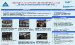
Ankit Mehta 1 Tyson R Finlinson 1 Bradley Ritland 1 Lisa H Lowe 12 Kay L North 12 1 UMKC School of Medicine Kansas City MO 2 Childrens Mercy Hospitals and Clinics Kansas City MO ID: 654931
Download Presentation The PPT/PDF document "NEONATAL SPINAL SONOGRAPHY: CASE BASED R..." is the property of its rightful owner. Permission is granted to download and print the materials on this web site for personal, non-commercial use only, and to display it on your personal computer provided you do not modify the materials and that you retain all copyright notices contained in the materials. By downloading content from our website, you accept the terms of this agreement.
Slide1
NEONATAL SPINAL SONOGRAPHY: CASE BASED REVIEW OF NORMAL VARIANTS
Ankit Mehta
1
, Tyson R
Finlinson1, Bradley Ritland1, Lisa H Lowe1,2, Kay L North1,21UMKC School of Medicine, Kansas City, MO 2Children’s Mercy Hospitals and Clinics, Kansas City, MO
INTRODUCTIONSpinal US is best done < 6 months of age. Recent advances in US have allowed sufficient characterization of nearly all spinal anomalies within the first months of life with a diagnostic value equal to MRI [1].Neonatal spine US is indicated if a skin stigmata is observed, including any dimple above the gluteal crease. Refer to an MRI if draining fluid is observed from any dysraphism [1,6]. There are an array of normal variants to be familiar with in order to prevent unnecessary imaging, referral, or worry.
METHODS
Images were obtained from Children's Mercy Hospital records with consent. They were obtained in the longitudinal and transverse planes using a linear 5–12-MHz transducer.
RESULTS
CONCLUSION
Ultrasound plays a critical role in the
characterization of nearly all spinal anomalies in the 1st month of life. Several normal variants may be confused with pathology on lumbar US including ventriculus terminalis, prominent filum terminale, positional cauda equina pseudomass, pseudosinus tract of coccyx & dysmorphic coccyx. It is important to identify imaging features of normal variants to prevent unnecessary intervention or further evaluation.
REFERENCESDick EA. Spinal ultrasound in infants. Br J Radiol 2002; 75:384–92 Barkovich AJ. Normal development of the neonatal and infant brain, skull, and spine. In: Barkovich AJ. Pediatric neuroimaging, 3rd ed. Philadelphia, PA: Lippincott Williams & Wilkins, 2002:50–4 Gamble HJ. Electron microscope observations upon the conus medullaris and filum terminale of human fetuses. J Anat 1971; 110:173–79 Malas MA. An investigation of the conus medullaris termination level during the period of fetal development to adulthood. Kaibogaku Zasshi 2001; 76:453–59Sadler T. Langman’s medical embryology, 5th ed. Baltimore, MD: Lippincott Williams & Wilkins, 1985:334–37, 343–45 Unsinn KM. US of the spinal cord in newborns: spectrum of normal findings, variants, congenital anomalies, and acquired diseases. RadioGraphics 2000; 20:923–38 Kriss VM. Occult spinal dysraphism in neonates: assessment of high-risk cutaneous stigmata on sonography. AJR 1998; 171:1687–92 Coleman LT. Ventriculus terminalis of the conus medullaris: MR findings in children. Am J Neuroradiol 1995; 16:1421–26
V
entriculus
terminalis – An incomplete fetal regression of the embryonic terminal ventricle in the conus medullaris [6,8].
Prominent filum terminalis
-
It is distinguished as normal by its thickness and typical midline course, <1-2 mm [1].
Filar
Cyst
– CSF between nerve roots of the cauda equine mimicking a cyst; No clinical significance. Unknown origin [4].
Note
: Arrowheads indicate the ventral and dorsal nerve root bundles
Positional Cauda
Equina
Pseudomass
- Found when infant is scanned in decubitus position. Resolves with prone imaging [7].
Pseudosinus
tract of coccyx
– Residual cordlike region composed of fibrous tissue extending from a skin dimple to coccyx [8].
Dysmorphic coccyx
– Tip of the coccyx can vary widely in shape. Can mimic mass on physical exam [6].