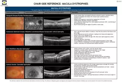
Southern General Hospital Glasgow Need to know Vitreous floatersasteroid hyalosis hge etc Posterior vitreous face tractionseparation epiretinal membrane Lesions in the retina exudates aneurysms ID: 919396
Download Presentation The PPT/PDF document "OCT and the Macula Dr William Wykes" is the property of its rightful owner. Permission is granted to download and print the materials on this web site for personal, non-commercial use only, and to display it on your personal computer provided you do not modify the materials and that you retain all copyright notices contained in the materials. By downloading content from our website, you accept the terms of this agreement.
Slide1
OCT and the Macula
Dr William Wykes
Southern General Hospital
Glasgow
Slide2Slide3Need to know:
Vitreous – floaters/asteroid
hyalosis
,
h’ge
etc
Posterior vitreous face – traction/separation
epiretinal
membrane
Lesions in the retina – exudates, aneurysms,
h’ge
, fluid/oedema
Lesions under the retina – sub-retinal fluid/
h’ge
/debris
Lesions of the RPE –
drusen
, atrophy
Lesions under the RPE – RPED/
h’ge
/scarring/fluid
Slide4Vitreous
Slide5Slide6Macular hole
Slide7Slide8Epiretinal membrane
Slide9Intraretinal
lesions
Slide10Diagnosis of diabetic macular oedema
CSMO
Colour photographs
FFA classification of DMO
Focal
Diffuse
Ischaemic
Mixed
Slide11OCT evaluation of DMO
Quantity of oedema
Location of the oedema
Morphological changes of DMO
Vitreo
-retinal interface changes
Hyper-reflective spots
Photoreceptor integrity
Slide12Location of DMO
OCT based Classification
Centre involving CSMO
(clinically significant macular oedema)
Non-centre involving CSMO
Non CSMO
No MO
Slide13Maculopathy Description
M1 observable
Lesions within a radius of >1 but<2 disc diameters of centre of fovea
Any hard exudates
Slide14Maculopathy Description
M2 Referable
Lesions within a radius of 1 disc diameter of centre of fovea
Any blot
haemorrhages
Any hard exudates
Slide15Slide16Retinal vein occlusion
Slide17Slide18The Retinal Pigment Epithelium
Slide19a) Intact ( Group1)
b) Disrupted (Group 2)
c) Absent(Group3)
OCT Scans showing the three groups
26/10/2014
19
Slide20Slide21Slide22Sub-RPE
Slide23Slide24Evaluation of OCT
Vitreous – normal/ adhesions/ detached
Inner retina- normal/ altered
Foveal
dip: normal/ absent/ altered
ELM: present/absent
IS-OS- present/ disrupted/ absent
RPE –normal/ absent
Fluid- layer
Deposits – present – which layer
24