/
HISTOLOGY OF TONGUE By Dr. Sobia Ibrahim HISTOLOGY OF TONGUE By Dr. Sobia Ibrahim

HISTOLOGY OF TONGUE By Dr. Sobia Ibrahim - PowerPoint Presentation

nicole
nicole . @nicole
Follow
373 views
Uploaded On 2022-06-07

HISTOLOGY OF TONGUE By Dr. Sobia Ibrahim - PPT Presentation

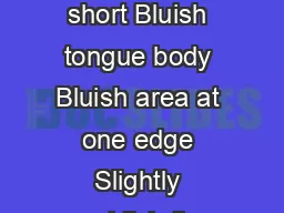
Assistant Professor Anatomy KEMU Tongue is a muscular organ projecting into the oral cavity from its inferior side Organ of speech Organ of taste It helps in swallowing a nd plays a role in digestion ID: 914511

papillae taste buds surface taste papillae surface buds tongue cells anterior dorsal mucous numerous terminalis organ sulcus muscles fibers

Share:

Link:

Embed:

Download Presentation from below link

Download Presentation The PPT/PDF document "HISTOLOGY OF TONGUE By Dr. Sobia Ibrahim" is the property of its rightful owner. Permission is granted to download and print the materials on this web site for personal, non-commercial use only, and to display it on your personal computer provided you do not modify the materials and that you retain all copyright notices contained in the materials. By downloading content from our website, you accept the terms of this agreement.


Presentation Transcript

Slide1

HISTOLOGY OF TONGUE

By Dr. Sobia Ibrahim

Assistant Professor Anatomy,

KEMU

Slide2

Tongue is a muscular organ projecting into the oral cavity from its inferior side

Organ of speech

Organ of tasteIt helps in swallowingand plays a role in digestion

Slide3

Three subdivisions

Root/base

BodyTip

Slide4

Tongue is basically a mass of striated

muscle

covered by mucous membraneLingual muscles are arranged in bundles that run in three

planes; longitudinal, transverse, vertical

Muscles fibers firmly adherent to

mucous membrane.

Between muscle fibers are gland

Mucous- at base

Serous- in body

Mixed- near tip

Slide5

It has two surfaces

Dorsal (superior)

Ventral (inferior)Dorsal is roughened bcz of papillaeVentral is smooth Grossly dorsal surface is divided into anterior 2/3 and posterior 1/3 by sulcus terminalis

Slide6

PAPILLAE

Mucosal irregularities and elevations in different forms and functions

Present anterior to sulcus terminalis on dorsum of tongueThese papillae and associated taste buds constitute the specialized mucosa of oral cavityFilliform Fungiform

Circumvallate

Foliate

Slide7

Papillae of tongue

Slide8

FILLIFORM PAPILLAE

Smallest and most numerous in humans

Conical

Heavily keratinized

Lacks taste buds

Distributed over entire anterior dorsal surface

Provides rough surface

Slide9

FUNGIFORM PAPILLAE

Less numerous

Mushroom shaped

Lightly keratinized

Covering epithelium thin

CT core with blood vessels

Taste buds on upper surface

Rich blood supply give its red color

Slide10

Vallate

(

circumvallate) papillae

Least numerous

Largest in size

Abundance of taste buds

Size = 1-3 mm & number is 8-12

Lie anterior to sulcus terminalis

Surrounded by groove

Ducts of deeply located serous glands (von

Ebner’s

) open into groove

Slide11

FOLIATE PAPILLAE

Poorly developed in humans

Forms furrows on posterolateral aspect of tongue

Contain taste buds

Slide12

Slide13

Slide14

TASTE BUDS

Receptors for taste sensation

Oval, pale staining bodiesExtends through thickness of epith

A small opening on epithelial surface at the apex of taste bud is Taste pore

Sensations

Sweet & salt- tip

Sour- sides

Bitter - back

Slide15

Consists of 50-75

cells

Three principal cellsNeuroepithelial/ Gustatory

cells

having

microvilli

Supportive cells

Stem cells

Rest on basal lamina

Gustatory cells supplied by sensory axon

Slide16

POSTERIOR 1/3

Irregular, nodular surface

Lingual tonsil (

Waldeyer’s

ring)

Crypts

Slide17

Slide18