/
Medical Surgical   Nursing-2/Theory Medical Surgical   Nursing-2/Theory

Medical Surgical Nursing-2/Theory - PowerPoint Presentation

nicole
nicole . @nicole
Follow
64 views
Uploaded On 2024-01-29

Medical Surgical Nursing-2/Theory - PPT Presentation

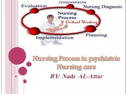
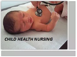
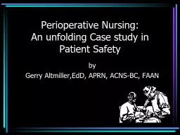
Unit One Nursing Care for Patients with Musculoskeletal Problems 22081439 Dr Nagwa M Ahmed Introduction A musculoskeletal system also known as the  locomotor system is an organ system that gives  human the ability to move using the muscular and skeletal systems ID: 1042624

1439dr nagwa client ahmed nagwa 1439dr ahmed client system joint skeletal musculo nursing patient muscle bone pain bones inflammatory

Share:

Link:

Embed:

Download Presentation from below link

Download Presentation The PPT/PDF document "Medical Surgical Nursing-2/Theory" is the property of its rightful owner. Permission is granted to download and print the materials on this web site for personal, non-commercial use only, and to display it on your personal computer provided you do not modify the materials and that you retain all copyright notices contained in the materials. By downloading content from our website, you accept the terms of this agreement.


Presentation Transcript

1. Medical Surgical Nursing-2/TheoryUnit OneNursing Care for Patients with Musculoskeletal Problems.22/08/1439Dr: Nagwa M., Ahmed

2. Introduction:A musculoskeletal system (also known as the locomotor system) is an organ system that gives  human the ability to move using the muscular and skeletal systems.The musculoskeletal system provides form, support, stability, and movement to the body.It is made up of the:Body's bones (the skeleton). Muscles.Cartilage.Tendons.Ligaments.Joints.Connective tissue, that supports and binds tissues and organs together.22/08/1439Dr: Nagwa M., Ahmed

3. Introduction:The musculoskeletal system's primary functions include supporting the body, allowing motion, and protecting vital organs.The skeletal portion of the system serves as the main storage system for calcium and phosphorus and contains critical components of the hematopoietic system.Bones are connected to other bones and muscle fibers via connective tissue such as tendons and ligaments. 22/08/1439Dr: Nagwa M., Ahmed

4. Introduction:The bones provide the stability to a body in analogy to iron rods in concrete construction. Muscles keep bones in place and also play a role in movement of the bones. To allow motion, different bones are connected by joints. Cartilage prevents the bone ends from rubbing directly on to each other. Muscles contract (bunch up) to move the bone attached at the joint.22/08/1439Dr: Nagwa M., Ahmed

5. Introduction:Human Skeleton:The Skeletal System serves many important functions:It provides the shape and form for the body.Supporting and protecting. (brain is protected by the skull and the lungs are protected by the rib cage)Allowing bodily movement.Producing blood for the body.Storing minerals (as calcium and phosphorus)22/08/1439Dr: Nagwa M., Ahmed

6. Human Skeleton:Introduction:22/08/1439Dr: Nagwa M., Ahmed

7. Introduction:Human Skeleton:Humans are born with about 300 to 350 bones; however, many bones fuse together between birth and maturity. As a result an average adult skeleton consists of 206 bones. There are five general classifications of bones:Long bones.Short bones.Flat bones.Irregular bones.Sesamoid bones. 22/08/1439Dr: Nagwa M., Ahmed

8. Introduction:Bone Marrow:Located in long bonesTwo distinctions of bone marrow (yellow and red).The yellow marrow:Has fatty connective tissue and is found in the marrow cavity.During starvation, the body uses the fat in yellow marrow for energy.The red marrow:Is an important site for blood cell production (all erythrocytes, platelets, and most leukocytes), approximately 2.6 million red blood cells per second are produced in order to replace existing cells that have been destroyed by the liver.22/08/1439Dr: Nagwa M., Ahmed

9. Introduction:Muscle:Types of muscle and their appearance:Cardiac.Skeletal.Smooth. Smooth muscles:Used to control the flow of substances within the lumens of hollow organs.Are not consciously controlled. 22/08/1439Dr: Nagwa M., Ahmed

10. Introduction:Muscle:Skeletal muscles: Have striations that are visible under a microscope due to the components within their cells. Only the skeletal muscles can move the body. Are attached to bones and arranged in opposing groups around joints.Cardiac muscles:Have striations that are visible under a microscope due to the components within their cells. Are found in the heart and are used only to circulate bloodAre not under conscious control.Only skeletal and smooth muscles are part of the musculoskeletal system. 22/08/1439Dr: Nagwa M., Ahmed

11. Introduction:Tendon:A tough, flexible band of fibrous connective tissue that connects muscles to bones.As muscles contract, tendons transmit the forces to the rigid bones, pulling on them and causing movement. Tendons can stretch substantially, allowing them to function as springs during locomotion, thereby saving energy.22/08/1439Dr: Nagwa M., Ahmed

12. Introduction:Joints:Joints are structures that connect individual bones and may allow bones to move against each other to cause movement. Synovial joints:Joints that are not directly joined.Are lubricated by a solution called synovial Fluid that is produced by the synovial membranes. This fluid lowers the friction between the articular surfaces and is kept within an articular capsule, binding the joint with its taut tissue.22/08/1439Dr: Nagwa M., Ahmed

13. Introduction:Synovial joints:Joints that are not directly joined.Are lubricated by a solution called synovial Fluid that is produced by the synovial membranes. This fluid lowers the friction between the articular surfaces and is kept within an articular capsule, binding the joint with its taut tissue22/08/1439Dr: Nagwa M., Ahmed

14. Introduction:Ligaments:A small band of dense, white, fibrous elastic tissue.Ligaments connect the ends of bones together in order to form a joint.Most ligaments limit dislocation, or prevent certain movements that may cause breaks. Since they are only elastic they increasingly lengthen when under pressure. When this occurs the ligament may be susceptible to break resulting in an unstable joint.22/08/1439Dr: Nagwa M., Ahmed

15. Introduction:Bursa:A small fluid-filled sac made of white fibrous tissue and lined with synovial membrane. Bursa may also be formed by a synovial membrane that extends outside of the joint capsule. It provides a cushion between bones and tendons and/or muscles around a joint; bursa are filled with synovial fluid and are found around almost every major joint of the body.22/08/1439Dr: Nagwa M., Ahmed

16. Assessment:Nursing history:Determine if client is involved in competitive sports and physical condition.History for alcohol use, cigarette smoking, diet, calcium intake < 500 mg daily, age 45, and family history of osteoporosis or other diseases.Ask client to describe history of alteration in bone, muscle, or join function.Assess for Hx of pain.Assess normal activity pattern (type of exercise routinely performed).Determine how alteration influences ability to perform activities of daily living (bathing, feeding) and social functions (work, recreation, sexual activities).Assess height loss (first clinical sign of osteoporosis).22/08/1439Dr: Nagwa M., Ahmed

17. Assessment:Nursing Assessment:The assessment focuses on:Determining range of joint motion.Muscle strength and tone.Joint and muscle condition.Notes in assessment:Assess patient when he walks, moves in bed, or do any type of activity. Muscular disorders are manifestations of neurological disease (neurological assessment is performed at the same time).The examination uses inspection and palpation, muscles and joints should be exposed and free to move.Depend on the muscle assessed, the client placed in a sitting, supine, prone, or standing position.22/08/1439Dr: Nagwa M., Ahmed

18. Assessment:Inspection for:Gait (a way of a walking). Posture (anterior, posterior, and lateral):Normal standing posture is an upright stance and parallel alignment of the hips and shoulders.Common postural abnormalities include:Lordosis (increased lumbar curvature).Kyphosis (increased thoracic curvature, older adult).Scoliosis (lateral spinal curvature).22/08/1439Dr: Nagwa M., Ahmed

19. Assessment:Lordosis (increased lumbar curvature).Kyphosis (increased thoracic curvature, older adult).Scoliosis (lateral spinal curvature).22/08/1439Dr: Nagwa M., Ahmed

20. Assessment:Inspection for:Inspect extremities for size, gross deformity, bony enlargement, alignment, and symmetry (should be bilateral symmetry).22/08/1439Dr: Nagwa M., Ahmed

21. Assessment:Palpation:Apply gentle palpation to all bones, joints, and a rounding muscle in a complete examination noting:Heat.Tenderness.Edema.Resistance to pressure.22/08/1439Dr: Nagwa M., Ahmed

22. Assessment:Range of motion (ROM): All ROM must be performed and note any limitation in movement. A goniometer instrument used to measure the degree of motion of the particular joint 22/08/1439Dr: Nagwa M., Ahmed

23. Assessment:Range of motion (ROM): Normal: no discomfort, firm muscles, no hotness or edema. Full range of motion without any discomfort.22/08/1439Dr: Nagwa M., Ahmed

24. Assessment:Muscle tone: Muscle tone: Slight muscular resistance felt by the examiner as relaxed extremity is passively moved through its range of motion. Normal tone is mild.Increase muscle tone (resistance increased) is called hypertonicity.Hypotonicity: a decrease in muscle tone.22/08/1439Dr: Nagwa M., Ahmed

25. Assessment:Muscle Strength:Method of assessment of muscle groups strength:Muscle groupMethod of assessmentNeck(sternnocleidomastoid)Place hand firmly against client's upper jaw. Ask client to turn head laterally against resistance.Shoulder (trapezius)Place hand over midline of client's shoulder, exerting firm pressure.Have client raise shoulders against resistance.22/08/1439Dr: Nagwa M., Ahmed

26. Muscle groupMethod of assessmentElbow :Biceps:triceps:Pull down on forearm as client attempts to flex arm.As client's arm is flexed, apply pressure against forearm. Ask client to straighten arm.Hip:quadriceps:gastrocnemius:When client is sitting, apply downward pressure to the thigh. Ask client to raise leg up from the table.Client sits, holding shin of flexed leg. Ask client to straighten leg against resistanceAssessment:Muscle Strength:Method of assessment of muscle groups strength:22/08/1439Dr: Nagwa M., Ahmed

27. Assessment:Muscle Strength:Muscle strength. Grading scale.Muscle function levelGradeNo evidence of contractility0Slight contractility, no movement1Full range of motion, passive movement2Full range of motion with gravity3Full range of motion against gravity (some resistance)4Full range of motion against gravity (full resistance)522/08/1439Dr: Nagwa M., Ahmed

28. Laboratory Tests:Antinuclear antibodies: Result either positive or negative (normal) and if positive indicate autoimmune disease.C-reactive protein: Protein appears in inflammatory cases, require fasting and note any medication used. Normal Result is negative. 22/08/1439Dr: Nagwa M., Ahmed

29. Laboratory Tests:Erythrocyte sedimentation rate (ESR): Increased in inflammatory, infections, and cancerous conditions.Performed within 3 hours after blood drawing and you should note any medication and pregnancy or menstruation. Normal in male up to 20 mm/h and in female up to 30 mm/h.22/08/1439Dr: Nagwa M., Ahmed

30. Laboratory Tests:Rheumatoid factor: IgM antibody developed against IgG. Normally test is negative.Uric acid: Elevated in gout.Serum: male 2.1-8.5 mg/dl. Female: 2-6.6 mg/dlUrine: 250-750mg/24h.22/08/1439Dr: Nagwa M., Ahmed

31. Radiologic Studies:Computed tomography (CT scan).Magnetic resonance imaging (MRI).X-ray.Arthrography/Arthrogram: Test includes injection of radiopaque dye into the joint then visualization of the joint taken place.22/08/1439Dr: Nagwa M., Ahmed

32. Radiologic Studies:Arthrography/Arthrogram: Nursing responsibility:Gathering the equipment and needed materials (sterile equipments and local anesthesia)Explain the procedure to the client.Obtain consent form.Check the site for edema and increased pain.Administer analgesia.Apply elastic bandage.22/08/1439Dr: Nagwa M., Ahmed

33. Electromyography: Measuring the muscle activity at rest and during voluntary muscle contraction by administering a needle electrode into the muscle. Used to detect primary muscular disorders. Test will take 1 h.Nursing responsibility:Explain the procedure to the client.Obtain consent form.Prohibit smoking and caffeine at least 3 h before test.Observe the site for hematoma and inflammation.22/08/1439Dr: Nagwa M., Ahmed

34. Arthroscopy: Direct visualization of the joint.Done in operation room under local or general anesthesia.Nursing responsibility:After test frequently check the neurovascular of the examined part.Elevate the client leg.Apply compression dressing.Administer analgesia.22/08/1439Dr: Nagwa M., Ahmed

35. Arthrocentesis: Sterile aspiration of the fluid found in the joint after local anesthesia. Used to administer medication and detect infection or inflammation.Nursing responsibility:Explain the procedure to the client.Obtain consent form.Apply pressure and ice.Check the site for edema and increased pain.22/08/1439Dr: Nagwa M., Ahmed

36. Musculoskeletal Changes Associated with Age:The following changes occur with age:Decreased muscle mass and strength.Bone demineralization (more pronounced in women).Shortening of trunk as result of intervertebral space narrowing.Decreased joint mobility and range of joint motion.22/08/1439Dr: Nagwa M., Ahmed

37. Musculoskeletal Changes Associated with Age:Nursing care:Help in self-care procedure and other activity.Prevent fractures by ensuring safe hospitalization, home, and community places.Teach client use of the walking aides.Provide psychological support.Educate client range of motion and other suitable exercises with adequate period of rest.Educate client about the suitable diet.22/08/1439Dr: Nagwa M., Ahmed

38. Surgical Intervention for Problems of Musculo-skeletal System:Joint Replacement(Arthroplasty): Is total or partial replacement of the joint with a synthetic prosthesis. Used mainly in severe chronic arthritis and extensive joint trauma to:Restores mobility and stability. Relieves pain. Increased sense of independence and self-worth.22/08/1439Dr: Nagwa M., Ahmed

39. Surgical Intervention for Problems of Musculo-skeletal System:Joint Replacement(Arthroplasty): Complications:Infection (removed).Hypovolemic shock.Fat embolism.Thromboembolism.Pulmonary embolism (most common cause of death) and pneumonia. Compression of the nerves and vascular necrosis.Dislocation or loosening of the prosthesis.22/08/1439Dr: Nagwa M., Ahmed

40. Surgical Intervention for Problems of Musculo-skeletal System:Joint Replacement(Arthroplasty): Nursing interventions:Before surgery:Preparation which include testing, examination, and teaching (mainly by doctor).Prepare the patient for an extended period of rehabilitation (physical therapist).Inform patient that he experience pain and may worsen for several weeks (use of analgesics).Ensure that the patient or family member has signed a consent form.Ensure that other requirements of operation are ready such as prosthesis and blood if needed.Provide psychological support.22/08/1439Dr: Nagwa M., Ahmed

41. Surgical Intervention for Problems of Musculo-skeletal System:Joint Replacement(Arthroplasty): Nursing interventions:After surgery:Keep client on bed rest for the prescribed period.Maintain the affected joint in proper alignment.Assess the patient's level of pain and provide analgesics as ordered.Monitor for complications of joint replacement (assess vital signs, neurovascular and motor status distal to the site of joint replacement and skin frequently).Watch for signs of a fat embolism (fever, disorientation, dyspnea, tachypnea, sleepiness, agitation, coma, and death may occur).22/08/1439Dr: Nagwa M., Ahmed

42. Surgical Intervention for Problems of Musculo-skeletal System:Joint Replacement(Arthroplasty): Nursing interventions:After surgery:Change position the patient (to enhance comfort and prevent pressure sores).Encourage coughing and deep breathing and adequate fluid intake.Administer prophylactic antibiotics.Patient will be out of bed the 1st or 2nd day after surgery (follow the surgeon instructions).Encourage client to follow the physical therapists instructions.Encourage client to take the prophylactic antibiotic in home.22/08/1439Dr: Nagwa M., Ahmed

43. Surgical Intervention for Problems of Musculo-skeletal System:Amputation:Amputation is the removal of the limb or part from it. Used mainly in gangrene, cancer, trauma, and vascular diseases.Purpose:Preserve function in a remaining part.Prevent death.22/08/1439Dr: Nagwa M., Ahmed

44. Surgical Intervention for Problems of Musculo-skeletal System:Amputation:Complications:Infection.Contractures in the remaining limb part.Skin breakdown.Phantom pain is a sensation of pain (itching, or numbness in the area of amputation) develops after 2 to 3 months.Depression.22/08/1439Dr: Nagwa M., Ahmed

45. Surgical Intervention for Problems of Musculo-skeletal System:Amputation:Nursing interventions:Before surgery:Answer any questions the patient may have.Prepare the patient for surgery and providing care and instruction that will be taken after the amputation.Provide emotional support (loss of a body part and dependence).Arrange for support group (with same problem) meeting.Explain that the patient may "feel" phantom pain.As ordered, administer antibiotics and premeditations.Consent form and other preparations.22/08/1439Dr: Nagwa M., Ahmed

46. Surgical Intervention for Problems of Musculo-skeletal System:Amputation:Nursing interventions:After surgery:Monitor vital signs.Check dressings frequently and change them as ordered. Assess for bleeding (through the dressing).Elevate the limb on a pillow or other support (If ordered).Assess drain patency (working) and note the amount and character of drainage.22/08/1439Dr: Nagwa M., Ahmed

47. Surgical Intervention for Problems of Musculo-skeletal System:Amputation:Nursing interventions:After surgery:Keep drainage bag below the level of the body and empty it after it become filled with fluid and after informing physician. Note amount, color, consistency.Inform physician if drainage system drain large fresh blood and if oozing. Assess for pain and provide analgesics (give analgesics about 30 minutes before scheduled exercises or ambulation).Care for cast (if applied).Maintain proper body alignment and regular physical therapy (passive ROM exercises).22/08/1439Dr: Nagwa M., Ahmed

48. Surgical Intervention for Problems of Musculo-skeletal System:Amputation:Nursing interventions:After surgery:Teach activities to "strengthen" the residual limb.give the patient information about available prosthesesEncourage the patient to adopt a positive outlook (spiritual counselor).Instruct the patient to examine the limb daily for swelling, redness, or excessive drainage, skin changes, and pain.Instruct the patient about good hygiene (wash the limb daily at night with mild soap and water and then to rinse and to gently dry it) and bandage it when dry.22/08/1439Dr: Nagwa M., Ahmed

49. Traumatic Injuries:Contusion (bruise): كدمة Contusion is the most common and is the result of an injury to the soft tissues. The injury causes a rupture of small blood vessels and subsequent bleeding into tissues → ecchymosis (discoloration: black-and-blue) or hematoma.Treatment:Elevation of the affected part.Application of cold immediately and if no bleeding after 24 hours, heat may be applied to relieve muscle soreness.22/08/1439Dr: Nagwa M., Ahmed

50. Traumatic Injuries:Sprains: التواء Sprain involves ligaments, tendons, and muscles and occurs as the result of twisting of a joint beyond its normal range of motion →Ligaments are torn.Tendons may be pulled from the bone.Blood vessels are ruptured, which causes contusions with ecchymosis →Pain related to pressure on nerve endings.Disturbance of circulation and lymph drainage→ muscle spasm & edema.22/08/1439Dr: Nagwa M., Ahmed

51. Traumatic Injuries:Sprains:Treatment:Immediately after the injury:Avoid weight bearing or other pressure on the joint (to avoid further bleeding into soft tissues).The involved extremity should be elevated to promote drainage away from the injury.Apply Ice bags for the first 24 hours to constrict blood vessels and to ensure control of further bleeding.22/08/1439Dr: Nagwa M., Ahmed

52. Traumatic Injuries:Sprains:Treatment:After 24 hours, mild heat may be applied to encourage circulation and healing and to relieve soreness in the area.Immobilize the joint by a splint, bandage or a cast.22/08/1439Dr: Nagwa M., Ahmed

53. Traumatic Injuries:Dislocations:Dislocation is the displacement of a bone from its normal position within a joint. It is accompanied by a stretching and tearing of ligaments and tendons, and fracture may occur.Causes of dislocation:Congenital: Congenitally Dislocated Hip (CDH).Disease process in the joint.Trauma.22/08/1439Dr: Nagwa M., Ahmed

54. Traumatic Injuries:Dislocations:Signs and symptoms:Severe pain.Deformity.Loss of function.Treatment:The displaced parts are manipulated manually into normal position (usually done with the patient under sedation or general anesthesia).Then, the joint is immobilized in splints, bandages, or casts until the injured tissues have healed.A fluoroscope may be used to assist the surgeon.22/08/1439Dr: Nagwa M., Ahmed

55. Traumatic Injuries:Dislocations:Nursing care include:Assistance in the manipulation.Observe for signs of impaired circulation/ such as pain, tingling, numbness, or loss of sensation.Apply cast or other supporting modalities and provide their care.22/08/1439Dr: Nagwa M., Ahmed

56. Traumatic Injuries:Fractures:Fracture possibility increase in:Increase age related to decalcification and osteoporosis.Cancer.Fractures are classified in 3 categories:Fractures described according to the skin injury.Open fractures: Fracture protrudes through a break in the skin. It is more serious and accompanied by soft-tissue damage, require surgical repair.Closed fractures:No skin injury.May be realigned by external manipulation only.22/08/1439Dr: Nagwa M., Ahmed

57. Traumatic Injuries:Fractures:Fractures described according to their appearance:Greenstick: Incomplete fracture.Common in children because their bones are softer and more flexible than adult bones.Bone is broken and bent but still secured on one side.Complete: Bone is completely broken through and severed on all sides.Comminuted: Bone is splintered into three or more fragments at the site of the break.22/08/1439Dr: Nagwa M., Ahmed

58. Traumatic Injuries:Fractures:Fractures described according to their appearance:Transverse fracture: Break runs directly across the bone.Oblique fracture: Break runs along a slant the length of the bone.Spiral fracture: Break coils around the bone.22/08/1439Dr: Nagwa M., Ahmed

59. Traumatic Injuries:Fractures:Fractures are described according to their location on the bone:Proximal.Shaft.Distal.22/08/1439Dr: Nagwa M., Ahmed

60. Traumatic Injuries:Fractures:Signs and symptoms:Pain and Tenderness.Swelling, deformity, and loss of function.Muscle spasm.Unconsciousness and severe shock (in skull and spinal cord fracture).Diagnosis: By x-ray examination.22/08/1439Dr: Nagwa M., Ahmed

61. Traumatic Injuries:Fractures:Complications of fractures:Neurological complications.Circulatory problems.Avascular necrosis: death of bone due to lack of blood supply.Infection.22/08/1439Dr: Nagwa M., Ahmed

62. Traumatic Injuries:Fractures:Treatment:Immobilization of the fracture to prevents further injury.Reduction (Realignment of a), closed or open.Closed Reduction: External manipulation of the bones into position and immobilization with external devices, such as casts, splints, or traction.22/08/1439Dr: Nagwa M., Ahmed

63. Traumatic Injuries:Fractures:Treatment:Open Reuction (internal fixation): The bones are exposed and aligned through a surgical incision and may be fixed in position with wires and plates. 22/08/1439Dr: Nagwa M., Ahmed

64. Traumatic Injuries:Fractures:Treatment:Open Reuction (internal fixation): Drainage (Redivac drainage system) may be applied to drain blood.Postoperative/reduction radiograph to ensure that the normal position is maintained. Radiographs then taken periodically to determine the healing how it is occurring and the bone is remains in the correct alignment. 22/08/1439Dr: Nagwa M., Ahmed

65. Traumatic Injuries:Fractures:In open fracture: the wound should be irrigated to remove gross contamination and dressed with saline and iodine soaks and gives client anti-tetanus as ordered.Fracture healing depend on:Type of fracture.Bone involved.Age.Condition of the patient (infection, illnesses, poor circulation, and nutrition).22/08/1439Dr: Nagwa M., Ahmed

66. Traumatic Injuries:Fractures:Nursing Intervention:Provide skin care.Turn patient and encourage him to cough and deep breathe every 2 hours.Maintain affected extremity in alignment.Mobilize patient and weight bearing as physician's order and patient tolerance.Request occupational therapy consultation for help with activities of daily living and physical therapy consultation for transfer techniques and use of mobility aids.22/08/1439Dr: Nagwa M., Ahmed

67. Traumatic Injuries:Fractures:Nursing Intervention:Perform range of motion exercises on all joints except affected leg.Administer intravenous and antibiotic fluids as ordered.Administer prophylactic anticoagulants as ordered.Apply antiembolism stockings as ordered.Encourage dorsiflexion and plantar flexion exercises.Provide high-protein, high-roughage diet.Administer analgesics as prescribed.Provide comfort measures (massage and relaxation).Maintain aseptic technique with dressing changes.Encourage participation in self-care activities.22/08/1439Dr: Nagwa M., Ahmed

68. Traumatic Injuries:Fractures:Nursing Intervention:Encourage balance between rest and activities.Provide cast or other devices care.Teach the patient and family the signs and symptoms of infection, cast care, pin care, and/or wound care.Teach the patient and family the nutritional needs of bone healing and de-creased mobility.Teach the patient and family the isometric exercises and the activity level of the patient.Provide care for drainage if applied (Drain output).22/08/1439Dr: Nagwa M., Ahmed

69. Traumatic Injuries:Fractures:Nursing Intervention:Assess vital signs, dressing for evidence of bleeding, wound for healing and evidence of infection, tissue perfusion to affected extremity, and respiratory status.Assess level of comfort, elimination patterns and bowel sounds, level of consciousness, orientation, and signs of dislocation.Assess peripheral pulses, circulation, and nerves.Help in providing treatment as needed.22/08/1439Dr: Nagwa M., Ahmed

70. Traumatic Injuries:Hip Fracture:Clinical manifestations:External rotation.Muscle spasms.Shortening of the affected ext.Severe pain.Tenderness to the region of the fracture site.Displaced femoral neck fractures cause serious disruption of the blood supply to the femoral head, which can result in avascular necrosis.22/08/1439Dr: Nagwa M., Ahmed

71. Traumatic Injuries:Hip Fracture:Treatment:Surgical repair:Is the preferred method. (Surgery may be delayed if the patient has DM, cardiovascular issues untill the patient is stabilized)Traction:Treatment with traction requires 12-16 weeks of immobilization for healing to occur, even if the blood supply to the region is intact.Traction can relieve muscle spasms.22/08/1439Dr: Nagwa M., Ahmed

72. Traumatic Injuries:Hip Fracture:Nursing management:Analgesics or muscle relaxants (control muscle spasms)Teach patient to use trapeze and show them the exercise prior to surgery.Practice getting in and out of bed.Post surgery nursing care:Vitals.I &O.Analgesics.Check dressing for signs of bleeding and infection.22/08/1439Dr: Nagwa M., Ahmed

73. Traumatic Injuries:Hip Fracture:Nursing management:Post surgery nursing care:Access the toes for neuro-vascualr assessment.Ambulation usually begins on the first day by P/T. Either a walker or crutches are used.Do not forces hip into greater than 90 degrees of flexion.Do not force into adduction.Do not force hop into internal rotation.Do not cross legs.Do not put on shoes or stockings until 8 weeks after surgery without adaptive device.22/08/1439Dr: Nagwa M., Ahmed

74. Traumatic Injuries:Hip Fracture:Nursing management:Post surgery nursing care: Do not sit on chairs without arms to aid rising to a standing position. Use toilet elevator on toilet seats.Place chair inside shower or tub and remain seated while you wash.Use pillow between legs for 8 weeks when lying on the good side or when supine.Keep hip in neutral , straight position when sitting, walking or lying.Notify surgeon of severe pain, deformity or loss of function.22/08/1439Dr: Nagwa M., Ahmed

75. Inflammatory Conditions of the Musculo-Skeletal System:Gout: Gout is a metabolic disease that's marked by uric acid deposits that cause swollen and acutely painful joints. It may affect any joint but mostly affects those in the feet, especially the big toe and ankle.Types of gout:Primary gout: inherited.Secondary: related to other disease and use of drugs.Idiopathic: unknown cause.22/08/1439Dr: Nagwa M., Ahmed

76. Inflammatory Conditions of the Musculo-Skeletal System:Gout: Causes:Diseases:Decreased renal excretion of uric acid.Obesity.Diabetes mellitus.Hypertension.Leukemia.Sickle cell anemia.Drugs such as hydrochlorothiazide.Genetic defect.22/08/1439Dr: Nagwa M., Ahmed

77. Inflammatory Conditions of the Musculo-Skeletal System:Gout: Signs and symptoms: May be asymptomatic.Pain in the joints.Chills and mild fever.Swollen, tender, and red joint with limited movement.Tophi (A Tophus (from the Latin for stone) is a deposit of crystallized uric acid normally found just under the skin. Commonly, they occur on the ear, fingers and toes and around the ankle and elbow). The skin over the tophi may ulcerate and release a white exudates or pus.22/08/1439Dr: Nagwa M., Ahmed

78. Inflammatory Conditions of the Musculo-Skeletal System:Gout: Signs and symptoms: Tophi (A Tophus (from the Latin for stone) is a deposit of crystallized uric acid normally found just under the skin22/08/1439Dr: Nagwa M., Ahmed

79. Inflammatory Conditions of the Musculo-Skeletal System:Gout: Diagnostic tests:Needle aspiration of synovial fluid (arthrocentesis) or of tophaceous material.Serum uric acid levels may be normal or more than 10 mg/dl. Normal is 2.5-8 mg/dl. Urine uric acid levels are high in about20% of gout patients.X-ray studies initially produce normal results. But in chronic gout it may show damage to the articularcartilage and subchondral bone.22/08/1439Dr: Nagwa M., Ahmed

80. Inflammatory Conditions of the Musculo-Skeletal System:Gout: Complications:Renal disorders: renal calculi.Circulatory problems, such as atherosclerotic disease, cardiovascular lesions, cerebrovascular accident, coronary thrombosis, and hypertension.Infection.22/08/1439Dr: Nagwa M., Ahmed

81. Inflammatory Conditions of the Musculo-Skeletal System:Gout: Goals of Treatment:Terminate the acute attack.Reduce urine uric acid levels to less than 6.5 mg/d.Prevent recurrent gout.Treatment:Bed rest, immobilization and protection of the inflamed and painful joints.Local application of cold.Analgesics and NSAID.Intra-articular corticosteroids.22/08/1439Dr: Nagwa M., Ahmed

82. Inflammatory Conditions of the Musculo-Skeletal System:Gout: Treatment:Medication:Allopurinol.Probenecid or sulfinpyrazoneColchicine: prevents acute gout attacks (doesn't affect uric acid levels).Adjunctive therapy:Avoiding alcohol (especially beer and wine).Careful use of urate-rich foods, such as fish, liver, and lentils.Weight-loss.22/08/1439Dr: Nagwa M., Ahmed

83. Inflammatory Conditions of the Musculo-Skeletal System:Gout: Nursing interventions:Give pain medication as needed.Apply cold compresses.Administer medication as ordered.Encourage client to use techniques that promote relaxation.Educate client about the prescribed food.Provide emotional support.Allow client to express his feelings.Encourage minimal of self-care if allowed with period of rest.22/08/1439Dr: Nagwa M., Ahmed

84. Inflammatory Conditions of the Musculo-Skeletal System:Gout: Nursing interventions:Educate and support client during diagnostic and treatment procures.Monitor client for condition and for complication (infection) after joint aspiration.Monitor client for side effect of medication especially anti-inflammatory medication.Monitor intake and output and serum uric acid level.22/08/1439Dr: Nagwa M., Ahmed

85. Inflammatory Conditions of the Musculo-Skeletal System:Gout: Nursing interventions:Encourage client to drink plenty of water (prevent renal calculi).Inform client not to take probenecid or sulfinpyrazone with aspirin or salicylic acid (cause uric acid retention).Inform client about side effect of medication: nausea, vomiting, dizziness, urinary frequency, and dermatitis.Inform client to inform physician about other treatment especially HTN medication.22/08/1439Dr: Nagwa M., Ahmed

86. Inflammatory Conditions of the Musculo-Skeletal System:Rheumatoid Arthritis (RA):Is a chronic, systemic, recurrent, symmetrical inflammation that attacks peripheral joints, surrounding muscles, tendons, ligaments, and blood vessels. Rheumatoid arthritis requires lifelong treatment.22/08/1439Dr: Nagwa M., Ahmed

87. Inflammatory Conditions of the Musculo-Skeletal System:Rheumatoid Arthritis (RA):Causes: Not known but there are factors that influence the occurrence of disease such as:Viral and bacterial infections.Hormonal factors.IgM antibody developed against IgG (rheumatoid factor).22/08/1439Dr: Nagwa M., Ahmed

88. Inflammatory Conditions of the Musculo-Skeletal System:Rheumatoid Arthritis (RA):Complication:Bone ankylosis (a stiffness of a joint due to abnormal adhesion and rigidity of the bones of the joint), soft tissue contractures, and joint deformities.Vacuities (a heterogeneous group of disorders that are characterized by inflammatory destruction of blood vessels) → ulcers and multisystem complications.May damage the vertebra.Carpal tunnel syndrome and osteoporosis.Necrosis of the hip, cardiac, pulmonary, and renal problems.22/08/1439Dr: Nagwa M., Ahmed

89. Inflammatory Conditions of the Musculo-Skeletal System:Rheumatoid Arthritis (RA):Signs and symptoms:Morning stiffness.Fatigue.Vague articualr symptoms (pain, paresthesia - sensation of tingling, burning, pricking, or numbness of a person's skin, and tenderness) at the beginning and then in specific place (bilateral) at rest and activity.Wight loss.Low grade fever.Weak and stiff muscles.Joint swelling, hotness, and deformity.Elbow nodules.22/08/1439Dr: Nagwa M., Ahmed

90. Inflammatory Conditions of the Musculo-Skeletal System:Rheumatoid Arthritis (RA):Diagnostic tests: No definite diagnostic tests.X-ray: bone demineralization, deformities, and erosions.Rheumatoid factors: positive.Synovial fluid analysis: increase volume, turbid, and increase WBC.Erythrocytes sedimentation rate: increase.MRI and CT scan: indicate extent of the damaged.22/08/1439Dr: Nagwa M., Ahmed

91. Inflammatory Conditions of the Musculo-Skeletal System:Rheumatoid Arthritis (RA):Treatment:Salicylates such as aspirin: decrease inflammation and reduce pain.Other NSAIDs such as indomethacin and ibuprofen.Immunosuppressant: methotrexate.Penicillin.Bed rest.Ice pack during acute episodes and others apply heat.Physiotherapy and joint protection devices.Arthroplasty.22/08/1439Dr: Nagwa M., Ahmed

92. Inflammatory Conditions of the Musculo-Skeletal System:Rheumatoid Arthritis (RA):Nursing intervention:Administer medication as prescribed assess for their effectiveness and side effect.Give skin care.Use low instrument of daily living activity.Coordinate with physiotherapy department as ordered.Assess for signs and symptoms of disease and complication and intervene to decrease them.22/08/1439Dr: Nagwa M., Ahmed

93. Inflammatory Conditions of the Musculo-Skeletal System:Rheumatoid Arthritis (RA):Nursing intervention:Prepare patient for diagnostic and treatments procedures.Observe the duration of morning stiffness.Educate client about the disease, diagnostic test, treatment, and encourage him to comply with prescribed treatment.Encourage client to take hot showers at bed time or in the morning.22/08/1439Dr: Nagwa M., Ahmed

94. Inflammatory Conditions of the Musculo-Skeletal System:Osteomyelitis:Osteomyelitis is a sever bone infection.Etiology: Trauma.Secondary infection.Clinical Manifestations:Localized bone pain.Tenderness, heat, and oedema in the affected area.Restricted movement in affected area.Fever, and generalized weakness22/08/1439Dr: Nagwa M., Ahmed

95. Inflammatory Conditions of the Musculo-Skeletal System:Osteomyelitis:Diagnostic Evaluation:Increase in WBCs.Increase in ESR.Positive blood culture.Radiograph and bone scan.Nursing Intervention:Administer antibiotics and analgesics.Provide ROM.No weight bearing on affected limb.Provide sterile dressing.22/08/1439Dr: Nagwa M., Ahmed

96. Connective Tissue Disorder: Lupus Erythematosus: الحمى الذؤابية Lupus erythematosus is a recurrent inflammatory autoimmune disorder affect the connective tissue.Affects females 8-15 times more than males.Forms of Lupus erythematosus:Discoid Lupus erythematosus (DLE):Affect only the skin Cause skin eruptions and may developed to Systemic Lupus erythematosus.Systemic Lupus Erythematosus (SLE). Affect multiple organs including skin (fatal).22/08/1439Dr: Nagwa M., Ahmed

97. Connective Tissue Disorder: Lupus Erythematosus: Causes: Causes are unknown but may be related to:Autoimmune.Genetic.Environmental and hormonal factors.Risk factors:Stress.Streptococcal or viral infections.Exposure to sunlight.Abnormal estrogen metabolism.22/08/1439Dr: Nagwa M., Ahmed

98. Connective Tissue Disorder: Lupus Erythematosus: Signs and symptoms:Fever, anorexia, weight loss, headache, and malaise.Abdominal pain, diarrhea, and constipation.Skin rashes mainly in the face (called butterfly rash).Affect ovary: amenorrhea or irregular menstruation. Joint involvement as in RA.Skin ulcers, gangrene, and rashes.Mucous membrane for ulcers.Lymph node enlargement.22/08/1439Dr: Nagwa M., Ahmed

99. Connective Tissue Disorder: Lupus Erythematosus: Complications:Pulmonary abnormalities: infections, plural effusion, and pulmonary hypertension.Cardiac: pericarditis, myocarditis, and coronary atherosclerosis.Renal: renal failure.Seizure and mental disability (neurologic damage).22/08/1439Dr: Nagwa M., Ahmed

100. Connective Tissue Disorder: Lupus Erythematosus: Diagnostic tests:Complete blood count: decreased Hb, WBC, and Platelets.Anti-nuclear antibody (ANA), anti-deoxyribonucleic acid (ADA), and Lupus erythematosus cell test: positive (all test are not specific).Urine analysis: protein in urine, RBC and WBC.22/08/1439Dr: Nagwa M., Ahmed

101. Connective Tissue Disorder: Lupus Erythematosus: Treatment:NSAIDs: control joint pain.Protection from exposure to sun.Topical medications including corticosteroids.Anti-malarial drugs (chloroquine): control skin eruptions.Corticosteroids.Provide high protein, vitamins, and iron diet.22/08/1439Dr: Nagwa M., Ahmed

102. Connective Tissue Disorder: Lupus Erythematosus: Nursing intervention:Check vital signs.Monitor intake and output.Weight client daily.Provide emotional support for the changes that occurs related to body image (skin changes).Provide the prescribed diet.Encourage bed rest.22/08/1439Dr: Nagwa M., Ahmed

103. Connective Tissue Disorder: Lupus Erythematosus: Nursing intervention:Educate client about his disease, diagnostic tests required, and encourage him to comply with treatment.Apply heat packs to relief joint pain and swelling.Encourage regular exercise especially range of motion.Continuous assess for complication.Educate client to avoid any infections.Advise client to notify physician if fever, cough, rashes occurs or if chest, abdominal, and joint pain increases markedly.22/08/1439Dr: Nagwa M., Ahmed

104. Connective Tissue Disorder: Lymes Disease: Lyme disease affects multiple body systems. It is typically begin with the classic skin lesion (erythema), weeks or months later, cardiac, neurologic, and joint abnormalities develop.Causes:Lyme disease is caused by the spirochete Borrelia burgdorferi carried by the small tick from the Ixodidae family. After incubating for 3 to 32 days, the spirochetes migrate outward on the skin, causing a rash and distribute to other skin sites or organs by the bloodstream or lymph system. 22/08/1439Dr: Nagwa M., Ahmed

105. Connective Tissue Disorder: Lymes Disease: Complications:Myocarditis, pericarditis, arrhythmias, and heart block. meningitis, encephalitis, cranial or peripheral neuropathies, and arthritis.S&S:Fatigue, malaise, and migratory myalgias and arthralgias. Cardiac symptoms (palpitations and mild dyspnea).Severe headache and neck stiffness (meningeal irritation).Neurologic symptoms (memory loss).Rash. 22/08/1439Dr: Nagwa M., Ahmed

106. Connective Tissue Disorder: Lymes Disease: S&S:Lesions (not seen in all patients) are: Bright red outer rims and white centers. Usually appear on the axillae, thighs, and groin. Large as 5 cm in diameter. The patient may describe the lesion as hot and pruritic.Arthritis: joint swelling, redness, and limited movement (large joints).Pulse: may be tachycardia or irregular heartbeat. Lymphoadenopathy.If the patient has neurologic involvement, Kernig's and Brudzinski's signs usually aren't positive.22/08/1439Dr: Nagwa M., Ahmed

107. Connective Tissue Disorder: Lymes Disease: Diagnostic tests:Blood tests:Antibody titers. Enzyme-linked immunosorbent assay (ELISA).Mild anemia and increase ESR and WBC.Lumbar puncture.Treatment: A 10- to 20-day course of antibiotics is the treatment of choice (tetracycline or doxycycline; penicillin and erythromycin are alternatives).22/08/1439Dr: Nagwa M., Ahmed

108. Connective Tissue Disorder: Lymes Disease: Nursing interventions:Plan care to provide adequate rest.Administer analgesics, antibiotics (ask for allergy), and antipyretics as ordered.Help patient with range-of-motion.Protect the patient from sensory overload and reorient him if needed.Encourage patient to express his feeling and concerns about disease and complication. 22/08/1439Dr: Nagwa M., Ahmed

109. Connective Tissue Disorder: Lymes Disease: Nursing interventions:Monitor the patient's vital signs (temperature).Watch for signs and symptoms of complications.Monitor the effectiveness of administered medication.Inform the patient, his family, and other caregivers about ways to prevent Lyme disease22/08/1439Dr: Nagwa M., Ahmed

110. Degenerative Disorder: Osteoarthritis:Osteoarthritis (common form of arthritis) is a slowly progressive, degenerative joint disease characterized by variable changes in weight- bearing joints due to deterioration of the joint cartilage and formation of new bone at the margins. It may cause disability that range from minor limitation to immobility. Affects both sexes, with onset usually after age 4o.22/08/1439Dr: Nagwa M., Ahmed

111. Degenerative Disorder: Osteoarthritis:Predisposing Factors: Obesity.Aging.Trauma.Congenital abnormalities.Genetics. Complication:Nerve root compression.Flexion contractures, subluxation, and deformity.Gross bony overgrowth.Ankylosis.Central cord syndrome (affecting cervical spine).22/08/1439Dr: Nagwa M., Ahmed

112. Degenerative Disorder: Osteoarthritis:Signs and symptoms: (gradually developed)Deep joint pain that relived by rest especially during weather changes.Pain and muscle spasms (after exercises, at night, in the early morning)Joint swelling and deformity.Muscle atrophy.Hard nodes in joints (painless then become red, swollen, and tender).Warmth of the joint.Limited motion in affected joints.22/08/1439Dr: Nagwa M., Ahmed

113. Degenerative Disorder: Osteoarthritis:Diagnostic tests:X-rays: in the late stages.Synovial fluid analysis: indicate inflammation.Arthroscopy: soft tissue swelling.CT scan: development of disease.22/08/1439Dr: Nagwa M., Ahmed

114. Degenerative Disorder: Osteoarthritis:Treatment:Medication:Aspirin.NSAIDs: indomethacin.Corticosteroid injection in the joint (very 4-6 months): delay development of disease.Rest: adequate rest with balanced activity.Physical therapy:Massage.Moist heat.Exercise.22/08/1439Dr: Nagwa M., Ahmed

115. Degenerative Disorder: Osteoarthritis:Treatment:Assistive mobility devices.Weight reduction.Surgery:Arthroplasty: replacement of the affected joint (completely or partially).Arthrodesis: fusion of bones.Osteoplasty: scarping and lavage of deteriorated bones form the joint.Osteotomy: cutting of the edges of the deteriorated bone.22/08/1439Dr: Nagwa M., Ahmed

116. Degenerative Disorder: Osteoarthritis:Nursing intervention:Administer medication as prescribed and observe their effectiveness and side effects.Provide emotional support.Encourage pt. to perform self-care activity as he can with periods of rests.Use methods of pain management.Provide comfort devices.Teach client about the range of motion exercises.Teach client how to use assistive devices.Collaborate with physical therapy for treatment.Educate client about diagnostic test and treatment modalities.22/08/1439Dr: Nagwa M., Ahmed

117. Degenerative Disorder: Osteoporosis:Osteoporosis is a disorder of bone metabolism in which there is a reduction of total bone mass, making bones abnormally prone to fracture.It affects one fourth of all older adults, and the greatest incidence occurs among white females between the ages of 50 and 70 years.8 times more common in women than men.Predisposing Factors:Postmenopausal status.Long-term corticosteroid use.Prolonged immobilization.Nutritional deficiency.22/08/1439Dr: Nagwa M., Ahmed

118. Degenerative Disorder: Osteoporosis:Clinical Manifestations:Fractures.Pain.Visible deformity (e.g., kyphosis)Loss of height.Constipation.Diagnostic Evaluation:Radiographic and bone density studies. (Cannot be detected by x-ray until more than 25%-40% of calcium in the bone is lost)Serum calcium, phosphorus, and alkaline phosphates (levels are within normal ranges). 22/08/1439Dr: Nagwa M., Ahmed

119. Degenerative Disorder: Osteoporosis:Nursing Interventions:Administer analgesics as prescribed to relieve pain.Assist patient with putting on back brace.Encourage walking daily for strong bone remodelling.Encourage young women at risk to maximize bone mass through good nutrition (contain calcium) and exercises.Suggest that perimenopausal women confer with the physician concerning need for calcium supplements and oestrogen therapy. Instruct patient to avoid ant trauma or falls.22/08/1439Dr: Nagwa M., Ahmed

120. Rehabilitation of client with Musculo-skeletal system:Drug therapeutics: NonSteroidal Anti-inflammatory Drugs (NSAIDs): Ibuprofen and diclofenac sodium.Side effect:GI tract irritation.Dizziness.Headache.Liver and kidney toxicity.Aggravate bronchial asthma.CNS and teratogenic side effect.22/08/1439Dr: Nagwa M., Ahmed

121. Rehabilitation of client with Musculo-skeletal system:Drug therapeutics: NonSteroidal Anti-inflammatory Drugs (NSAIDs): Uses and action:Decrease inflammation: decrease edema, decrease fever, and other inflammatory response.Immunosuppressant in organ transplantation and rheumatoid arthritis.NSAID used as analgesia.Contraindication (CI):Hypertension, HF.Depressive psychosis.Osteoporosis.DM.22/08/1439Dr: Nagwa M., Ahmed

122. Rehabilitation of client with Musculo-skeletal system:Drug therapeutics: NonSteroidal Anti-inflammatory Drugs (NSAIDs): Used in caution for patients with: Peptic ulcer.Thrombophlebitis.Adrenal suppression.Mood swing.Infection cases (may be contraindicated in serious infections because it masks infections).22/08/1439Dr: Nagwa M., Ahmed

123. Rehabilitation of client with Musculo-skeletal system:Casting: Cast: is used to fix the fracture.Types:A plaster cast: become rigid in 24 - 48 hours. Synthetic cast: become rigid in 30 minutes, and are much lighter and stronger.Nursing Care:Before application of cast: The skin is cleansed and examined for any contusions or abrasions.Over padding is used over bony prominences.Check that any jewellery and ring has been removed.22/08/1439Dr: Nagwa M., Ahmed

124. Rehabilitation of client with Musculo-skeletal system:Traction :Traction is applying of a force along the long axis of the bone distal to the fracture.Traction is applied by the use of a weight and pulley system. Traction used to:Correct deformity and fractures.Relieve pressure on a spinal nerve.Prevent contractures.22/08/1439Dr: Nagwa M., Ahmed

125. Rehabilitation of client with Musculo-skeletal system:Traction :Types of tractions:Skin traction: Application of adhesive or non-adhesive tapes to the medial and lateral surfaces of the limb. These are secured by a firm encircling crepe or elasticated bandage. Skin traction applied in patient room.Skin traction may cause skin or vascular complications (must not be applied tightly around the limb).22/08/1439Dr: Nagwa M., Ahmed

126. Rehabilitation of client with Musculo-skeletal system:Traction :Types of tractions:Skeletal traction:Is a metal wire or pin inserted through the bone distal to the fracture.22/08/1439Dr: Nagwa M., Ahmed

127. Rehabilitation of client with Musculo-skeletal system:Traction :Potential problems for the patient in traction or cast especially for limbs.Infection at the pin site.Muscle wasting due to immobility.Skin breakdown due to traction and immobility.Constipation and urinary retention due to immobility and embarrassment.Depression due to enforced immobility.22/08/1439Dr: Nagwa M., Ahmed

128. Rehabilitation of client with Musculo-skeletal system:Assistive devices:Cane.Crutches.Walkers.Trapeze devices: found in the superior part of the orthopedic bed and help client in his movement in the bed.22/08/1439Dr: Nagwa M., Ahmed

129. Rehabilitation of client with Musculo-skeletal system:Physical Therapy:Is the function of the physiotherapists but nurse must have a knowledge and skill for physical therapy.Purpose of physical therapy is to prevent complication of immobility, enhance healing, and educate client how to walk and use of assistive devices.Physical therapy include:Passive exercise.Active exercise.Resistance exercise.Walking after trauma and treatment.22/08/1439Dr: Nagwa M., Ahmed