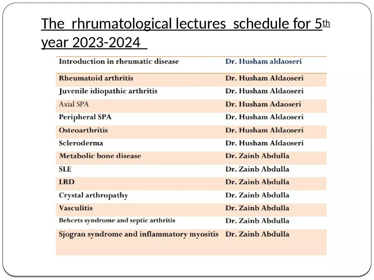
th year 20232024 INTRODUCTION TO RHEUMATOLOGY What is Rheumatology A medical science devoted to the study of rheumatic diseases and musculoskeletal disorders Approximately 30 of the US population has arthritis andor back pain ID: 1039537
Download Presentation The PPT/PDF document "The rhrumatological lectures schedul..." is the property of its rightful owner. Permission is granted to download and print the materials on this web site for personal, non-commercial use only, and to display it on your personal computer provided you do not modify the materials and that you retain all copyright notices contained in the materials. By downloading content from our website, you accept the terms of this agreement.
1. The rhrumatological lectures schedule for 5th year 2023-2024
2. INTRODUCTION TO RHEUMATOLOGY
3. What is Rheumatology A medical science devoted to the study of rheumatic diseases and musculoskeletal disorders.
4. Approximately 30% of the U.S. population has arthritis and/or back pain.One out of every five office visits to a primary care provider and 10% of all surgeries are for a musculoskeletal problem.Arthritis/back pain is the second leading cause of acute disability, the number one cause of chronic disability. Most common reason for social security disability payments.
5. Sutures B. SyndesmosisPri. Cart. joints (Synchondrosis) B. Sec.cart. Joints (Symphysis)1. Plane2. Hinge3. Pivot 4. Bicondylar5. Saddle 6. Ball and socketSynovialFreely movable(Diarthrosis)CartilaginousSlightly movable(Amphiarthrosis)FibrousFixed(Synarthrosis)Classification of Joints
6. SYNDESMOSISFibrous connection between bones
7. CARTILAGINOUS JOINT 1. Primary Cartilaginous JointAlso called as synchondrosis
8. CARTILAGINOUS JOINT 2. Secondary Cartilaginous JointsAlso called as symphysis
9. SYNOVIAL JOINTMost evolved joint.Freely movable joint.Possess a joint cavity that consists of synovial fluid.
10. Approach to the Patient with Joint SymptomsClinical Presentation A careful history and detailed physical examination should be directed at answering the following five key diagnostic questions: Is the process truly articular? Is the process inflammatory or mechanical? What is the pattern of joint involvement? Are there extra-articular manifestations? Who is the host?
11. Features that suggest an articular process Symptoms (pain) localized to the joint(s) Physical findings (swelling, erythema, heat, or tenderness) Joint range of motion is painful Joint range of motion is restricted
12. Features that suggest an inflammatory processMorning stiffness longer than 60 minutes (versus worsening in the evening in mechanical processes) Gel phenomenon (stiffness after prolonged inactivity) Symptoms improve with use (versus worsening with use in mechanical processes)Joint swelling, erythema, heat, or tenderness Active constitutional manifestations (e.g., fever, malaise, anorexia or weight loss)
13. Relevant patterns of joint involvement Acute onset (e.g., microcrystalline) versus insidious onset (e.g., osteoarthritis ) Episodic (e.g., microcrystalline) versus migratory (e.g., disseminated gonococcal infection and acute rheumatic fever) versus additive (e.g., rheumatoid arthritis ) Monoarticular (e.g., microcrystalline and septic)versus oligoarticular (e.g.,Peripheral SPA) versus polyarticular (e.g., RA) Symmetrical (e.g., RA) versus asymmetrical e.g., Peripheral SPA
14. Relevant extra-articular features Constitutional symptoms and signs (i.e., presence of a fever) Mucocutaneous (e.g., photosensitive and other cutaneous eruptions, alopecia, mucosal aphthous ulcers, and Raynaud’s phenomenon) Ocular (e.g., conjunctivitis, episcleritis, scleritis, and uveitis) Renal (e.g., glomerulonephritis, renal tubular acidosis, and nephrolithiasis)
15. Neurologic (e.g., focal central and peripheral nervedisease, seizures, and cognitive and psychiatric disorders) Pulmonary (e.g., nodules, infiltrates, interstitial fibrosis,pulmonary embolism and hypertension, alveolarhemorrhage, bronchiolitis, and pleuritis) Gastrointestinal (e.g., inflammatory bowel disease andautoimmune liver disease) Hematologic Anemia (e.g., anemia of “chronic disease” and hemolytic anemia) Leukopenia (e.g., neutropenia in Felty syndrome and lymphopenia in systemic lupus erythematosus (SLE) Thrombocytopenia (e.g., SLE with antiphospholipid antibodies)
16. Relevant host factors Age Gender Race Comorbidities Occupational exposures Family history
17. Diagnosis and Evaluation
18.
19. Investigation of Musculoskeletal disease Obtain relevant laboratory studies as indicated Kidney and liver function Complete blood count with leukocyte differential Urinalysis Erythrocyte sedimentation rate and C-reactive protein Thyroid function Autoantibodies
20. Obtain and evaluate synovial fluid as indicated Check synovial fluid leukocyte cell count, Gram stain and culture, and polarized microscopy for crystals White blood cell (WBC) count less than 200/mm3 (normal), greater than 2000/mm3 (inflammatory), and greater than 100,000/mm3 (presumptively septic)
21.
22.
23. Obtain relevant imaging studies Conventional radiography is the starting point in imaging studies due to widespread availability, low cost, and high resolution, but may reveal only nonspecific soft tissue swelling in early inflammatory disease Computed tomography (CT) is widely available and a good choice for assessment of the spine and pulmonary parenchyma Magnetic resonance imaging (MRI) has replaced CT in many situations (e.g., disc herniation, sacroiliac joints, osteonecrosis, and synovitis)
24. Arthrography (e.g., evaluation for rotator cuff tears) Angiography (e.g., evaluation for vasculitis) Both mechanical and inflammatory joint disease cause increased tracer uptake so that nuclear medicine scans have a limited role in the evaluation of arthritis Ultrasound is useful in differentiating thrombophlebitis from pseudothrombophlebitis (ruptured Baker cyst), with an expanding role in the assessment of synovitis and erosive joint disease
25. Dual energy X-ray absorptiometry (DXA)Bone mineral density (BMD) measurements play a key role in the diagnosis and management of osteoporosis.
26.