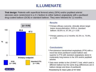
/
Balloon Angioplasty & Catheterization Balloon Angioplasty & Catheterization

Balloon Angioplasty & Catheterization - PowerPoint Presentation

norah
norah . @norah
Follow
0 views
Uploaded On 2024-03-13

Balloon Angioplasty & Catheterization - PPT Presentation

الدكتور مهند احمد صاحب الأستاذ مرتضى عبد الامير محمد Al Mustaqbal University Special radiological procedure 2 Lecture 10 ID: 1048133

guide balloon vessel wire balloon guide wire vessel procedure artery blood coronary diameter arteries angioplasty wires stenosis heart catheter

Share:

Link:

Embed:

Download Presentation from below link

Download Presentation The PPT/PDF document "Balloon Angioplasty & Catheterizatio..." is the property of its rightful owner. Permission is granted to download and print the materials on this web site for personal, non-commercial use only, and to display it on your personal computer provided you do not modify the materials and that you retain all copyright notices contained in the materials. By downloading content from our website, you accept the terms of this agreement.


Presentation Transcript

1. Balloon Angioplasty & Catheterization الدكتور/ مهند احمد صاحب الأستاذ / مرتضى عبد الامير محمد Al-Mustaqbal UniversitySpecial radiological procedure /2 Lecture 10اعداد

2. Catheterization (Seldinger technique)is a procedure in which a thin, flexible tube (catheter) is guided through a blood vessel to the heart to diagnose or treat certain heart conditions, such as clogged arteries or irregular heartbeatsThe Swedish Radiologist

3. AngioplastyAngioplasty is a procedure to improve blood flow in coronary arteries that have become narrow or blocked. Your coronary arteries supply oxygen-rich blood to the heart. If you have coronary artery disease, a sticky material called plaque builds up in the coronary arteries. Plaque is made of cholesterol, calcium, and other substances in your blood. Over time, it can narrow your arteries or fully block them. When this happens, some parts of your heart don't get enough blood.Angioplasty widens the blocked part of the artery so more blood can get through. It is also called balloon angioplasty  and percutaneous coronary intervention (PCI).

4. Indications localized vascular stenosis, mainly of the renal, iliac, lower limb and coronary arteriesoccluded segments of vessels in selected cases.

5. Equipment Digital fluoroscopy unit with C-arm capable of angiographyArterial pressure measuring equipment. Catheters may be straight to enable better control over the direction of the guide wire.

6. 4. Guide wires:(a) 0.035-inch diameter wires, typically 145 cm long; guide wires may be helpful for crossing stenosis.(b) 0.018-inch and 0.014-inch diameter wires may be used for smaller vessels .(c) guide wires have a specially stiffened tip . These can be useful for crossing occlusions particularly in small vessels and allows the wire to stay within the vessel lumen .

7. 5. Angioplasty balloons:(a) Plain balloons are used for . Those that can pass over a smaller diameter guide wire are of lower crossing profile and are particularly useful in very tight stenosis.(b) Drug coated balloons (DCB) are available, in which the drug is bonded to the balloon surface . When the balloon is inflated, the drug passes into the media of the vessel and eventually into the local tissues. (c) High pressure balloons may be used to treat resistant stenosis and are more often used in the venous system.

8. Technique1. Heparin 3000–5000 units i.v. should be administered.2. The guide wire must be advanced across the lesion (atheroma) to be treated. A hydrophilic wire is pushed up to the occlusion. The wire usually breaks back into the true lumen distal to the occlusion and this is confirmedly passing the catheter over the guide wire.3. The balloon is advanced over the guide wire across the lesion to be treated and inflated using a pressure inflation manometer. The size of balloon should be chosen according diameter of the vessel, measured with the pre-procedure imaging or from the catheter angiogram. It is important not to oversize the balloon, because this may cause vessel rupture. If the patient experiences pain during balloon inflation, it indicates that the balloon diameter may be too large, stretching the vessel adventitia. The balloon should be deflated immediately and angiography performed to make sure the vessel has not been ruptured, and then a smaller diameter balloon should be selected for further angioplasty.

9. 4. Check angiography is performed to ensure that the angioplasty has been successful.(a) Renal arteries: A J-guide-wire is positioned in the renal artery distal to the stenosis, from either the femoral or high brachial artery approach.(b) Iliac artery: A 3-mm J-guide-wire is advanced carefully through the lesion.(c) Superficial femoral : A 15-mm J-guide-wire is used to select the required branch.

10.

11. ContraindicationsSpecific contraindications depend upon the type of angiography being performed. In general, angiography is contraindicated if the diagnostic information obtained will not alter clinical management or obtainable information via a less invasive procedure. Contrast sensitivity, hypotension, coagulopathy, renal disease, and heart failure are relative contraindications that should be considered before the intervention.

12. Complications1. Dissection—inflate the balloon again and leave it inflated for about aminute and then reimage.2. Perforation of artery leading to retroperitoneal hemorrhage3. Embolization of clot or atheroma distally down either leg. This may be removed by surgical embolectomy.4. Occlusion of main artery5. Major groin hematoma formation, which may suddenly develop several hours after the procedure is completed6. False aneurysm ( pseudoanurysm) formation at the puncture site7. Cholesterol embolization.

13. Balloon angioplasty. X-ray of a balloon catheter