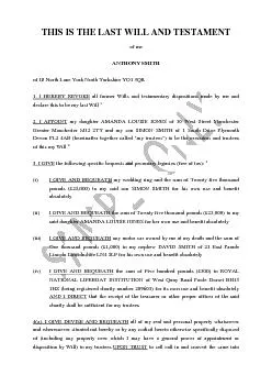
/
ChennaiApril 201DECLARATION BY THE CANDIDATEI hereby declare that this ChennaiApril 201DECLARATION BY THE CANDIDATEI hereby declare that this

ChennaiApril 201DECLARATION BY THE CANDIDATEI hereby declare that this - PDF document

norah
norah . @norah
Follow
342 views
Uploaded On 2022-10-14

ChennaiApril 201DECLARATION BY THE CANDIDATEI hereby declare that this - PPT Presentation

CERTIFICATE BY THE GUIDEThis is to certify that the dissertation titled Comparison study between open transinguinal Preperitoneal hernia repair and Lichtenstein146s repair148a bonafide research ID: 960075

inguinal hernia mesh repair hernia inguinal repair mesh pain preperitoneal patients 146 femoral operative sac cord recurrence post ring

Share:

Link:

Embed:

Download Presentation from below link

Download Pdf The PPT/PDF document "ChennaiApril 201DECLARATION BY THE CANDI..." is the property of its rightful owner. Permission is granted to download and print the materials on this web site for personal, non-commercial use only, and to display it on your personal computer provided you do not modify the materials and that you retain all copyright notices contained in the materials. By downloading content from our website, you accept the terms of this agreement.


Presentation Transcript

ChennaiApril 201DECLARATION BY THE CANDIDATEI hereby declare that this dissertation titled “COMPARISION STUDY BETWEEN OPEN TRANSINGUINAL PREPERITONEAL HERNIA REPAIR AND LICHENSTEIN’S HERNIA REPAIR”is a bonafide and genuine research work carried out by me under the guidance of Prof. Dr.P.N.Shanmugasundaram M.S, HOD, Department of General Surgery, Kilpauk Medical College, Chennai10.This dissertation is submitted to THE TAMILNADU DR. M.G.R. MEDICAL UNIVERSITY CHENNAIin partia

l fulfillment of the degree of M.S. General Surgery examination to be held in April 2016Date: Place: DR T.PAULIA DEVI CERTIFICATE BY THE GUIDEThis is to certify that the dissertation titled Comparison study between open transinguinal Preperitoneal hernia repair and Lichtenstein’s repair”a bonafide research work done by Dr.T.PAULIA DEVI, post graduate in M.S. General Surgery, Kilpauk Medical College, Chennai10 under my direct guidance and supervision in my satisfaction, in par

tial fulfillment of the requirements for the degree of M.S. General SurgeryDate:PROF.Dr. P.N.SHANMUGASUNDARAM. M.SPlace: Professor of General Surgery,Kilpauk Medical College, Chennai ACKNOWLEDGEMENTI am most pleased to acknowledge the Dean Prof. Dr. R. NARAYANA BABU. MD, DCH, of Kilpauk Medical College and Hospital for the opportunity to conduct this study in the Department of Surgery, Kilpauk Medical College.I would like to register my humble thanks to my guide Prof.Dr. P.N. Shanmugasun

daram, M.S, Head of the Department and Chief of Surgery, for his encouragement and advice and under whose supervision this study went on smoothly.This study would have not been possible without the support of my Associate Prof. Dr. V. Vijayalakshmi M.S, D.G.O and also my Unit Assistants, Dr. P. Mathusoothanan, M.S,Dr. K. SrideviS, Dr. M. Senthil Kumar M. S, and also my former Asst. Dr. P. Chelladurai, M.S, to whom I owe my surgical learnings.Place: ChennaiDate: Dr. T.PAULIA DEVI TABLE OF

CONTENTSSl. NoTopicPage No. Introduction History of treatment or Inguinal Hernia Review of LiteratureAnatomy of Inguinal CanalEtiology of Inguinal HerniasClinical FeaturesTreatmentComplications Aimof Study Materials and Methods Observations and Result. Summary Discussion and Conclusion Study Proforma Bibliography Master Chart ABTRACTCOMPARISON STUDYBETWEEN TRANSINGUINAL PRE PERITONEAL MESH REPAR AND LICHTENSTEIN’S REPAIR Department Surgery, IN KILPEUK MEDICAL COLLEGE ,CHENNA

I. Subject Category : SURGICAL TECHNIQUE The preperitonealrepair of an inguinal hernia (IH),performed by a classical anterior transinguinal approach is a simple and safe procedure, particularly prosthetic mesh anteriorly, or behind the conjoined tendon in the preperitoneal space(1). Generally the basic principles and indications of the preperitoneal procedure are Methods Our experience of preperitoneal inguinal prosthetic repair includes 25cases, operatedin the years jan 2

015june2015 out of a total number of 25operations for inguinal repair. There were 13 cases of indirect hernia, 12 direct.. All the operations have been performed monolaterallysurgery doneunder spinal anaesthesia. The followup was continued up to 3 month.we also performed lichtenstein’s repair for 25 cases Classical inguinal incision between the anterosuperior iliac spine and the pubic tubercle, dividingthe external oblique fascia and the external ring and mobilizing the cord. T

he ilioinguinal nerve is gently isolated from the posterior inguinal wall. The external oblique fascia is largely cleaved from the conjoined tendon. In case of indirect hernia, the sac, carefully separated from the cord well beyond the internal ring, is reduced in the peritoneal cavity. In case of firm adhesion with the tunica vaginalis, it can be transacted in its middle part, leaving open the distal. When present a direct hernia, its sac is trimmed off the TF. At this moment, the d

ecision for a preperitoneal repair is based on the conditions of the posterior inguinal wall: enlargement of the internal orifice, presence of a double IH, direct and indirect, global weakness of the TF In this case, the TF is opened from the internal orifice to the pubic tubercle, respecting the epigastric vessels. The subsequent dissection of the preperitoneum is extended laterally beyond the internal orifice, inferiorly to the Cooper’s ligament, and medially to the external

border of the rectus sheath. A synthetic mesh,usually of polypropylene, rectangular in shape and of about 15 x 7cm in size, is prepared to cover all the dissected preperitoneal area, including the Bogros’space and the Fruchaud’s myopectineal orifice . An adequate slit is made in its superior border, to create a new internal ring and allow free passage of the cord The mesh, placed underneath the conjoined tendon, is anchored medially to the rectus abdominis sheath, inferior

ly to the Cooper’s, and laterally to the inguinal ligament. In this way care is taken not to damage the iliohypogastric nerve in its possible intramuscular course. The two tails of the new created internal orifice in the prosthesis are crossed behind the cord and laterally sutured to the internal oblique muscle When possible, a new posterior musculoaponeurotic inguinal wall is constructed approximating the edge of the conjoined tendon to the inguinal ligament. It helps to cove

r and isolate the mesh, and to prevent adhesions with the spermatic cord. The external oblique fascia issutured to close the inguinal canal. In our experience we have not observed any recurrence; in only 1cases a transient inguinal pain was well controlled by a pharmacological treatment. Discussion The main characteristics of this technique are: adequateexposure of the preperitoneal space; hermetic closure, from inside the muscoloaponeurotic wall, of all the possible sites of recur

rence, inguinal,femoral and obturator; anchorage of the mesh to musculoaponeurotic structures, preventing in this way its islodgement or folding . We think that the construction of a new internal orifice avoids the long dissection of the cord, necessary in case of its parietalisation with subsequent risk of damage and entrapment of its nervous structures. The preperitoneal dissection is usually easy to accomplish, except in case of local scarring, as after iliac lymphadenectomy, or

vascular approach to the external iliac artery. These two conditions are a contraindication to this technique. Clearly, the above described procedure combines the advantages of the preperitoneal placement of the prothesis with the easy open anterior inguinal approach. Comparing this technique with others more commonly used and based on the anterior placement of the prosthesis, characteristically thatLichtenstein, some differences are evident: minor possibility of recurrence, inguin

al and especially femoral; the deep preperitoneal lodgement of the mesh protects against infections from the superficial planes of the wound; the abdominal pressure helps the mesh to adhere to the muscoloaponeurotic structures of the whole inguinal region, that constitute a strong barrier against its anterior bulging or displacement; the preperitoneal location of the prosthesis resumes the position and the unction the TF, also before its colonization by the newproduced connective ti

ssue; the TF, clearly weak, is completely replaced, rather than only reinforced, as in the anterior disposition of the prosthesis. The anterior placement of the mesh, typically the Lichtenstein’s operation, is applied more largely thanthe preperitoneal repair: . It requires a more limited dissection, also permitting a good reinforcement of the posterior inguinal wall through a simpleranterior approach, and finds its principal indication when the TF can be still recognized as

a preserved anatomical plane. Conclusion(s) The surgical correction of an IH requests a good anatomical dissection and an accurate evaluation of the conditions ofthe whole inguinofemoral region, in orderto choose the best technique of reconstruction compared with lichtenstein repair,pre peritoneal repair [using prolene mesh] is best method to deal about inguinal,femoral and obturator herniaIn our experience, the repair of groin hernias with Preperitoneal mesh(Prolene mesh through an ing

uinal incision) has resulted in greater patientcomfort with reduced post operative pain and also decreased number of complications.lthough there was no recurrence observed in my study,the follow up period was only minimal (average 3 month). The duration of stay in the hospital was reduced and the patients had a rapid return to work.Hence the transinguinal pre peritoneal mesh repair is an amazingsimplistic technique which gives an approach to inguinal, femoraland obturatoherniasand bears

the same anatomical relationship in TEP and TAPP approaches which gives a better understanding of the TEP and TAPP procedures.It is an easy technique with short learning curve.The risk of vessel injury is less in the hands of an expert. The contact of mesh with the cord structures and nerve is minimal which reduces the postoperative cord oedema, pain (Inguinodynia), orchitis and sensory loss. Reference(s) 1. Awad S.S., Fagan S.P. Current approach to inguinal hernia repair. Am Surg20

04;188(Suppl.6A):9S16S. 2. Horton M.D., Florence M.G. Simplified preperitoneal Marlex hernia repair. Am J Surg 1993; 165: 595599. 3. Fagan S.P., Awad S.S. Abdominal wall anatomy: the key to a successful inguinal hernia repair. Am J Surg 2004;188(Suppl.6A):3S8S. 4. Pelissier E.P. Inguinal hernia preperitoneal placement of a memoryring patch by anterior approach. Preliminary experience. Hernia 2006; 10:248252. 5. Pelissier E.P., Monek O., Blum D., Ngo P. The Polysoft patch:prospective

evaluation of feasibility, postoperative pain and recovery. Hernia 2007;11:229 234. 6. Berrevoet F., Maes L., Reyntjens K. et al. Transinguinal preperitoneal memory ring patch versus Lichtenstein repair for unilateral inguinal hernias. Langenbeck’s Arch Surg 2010;395:557562. 7. Berrevoet F.,Sommeling C., Gendt S. et al. The preperitoneal memoryring patch for inguinal hernia: a prospective multicentric feasibility study. Hernia 2009;13:243249. 8. Amid P.K. Lichtenstein tensionfre

e hernioplasty: its conception, evolution and principles. Hernia 2004;8:1 9. Muldoon R.L., Marchant K., Johnson D.D. et al. Lichtenstein anterior preperitoneal prosthetic mesh placement in open inguinal hernia repair: a prospective, randomized trial. Hernia 2004;8:98103. 10. Condon R.E. Surgical anatomy of the transversus abdominis and trasversalis fascia. Ann Surg 1971;173:1 INTRODUCTION100 years ago, Bassini described the first herniorrhaphy. They believed immobilization and bed

rest enhanced wound healing. But it turned out to be the culprit for genesis of fatal pulmonary embolism. Hence early ambulation was suggested.This study is done to prove the fact that Tans Inguinal Preperitoneal(TIPP)Hernia Repair(using prolene mesh)resulted in greater patient comfort with reduced post operative pain and also decreases the number of complications and recurrence rate and that it can be recommended for all primary unilateral Inguinal HerniaMore than 7 lakh inguinal hernia

repairs wereperformed each year in US in 1980. More than 70,000 patients developed recurrent hernia due to excessive tension repair which was then replaced with Lichtenstein’s tension free mesh repair. But due to chronic post operative pain, sensoryloss, cord oedema, TransInguinal Pre Peritonealrepair was tried which proved to be useful. HISTORY OF TREATMENT OF INGUINAL HERNIA(The History of hernia is the history of surgery)Since the beginning of surgical history, treatment of hern

ia has evolved through different stages.The Latin word hernia means rupture / tearThe Greek word hernia means bulge / buddingANCIENT TIMESThe oldest scientific book, The EDWIN SMITH SURGICAL PAPYRUS, thatdeals with surgery, during the Egyptian Kingdom (3000 2500 BC) contains observations on hernias:[2](When you see a swelling on the abdomen…………when come out ………….caused by coughing)Heliodorus (sun’s gift) who did the first hernia surgeryseparat

ed the hernial sac from the cord, but did not touch the tesicles.In 25 BC TO 50 AD Aulus Cornelius Celsus was the first medical writer. In his book he discussedgroin anatomy and the pathology and causes of herniasIn AD 200, Galen said that peritoneal tear is the etiology of herniation. MIDDLE AGES (DARK AGES)Period of time between the fall of the Roman Empire, the beginning of the Italian Renaissance (15Century).In this periodsurgery was performed by cutters, barbers scissorsIn the 4cent

ury Oribasius performed herniotomiesIn the 6century AD, hernia was treatedwith a plastic, a bandage and a prayer.AD (62590) Pool of Aegina, Egypt performed hernia surgery using double liganon & excision of thecord, sac & testicles.THE RENAISSANCEAmbroise pare describedthe uses of TRUSSES for the control of hernia.One of his greatest contributions was the ligature of vessels,which supplanted the method of hemostasis by the use of hot oil. In the 16century , in ceremony kasper stomayr was

a cutter of herniain 1589, he described the difference between Direct & indirect hernias.Castration was sanctioned in surgery of indirect hernia butnot fortheother types. In 16511714 Jacques performed �2000 hernia repairTHE POST RENAISSANCE ERAAntonica Scarpa(1752 1832) described the sliding hernia.In 1844, Cooper described the role of Superior Pubic Ligament(cooper’s ligament) and the fascia transversalis in the pathogenesis of hernias& 20CENTURIESEdoardo Bassini (1844 1924)

“Father of Modern Herniorrhaphy” Bassini’s repair consists of the higligation & removal of the hernia sac followed reconstruction of the floor using internal oblique, transversalisfascia, a triple layer triple layer offascia transversalis that is supported toinguinal ligament with the cordcoveredby external oblique aponeurosis.The shouldice repair is a modern revival of the original Bassini repair startedin 1953In the USMarcy 18371929, Halsted (18521922) and Fergusson des

cribed technique similar to the Bassini repair.In 1892, Marcy described the high ligation of hernia sac and closure of the dilated deep ring as important steps in the inguinalhernia repair. 18521922 Stowarr Halsted, reported 2 types of herniorrhaphy, In Halsted I the cord is under the skin and in Halsted II the cord is under the repair.In 1890 Halsted stated the modern relaxing incision over rectussheathIn 1898 Cooper’s ligament repair was described by George Lotheissen, which waspo

pularized by Mcvay (191187).CONTEMPORARY TIMESHarvey Cushing used local anesthesia for hernia using Cocaine Infiltration.In 1920, Cheatle performedthe first preperitonealprocedure.In 1936 Henrydid a high closure of the sac and fascial preperitoneal repair.In 1952 Irvine Lichtenstein described tension free meshrepair. In 1982 Ger performed an indirect inguinal hernia repair laparoscopially, closing the defect with Michel staple clips.In 1989 Lichtenstein and colleagues performed 1000 hern

ioplastis, in which reconstruction of the floor of inguinal canalwasby the synthetic mesh.In 1990 Popp placed a dual patch over the defect of an inguinal indict hernia. In 1991 Arregui introduced Trans Abdominal Preperitoneal repair (TAPP).CheatleHenry procedure which is a preperitoneal surgery for inguinofemoral herniation is also known as the Nyhus procedure. REVIEW OF LITERATUREANATOMY OF THE INGUINAL CANFigure:1ANATOMY OF THE INGUINAL CANDEFINITION: The inguinal canal is an oblique r

ift about 4cm long lying above the medial half of the inguinal ligament. It commences at the deep ring and ends at the superficial ring and transmits the spermatic cord (round ligament in female) and the ilio inguinal nerve. SSELBACH’S TRIANGLEIt is bounded laterally by inferior epigastric vessels, mediallythe lateral edge of the rectus abdominis scle and below by the inguinal ligament.DIRECT AND INDIRECT HERNIAHernial sac passing through the Hasselbach’s triangle is direct her

nia;hernial sac passing lateral to inferior epigastric arteryis called indirectinguinal hernia.Figure:3 DIRECT, INDIRECT AND FEMORAL HERNIA EMORAL CANALThisis a enclosed space just medial to the femoral vein,which containfat and some lymph node(node of Cloquet).FEMORAL HERNIAProtrusion of viscus or preperitoneasac through the femoral canal is termed femoral hernia.The walls of femoral herniare formed by the inguinal ligament anteriorly,the femoral vein laterally,the pubic bone covered by

the iliopectinealligament posteriorlyand the lacunarligament mediallyhis is a strong curved ligament witha sharp unyielding edge which impedes reduction of a femoral hernia.t is more common in lowweight, elderly female(becauseof the increase in size of femoral canal in female)Easily missed on examination50% of cases present as an emergency with high risk of strangulatioVariants of femoral herniaVelpeau hernia: hernia sac lies in front of the femoral vessels Narath’s hernia: hernia

lies behind the femoral vessels, usually associated with congenital dislocation of hip Serafini’s hernia: behind the femoral vessels.Figure : 4 VARIANT OF FEMORAL HERNIACloquet hernia: it lies under the fascia covering the pectineus muscle.Laugier hernia: hernia through lacunar ligamentBeclard hernia: sac through the saphenous opening carrying the cribriform fasciaIdeal timing for femoral hernia surgery as soon as possible.Principle of repair suturing of inguinal ligament to Pectine

al ligament after dealing with the sac It contains External iliac vesselsDeep circumflex iliac veinenital branch of genitofemoralnerve.During surgery, if these vessels are injured, the chance of mortality is high.TRIANGLE OF PAINFigure:TRIANGLE OF PAINIt is formed by the:Spermatic vessels medially Iliopobic tract laterallyIt contains genitofemoral nerve, the femoral nerveand the lateral femoral cutaneous nerve of thigh. Any staples placed caudal to the iliopubic tract and lateral to the

spermatic vessels result in neuralgiaCORONA MORTIS (DEATH CROWN)It refers tothevascular ring, which is formed bytheanstomis of an aberrantobturator artery and the normal obturator artery. During hernia surgeryinjury to this anastomosis causes torrential bleeding.FigureCORONA MORTIS NERVES IN THE INGUINAL REGIONFigure:NERVES IN THE INGUINAL REGIONILIOHYPOGASTRIC NERVE [T12L1]Emerge lateral to the psoas muscleurface marking2cm medial to anterior superior iliac spine. It piercthe internal o

blique muscle.ust above the external ringit pierces the external oblique muscle.Sensory innervationto supraic region.Motor supply to the transverse abdominis. ILIOINGUINAL NERVE [T12L1]temerges lateral to the psoas muscleSurface markingnear the iliac crestt piercethe transverse abdominis muscle,near the deep ring it lies between ILM and EOM.esalong the cord structureotor supply totheinternaloblique muscle,sensory innervationtheroot of penis,anteriorpartof the scrotum,mons pubis,labia maj

ora.NITO FEMORAL NERVE [L2]Passes on the medial border ofthepsoas muscle.rosses behind the ureterust above the inguinal ligament,it is divided intothefemoral and genital branch.ensory and motor components to the cremastric muscle[cremastricreflex]Femoral branchsensory innervations to the femoral triangle areaGenital branch enters into the inguinal canal through the deep ring.ensory to the skin of scrotum,mons pubis,labia majora LATERAL FEMORAL CUTANEOUS NERVE [L2L3]Passes onthelateral bo

rderof the psoas,then passesthe iliacusFinally it passes medial to the ASIS,then behind the iliopubic tract.ntrapment of this nerve is common in the region where it passes near inguinal ligament from abdomen to the thigh[meralgia paresthetica]ensory to the anterior and medialaspects of thethighFEMORAL NERVE (L2,L3,L4)t passes lateral to the iliac artery, deep totheinguinal ligament.Motor to iliacus,pectineus,artorius,quadriceps,hip and knee joint.Sensory to the anterior and medial aspect

of thigh, distal to genitfemoral nerve and medial tothelateral cutaneous nerve of thigh.Cutaneous branches are intermedial, medial and saphenous nerve.MECHANISM OF INGUINAL CANALFLAP VALVE MECHANISM:Increased intra abdominal pressure leads on to approximation of theanterior and posterior walls of the canal The superficial ring is protected bytheconjoint tendon inferiorly and the reflected part of the inguinal ligament superiorlySHUTTER MECHANISMAs the transversalis abdominis and the ingu

inal oblique muscular contract, the roof is approximated to the floor like shutter.BALL VALVE MECHANISMContraction of the cremaster helps Kcord to plug the deep ringSLIT VALVE MECHANISMContraction of the external oblique leads to approximation of 2 crura of the superficial ring. Hormones may play a role in maintaining the tone of the Inguinal Musculature.ETIOLOGY OF HERNIAPreformed Sac Patent processus vaginalis Congenital Indirect inguinal hernia.Repeated high abdominal pressure due to

constipation, excess coughing, prostatic symptoms and obesity. Weakening of body muscle and tissues due toa)Genetic weakness of collagenAgeing and pregnancyc)Structures entering and leaving the abdomen.Eg: Femoral vesselsemoral herniaObturator Nerve Obturator HerniaSciatic NerveSciatic Hernia.THE EUROPEAN HERNIA SOCIETY CLASSIFICATION [1]Primary or Recurrent (P or R)Lateral, Medial or Femoral (L,M or F)fect size in finger breadths assumed to the 1.5cm.Eg:A primary,direct, inguinal herni

a with a 3cm defect would be PM2.NYHUSCLASSIFICATION OF GROIN HERNIABased on the deep ring and posterior wall weaknessType 1: Indirect hernia with normal deep ringType 2: Indirect hernia with dilated Deep ringType 3: Posterior wall defect a)Direct herniaIndirect with posterior wall weakness (Pantaloon hernia)c)Femoral HerniaType 4: Recurrent herniaCLINICAL FEATURESSwelling in the inguinal region above the inguinal ligament, medial to pubic tubercle Inguinal herniaSwelling below the ingui

nal ligamentandlateral to the pubic tubercle femoral herniaAching (or) heavy feelingINVESTIGATIONPlain xrayabdomen isnot much usefulUSG low cost, very useful in the early post operative period to differentiate a hematoma or seroma from an early recurrence.MRIuseful in the diagnosis of sportsman’s groin, also useful to differentiate an occult hernia from an orthopaedic injury. Herniogram to identify occult hernia/not detectable clinically, may cause severe painLaparoscopy to diagnose

the occult contra lateral inguinal hernia.CT ScanON EXAMINATIONCheck for the:ReducibilityCough impulseTenderness and overlying skin colour changesContent of herniaDIFFERENTIAL DIAGNOSIS OF INGUINAL HERNIAAn encysted hydrocele of the cordSpermatoceleFemoral HerniaUndescended testis Vaginal hydroceleLipoma of the cordInfantile hydroceleEctopic testisPsoas abscessPsoas bursaEnlarged lymph nodeSaphena varixFemoral aneurysmIN FEMALESdrocele of the canal of NUCKFemoral herniaDIFFERENTIAL DIAGN

OSIS OF FEMORAL HERNIAInguinal herniaLymphadenopathySaphena Varix Femoral artery aneurysmPsoas abscessRupture of adductor longus with hematomaTREATMENTTRUSSESHernias can be controlled by suitably fitted trusses. But it may be uncomfortable and difficult to keep in place. It never prevents strangulation norcurethe hernia.ANESTHESIA IN HERNIA SURGERYGeneral Anesthesia INDICATIONSPatients with localized skin infection in the back who are not suitable for spinal / epidural, spine deformity.R

egional anaesthesia spinal and epidural anaesthesia offer a number of advantage for inguinal hernia repair. Postoperative nausea and vomiting are less common.patients are usually ready for discharge sooner after regional anaesthesia. Complications include urinary retention, post dural puncture headache, greater degree of muscle relaxation, pain.Local Anesthesia 1) Nerve block method2) Field block methodUsing alkalinisation of 1% lignocaine with sodium bicarbonate will decrease the pain

of injection. Infiltrated in area two finger breadth above and medial to ASIS, just proximal to the pubic bone using 0.5% lignocaine or 0.25% bupivacaine. The addition ofadrenaline help to reduce the plasma concentration of local anaesthesia.PRE OPERATIVE ASSESSMENT AND PREPARATIONClinical ExaminationLaboratory testsCardiovascular, pulmonary, renal functionLook for DM, HTStop smokingObese patients are advised to reduce weight before the operation.TYPES OF SURGERY IN HERNIAHerniotomy (Exc

ision of sac)HerniorrhaphyHernioplastyHERNIOTOMY IN CHILDREN MICHAELIS PLANK OPERATION Through inguinal approach, high ligation of hernia sac is performed.Before ligatingone should ensure that the contents are reduced.Cut the sac distal to the ligation. Repair is not needed in children.OTHER APPROACHESSuprainguinal Preperitoneal open methodTransperitoneal laparoscopic method.HERNIA SURGERY IN ADULT[3]Herniorrhaphy (Strengthening of the posterior wall of inguinal canalBassiniShouldiceDasa

rdaHernioplasty Strengthening of the posterior wall by mesh)a)Open flat mesh repair LichtensteinOpen Complex mesh repairPlugsHernia Systemsc)Open Preperitoneal Repair StoppaKugel’sLaparoscopic RepairTEPTAPPBASSINI (IN 1890) Sutures are placed between the conjoint tendon above and the inguinal ligament below using non absorbable interrupted prolene sutures extending from the pubic tubercle to the deep ring. Medial most stitch is taken from the periosteum of pubic tubercle

which is called astheKEY (or) BASSINI’S STITCH. In1887Bassini originally used silk as the suture materialMODIFIED BASSINIHere transversalis fascia is opened, triple layer of upper leaf which contains transversalis fascia, transversis abdominis and internal oblique muscle is uturetheouter shelving edge of inguinal ligament.DASARDA2cm strip of external oblique aponeurosis lying over the inguinal canal is isolated but left attached both medially and laterally which is sutured to the c

onjoint tendon and inguinal ligament, reinforcing the posterior wall of the inguinal canal. SHOULDICE REPAIR (1930)It was introduced by Shouldice in Toronto at Shouldice hernia clinic. It is a multilayered repair. Often cremasteric resection is done to have proper access to the posterior inguinal wall. After doing herniotomy, transversalis fascia is incised and raisedintotheupper and lower flap.FIRST SUTURE LINELower flap is sutured to posterior deep part of upper flap using continuous s

utures from pubic tubercle to the deep ring where it is tied at deep ring without cutting.SECOND SUTURE LINEsing same suture which is not cut upper flap is sutured to the shelving edge of the inguinal ligamentknot is placed over the pubic tubercle.Suture lineSuture is placed in between conjoin tendon and anterior to the shelving part of the internal ringto pubic tubercle and tied without cuttingSuture lineUsing same suture continuously between conjoined tendon and anterior fibersof the i

nguinal to reach deep ring. Lytle’s RepairDeep ring is narrowed by placing interrupted suture over the medialaspect of the ring to the transversalis fascia.Tanner SlideTo reduce the tension in the repair area, relaxing incision is muscle over the lower rectus sheath.LichtensteinTension free, simpleflap, polypropylene mesh repair for inguinal hernia.Skin is incised. Two layers of superficial fascia, outer Camper’s and inner Scarpa’s layer are incised.External oblique apo

neurosis is identified and incised.in the inguinal canal, cord is covered by Cremasteric muscle and internal spermatic fascia; external spermatic fascia covers the cord below the level of external ring.cremasteric muscle is opene. Medial dissection is done beyond the pubic tubercle.hernial sac is identified which is white in colour.sac is anterlateral in position with respect to ord in case of indirect sac. Sac is transfixed above the inguinal ring with 3vicryl after reducing contents. r

edundahernia sac is removed and Herniotomy completed. In case of direct hernia do not open the sac. Closethe medial defect 15*8cm sized prolene mesh is placed over the posterior wall, behind the cord and tail made at the deep ring where it encirclethe cord structures. Figure:1LICHTENSTEIN’S REPAIRMesh is placed beyond the pubic tubercle (beyond 2cm), superior margin(4cm), laterally beyond the deep ring (6cm). mesh is sutured below to inguinal ligament, medially to the pubic tubercl

e and above to the conjoint tendon mess cord EOA IS CLOSED Figure:1EOA IS CLOSED IN LRWound closed in layers and sterile dressing applied.Gilbert Mesh Repair (Patch & Plug)Here internal ring splugged with umbrella shaped piece of prolene mesh.Gilbert’s Prolene hernia System RepairOpen transinguinal approach to keepa mesh in both Preperitoneal (as inlay) and in front (as onlay) mesh repair.Stoppa’s (Giant prosthesis reinforcement of visceral sac)It is performed in bilatera

l hernias, recurrent and hernias & hernia with collagen disease. It is performed through lower midline subumblical incision as a posterior preperitonealspace which coversthe space of Bogros and space of Retzius.Preperitoneal Mesh RepairNyhus Preperitoneal onlay mesh repairIt is performed through supra inguinal horizontal incision above the pubic symphysis and deep ring through lateral border of rectus muscle Preperitoneum is approached. Mesh is placed deep to the conjoined tendon, cord a

nd transversalis fascia. Mesh is sutured to the Iliopectineal ligament using 23 interrupted non absorbable sutures. It also sutured transverse abdominis and fascia from deep.Modified Rives Preperitoneal Sublay Mesh RepairPreperitoneal mesh repair through transinguinal approach (anterior approach) here mesh is place in Preperitoneal space and sutured to Iliopectineal ligament using non absorbable suture material.Kugel’s Preperitoneal Mesh RepairIt is a tension freeand suturelessPrep

eritoneal or posterior abdominal wall groin hernia repair. It is performed through abdominal grid iron incision using fortified patch to reinforce the damaged transversalis fascia/ floor of the inguinal and femoral canal.TAPP (trans abdominal Preperitoneal mesh repair) Arregui 1991It is very much useful in large indirect hernia and irreducible inguinal hernia. 10mm umbilical port is used for laparoscope. Pneumoperitoneum is created.5mm ports on para rectal point just above the level of u

mbilicus one on each side. Hernia sac is dissected in the Preperitoneal plane by making incision at the upper part of the opening of the sac. Sac is reduced and Preperitoneal space is dissected to identify the pubic bone, cooper’s ligament, gonadal vessels, vas and inferior epigastric vessels. Sac is dissected and excised.Mesh is placed in the Preperitoneal space and fixed to pubic bone/ cooper’s ligament using tacks.TEP (totally extra peritonealrepair) Mckernon 1990Here we do

n’t open the peritoneal cavity. It is useful for bilateral recurrent hernias. 2 sub umbilicalhorizontal incisions (10mm). Extra peritonealspace is reached. After CO2 insufflations, another 5mm port is inserted 4 cm below the first port in the midline. 35mm port is inserted in the same line 4 cm below or in the right iliac fossa. Dissection is carried out downwards then medially up tothe pubic tubercle, Iliopectinealligament lateral to iliac vessels and inferior epigastricvessels. Sa

c is identified, dissectedand excised. Sac is transected at the internal ring byplacing aend loopon the proximal part of the sac. Distal part of the cut sac is left open without ligation. Mesh is placed and spread and sutured to the Iliopectineal ligament.Post operative complicationGeneral ComplicationPulmonary atelectasisPulmonary embolismPneumonia Thrombophlebitis: Most of them avoided by chest physiotherapy, Limb physiotherapy, early ambulation, good Pre operative preparation.Post o

perative urinary retentionCan be treated by temporary catheterizationLocal ComplicationWound infectionHydroceleHemotoceleDysejaculation syndromeTransectionof the vasNerve InjuriesIschemic OrchitisTesticular AtrophyInguinodyniaRecurrent HerniaHematoma CAUSES:Bleeding from superficial vessel During resection of cremaster, careless ligature of external spermatic artery.Injury to deep inferior epigastric vessels during division of transversalis fasciaBleeding from venous circulation within t

he space of BogrosBleeding from IliopubicarteryInjury to aberrant obturator artery.Injuryto femoral vein and artery.1. WoundInfectionWound infection depends on 4 variables Number of bacterial ContaminantsVirulence of bacteriaMicroenvironment of the woundIntegrity of the woundNumber of bacterialcontaminantsThreshold of bacteria willbe 1lakbacteria per gm of tissue beyond which infection will occur.Bacterial Virulence Staphylococcus aureus is most common pathogen in surgical site. It creat

es thick, turbid wound abscess.Staphylococcus epidermidisE.ColiB.FragilCorynebacterium acneMicro environment of the woundWound hematomaDead space leads to seroma which is Opsonin freeSuture materialBraided suture material eg.Silk.Abundant number of knots on monofilament materialMeshInfection will be more when there is a use of fine weave such as polytetraflouroethylene.Necrotic Tissue Excessive usage of cautery creates foci of dead tissue, which increases infection.The HostObesity increa

ses infection, because of the avascular character of the large subcutaneous tissue.Comorbidities eg. DM, Renal failure, alcoholism and malnutrition.Prevention of wound infectionPreoperative shower with soapScrubbing the surgical siteperative site should not be shaved the night before the procedure.Hair removal immediately prior to the operationPreoperative hospitalizationSurgical site is scrubbed with betadine, chlorhexidine (or) isopropyl alcohol. Isopropyl alcohol is discouraged if cau

tery is to be used.Minimal number of knots in the monofilament suture material. Avoid redundant and folded mesh causes dead spacefor the sequestration of bacteria. Crinkled and bunched mesh around the perimeter of the repair increases infection.Cefazolin 1Preoperative antibiotic of choice in hernia repair.Figure : 13 SURGICAL SITE INFECTION Local Drainage, Remove the Knots Remove the Redundant Mesh Wound Healed Wound Not Healed Complete Removal Mess Wound Healed Tracks

of the Mesh Surgical Site Infection hydroceleIt occurs if the distal sac is ligated.needle aspiration is a treatment of hydrocele .hematoceleIt occurs if the collection of blood in the distal sac in patientwith a patent processematocele presents within first 12 to 24hoursof early post operative period.surgical evacuation is the treatment of hematocele.dysejaculation syndromeIts occurs due to trauma to the vas during surgery, results in scarring of the lumen of the vas.transect

ion of the vasIts occurs during hernia surgery.it is treated by end to end anastomosis over a small stent using nylon suture.Ischemic orchitis and testicular atrophycausesvascular compromiseremoval of adherent distal sactight reconstruction of the deep ring by mesh Histology of testicular atrophyshows leydig’s cell and sertoli cells appeanormal and seminiferous tubules are absent. investigationdoppler studytreatment antibioticAnti inflammatory drugsteroidinguinodynia Causes of in

guinodynia:Injury to ilioinguional/iliohypogastric/genitofemoral nerve.Direct trauma to the pubic bone.Hidden/incarcerated hernia either missed during the initial procedure/ early recurrence following the procedure.Injury/entrapment/stretching/chronic irritation of the peripheral nerves.Laparoscopic staple causes injury to lateral femoral cutaneous nerve of thigh, femoral nerve, obturator nerve. Staple placement into the periosteum of the pubic bone (osteitis pubis).Fibroblastic response

caused by foreign body (mesh) resulin eventual cicatrix, which entraps nerve in the inguinal region resulting in neuralgia.JF Maillar, P.Vantournhoudt, G. PirerGerard, E.Mauel, Pelissier et al [8]TIPP groin hernia repair using a Preperitoneal mesh performed with a permanent memory ring; a good alternative to LR (20062008)n=145no infection of mesh, no clinical recurrence. There was aultrasound recurrence in 2%(n=3) of the asymptomatic patients and chronic pain in 4.8% of the patients.B

enefits of the anterior approach (easy technique, short learning curve, low cost) and the Preperitoneal placement of the mesh (less recurrence, less pain). This procedure is a good alternative to LR.Frederik berrevoetUGent,leander Maes UGent,Koen Reyntjens UGent,Xavier Rogiers UGent,Roberto trouser UGent and Bernard de hemptinne UGent [2010][9],did study to compare TIPP versus lichtensteins in relation to acute and chronic pain, post operative complication and recuunce rate.duration of

study was 18 monthhey observed mean operative time for TIPP is less than Lichtenstein,33 versus 44min,respectively (p=0.04).less post operative pain observed in the TIPP than Lichtenstein group.recurrence were observed less in TIPP than LICHTENSTEIN group respectively 2.8% versus 5.1%.pelissier and colleagues(2007)[5]described thatrecurrence rate is 2% and rate of chroin pain is 5in TIPP groupsmore recently berrevoet and his team[6]described a recurrence rate of3% and visual analoguepai

nscale of 0.2 1yr after TIPP.reason for lower rate of post operative pain 1.minimal dissection around the ilioinguinal and iliohypogastric nerve.2.no fibrosis of the mesh in contact with the inguinal nerve.koning GG ,Schipper HJP, Oostvogel HJM, Verhofstad MHJ,Gerritsen PG,Larrhoven KCJHM,Vriens PWHI[12]double blind RCT comparing Lichtensteins and TIPP (200910). Studied in 496 patients:225 TIPP and 271 LICHTENSTEINS. This study revealed no significantly better result forthe TIPP as com

pared to lichtensteins.Moldoon RL,Marchant k, J OHNSON dd,yoder GG,Read RC, HauerJensen M, RCT study of lichtenstien and TIPP Trial (2004)[10]described recurrence in the lichentenstein is 4.3% and in less than 1% recurrence in THE PRE PERITONEAL Read Rives. Both anterior repairs are associate with low post operative morbidity and recurrence rates.(p=0.21)Giel G.Koning : Patrick W.H.E Vriens 2011 , St Elizabeth hospital, The Netherlands[11]Anterior PPR of extremely large hernias. In extr

emely large hernias, the lateral side of the mesh can be insufficient to fully embrace the hernia sac. They describe the use of 2 preperitoneal placed meshes (Butterfly technique) to repair extremely largehernias. 2 inverted meshes to cover the deep ring both medial and lateral. Follow up was done at 6 months.n=689pts (20062008)mean age 69.9 years (63all U/L extremely large hernias 1% n=7recurrence did not occur after repair. Chronic pain wasnot reported.TEP no contact with nerves in the

inguinal canal.8. G.G.Koning , J.P de Schipper, H.J.M. Oostrigel, M.H.J. Verhofstad, G.P.Gerristen, C.J.H.M. Van Larrhova et al (2010)[14] n= 496, TIPP=225, LR=271TIPP MEAN AGE52.757.3 ASA CLASSIFICATION I54%51% ASA CLASSIFICATION II36%38% ASA CLASSIFICATION III5.311% GENDER M/F257/15210/14 COMPLICATIONS7.6% RECURRENCEn=1n=3 BLEEDING AND REOPERATIONn=4, 1 patientn=4, 3 patients CHRONIC PAIN10 patients (4.4%)10 patients(4%) PERSISTING SENSORY LOSS0.9%, 2 patients2.2% (6 patients) Table:1

9. Muldoon RL, Marchant K, Johnson DD, Yoder GG, Read RL, Haver[10]Jensi M PPR (n=121); LR(n=126)Read Rive’s 9min longer than LR.No wound infection.PPMR RECURRENCE(%) P=0.215 (4.3%) Table:2Jamal Akhavan Moghaddan, Shaban Mehrvarz, Hassan ali Mohabbi[13]Comparison of Read Rive’s and LR for treatment of unilateral inguinal hernia. RCT=126 patients. Read Rive’s 64 ; LREvaluated for early postoperative complications, duration of surgery and hospital tay, return

to normal activity, recurrence. Equal in both the groups. G.G.Koning, C.S.Andeweg, F.Keus, M.W.A. Van Tilburg, C.J.H.M. Van Larrhova, W.L. Akkers dijk.Laparoscopic hernia repair popularized the Preperitoneal mesh position due to promising result of less chronic pain. However, considering the proportions of severe adverse events, learning curve, added cost,erformed trans rectus sheath preperitoneal repair in 50 patients.They observed no technical problemin surgery, no recurrence and chro

nic pain after a mean follow up of 2 years. AIM OF THE STUDYThe prime cause of post operative pain in hernial surgeries is nerve entrapment by mesh,ostetis pubis,injury to a neural structure, injury to vas, injury to testis.many surgical techniques are there to treat inguinal herniapost operative pain has been reported in 1535% after lichtenstein’s repair.Classical anterior transinguinal Preperitoneal inguinal hernia prosthetic repair is a safe and simple procedure. Pre periton

eal placement of mesh protects against infections from the superficial planes of the woundabdominal pressures helps the mesh to adhere to musculoaponeuotic region in orderto provide a strong barrier against recurrence,inguinal,femoral and obturator hernia.The objectives of this study are:rospective study of 25 cases of transinguinal Preperitoneal mesh repair done between 2014 and 2015 in our institution. To assess the post operative pain in these patients by visual analogue pain score a

nd compare with those experienced in patients who underwent lichtensteins repair.To compare the duration of procedure between transinguinal Preperitoneal repair and lichtensteins repair procedures. To study the incidence of complications [like scrotal collection,seroma,cord oedema, wound infection,injury to vessels and injury to nerves] that occurred after pre peritoneal mesh repair.To look for any recurrence during the follow up period in the transinguinal Preperitoneal mesh repair. M

ATERIALS AND METHODSAbout 25 cases oftransinguinal pre peritoneal mesh repair was done in the period 2014 to 2015 at Kilpauk medical college hospital.cases were selected at random irrespective of the type of inguinal hernia, the age of the patient and the size of the defect.the material used for repair is monofilament polypropylene clear non absorbable synthetic knitted surgicalmesh available in our hospital as SURUMESH manufactured by SURU INTERNATIONAL PVT .LTD.These cses were follow

ed up in the immediate and post operative periods.post operative pain,scrotal collection,seroma, cord oedema and wound infection were looked for.they were asked to come for regular followup visit after discharge.during each followup visit,the patients were assessed for pain,surgical site infection and recurrence.POLYPROLENE MESHMonofilament mesh does not have any antibacterial properties..Its hydrophobic nature and monofilamentmicrostructure impede bacterial ingrowth.Provokes a fibrous

reaction leading to collagen contraction and stiffening. Mesh shrinkage is due to natural contraction of fibrous tissue embedded in the mesh reducing the area of mesh which leads to tissue reaction, pain and also hernia recurrence.Mesh shrinkage by upto 50%.Figure:14 PROLENE MESHMeshes with thinner strands and larger spaces between them are lightweight (40g/sq.m) Figure:1external oblique aponeurosis.IOAinternal oblique muscle.re peritoneal space is defined,dissection is extended lateral

ly beyond the deep ring,inferiorly to the cooper’s ligament and medially to the outer border of the rectus sheathFigure:1PPSpre peritoneal space EOA PPS CORD Figure:1MESH IN THE PRE PERITONEAL SPACE MESH Figure : 20 FASCIA CLOSED IN TIPPFigure:21 SKIN CLOSED IN TIPP SCARPA FASCIA SKIN SUTURED xternal obligue fascia is sutured.skin is closed.compressive dressing to be done at the end of the procedure.post operatively analgesic and antibiotics given.each patients disch

arged at 7post operative days.OBSERVATION AND RESULTSTypes of outcome measures:Primary outcomes: Preoperative period:Injury to peritoneumInjury to vesselsMean duration of operationEarly postoperative period:HemorrhageWound infectionScrotal collectionCord edema Acute pain, measured with the VAS pain score, was defined as any score above 0Duration of hospital stayReturn to sedentary workAt 1 month: Wound infectionScrotal collection Hernia recurrence Chronic postoperative pain (inguinodyni

a) measured using the VAS pain score.The cut off value for pain on the 100mm VAS pain score was 0.At 3month:Wound infectionScrotal collection Hernia recurrence Chronic postoperative pain (inguinodynia) measured using the VAS pain score at three months during followup. The cut off value for pain on the 100mm VAS pain score was 0. QUESTIONNAIREName:Age:Sex:Type of hernia:Co morbidities present: hypertension/ diabetes/ COPD/ prostatism/ constipationMean duration of operation:Duration o

f hospital stay:Return to sedentary work:Preoperative period:Yes No Injury to peritoneum Injury to vessels Early postoperative period: Hemorrhage Wound infection Scrotal collection Cord edema Acute pain, measured with the VAS pain score, was defined as any score above 0 At 1 month: Wound infection Scrotal collection Hernia recurrence Chronic postoperative pain (inguinodynia) measured using the VAS pain score.The cut off value for pain on the 100mm VAS pain score was 0. At 3m

onth: Wound infection Scrotal collection Hernia recurrence Chronic postoperative pain (inguinodynia) measured using the VAS pain score at three months during followup. The cut off value for pain on the 100mm VAS pain score was 0. Table:3 RESULTSAmong the 50 patients taken for the study, 25 patients were subjected to lichenstein’s hernia repair and 25 for the TIPP procedure. The mean age of the patients subjected to lichenstein repair was 53.84years and for TIPP it was 48

.76yrs. he baseline characteristics of both the group is shown in the table 4TIPPichenstein Total no. of patients ex Male Female Age 40yrs 60yrs �60yrs Type of hernia Indirect Direct Duration of surgery45mins45mins Table 4: Baseline Characteristics Figure : 22 AGE DISTRIBUTION OF PATIENTS SUBJECTED TO TIPP age distribution 20-40yrs 41-60yrs �60yrs Figure: 23 AGE DISTRIBUTION OF PATIENTS SUBJECTED TO LRThe duration of operation was more in the TIIP group and this was stati

stically significant. (p .05) Age distribution 20-40yrs 41-60yrs �60yrs The per operative complications encountered were injury to peritoneum, vesseland nerve as shown in figure:24Figure : 24 PER OPERATIVE COMPLICATIONS 05101525 lichenstein repairTIPP injury to peritoneum injury to vessels injury to nerves total The various early post operative complications encountered among the patients subjected to Lichenstein repair were seroma, surgical site infection, scrotal collection, c

ord edea and pain as shown in figure:25Figure : 25EARLY POSTOP COMPLICATIONS IN LRAmong the patients who underwent TIPP procedure the early post operative complications seen were pain and surgical site infection with only 8% of the patients experiencing it.(table 4Table 4early post operative complications in TIPP seromascrotal collectioncord edemacord edemapainEarly Postop complications in LR Early Post-opcomplications ComplicationsNumber Seroma Surgical site infection Scrotal collection

Cord edema ain Table 5EARLY POST OPERATIVE COMPLICATE IN TIPPFigure : 26 EARLY OPERATIVE COMPLICATE IN TIPP 0510152025 paincord edemascrotal collectionSSIseromaEarly post-op complication in TIPP Series2 Series1 During the first follow up of patients at one month it was noticed that 24% of patients who underwent linchenstein repair had complications of SSI, cord edema, recurrence and pain. On the other hand only one patients who underwent TIPP had paincomplications.Complications Lic

hensteinTIPP At 1 month Surgical site infection ecurrence Cord edema ain Loss of sensation Table: Figure :27 COMPARISON BETWEEN LICHESTEIN AND TIPPAt second follow upit was noticed that noone patient who underwenTIPP had complication like chronic pain,cord oedema,sensory loss and recurrence in my observation.on the other hand one patient who underwent LR had chronic pain and 2 patient who underwent LR had sensory loss 01234 lichensteintippinjury to nerves injury to nerves Complication a

t 3monthTIPP CHRONIC PAIN CORD ODEMA RECURRENCE SENSORY LOSS Table :Post operative complication at 3month.Total patients25 in LR,25 in TIPPFigure :28 POST OPERATIVE COMPLICATION AT 3MONTH. lichensteinTIPP 0.51.52.53.5 lichenstein TIPP Median pain score (using visual analog score) On day 1Day 7At 1 monthAt 3month Lichenstein repair TIPP Table:Figure: 29 MEDIAN PAIN SCOREOccurrence of pain at 3 rd month between thelichenstein and TIPP repair is not statiscally significant(p�0.05)

012356 day 1day 71st month3rd month lichenstein TIPP SUMMARYOf the 25 transinguinal pre peritoneal mesh repair was done 12 were indirect hernia versus 13 were direct hernia.most of them were European hernia societyclassification PM1.maximum number of patients belonged to 4555year age group.From the observation,it is clear that post operative pain is very much less with transinguinal pre peritoneal mesh repair using prolene mesh with most of the patients having minimal or no pain after 2

days.The majority of patients required only oral analgesic.Most of them required sedation only on the day of thesurgery.One patient developed wound infection at the end of 1st week,patient developed wound infection,cord oedema,recurrenceat the end of 1nthone patient who developed chronic pain at the end of 1monthin this group of patients. unrestrictedactivity was encouraged in these patients after discharge.Out of 25 patients for transinguinal pre peritoneal mesh repair. 23 patients cam

e for regular followup.the average follow up period was 3 month.during each followup visit, patients were assessed for pain, any restriction of physical activity,surgical site infection,mesh rejection and recurrences . 25patients underwent lichtensteins repair experienced more pain in the early post operative period.the intensity of pain was more in the early postoperative period, theintensity of pain was even more increased during coughing and during ambulation. Althoughthese patient

s experienced minimal pain at rest after 5 days the intensity was increased during coughing and ambulation. Thesepatients needed larger doses of analgesics and sedatives and most of them had restricted physical activities up to 1 month postoperatively.5 patients developed wound infection.8 patients developed cord oedema,at the end of 1week 9 patients developed pain over surgical site.althouth no recurrence was noted in these group, 1 patient developed pain at 3month. DISCUSSION ANDCONCL

USIONIn our experience, the repair of groin hernias with Preperitoneal mesh(Prolene mesh through an inguinal incision) has resulted in greater patientcomfort with reduced post operative pain and also decreased number of complications.lthough there was no recurrence observed in my study,the follow up period was only minimal (average 3 month). The duration of stay in the hospital was reduced and the patients had a rapid return to work.Hence the transinguinal pre peritoneal mesh repair is a

n amazingsimplistic technique which gives an approach to inguinal, femoraland obturatoherniasand bears the same anatomical relationship in TEP and TAPP approaches which gives a better understanding of the TEP and TAPP procedures.It is an easy technique with short learning curve.The risk of vessel injury is less in the hands of an expert. The contact of mesh with the cord structures and nerve is minimal which reduces the postoperative cord oedema, pain (Inguinodynia), orchitis and sensory

loss. STUDY PROFORMAName of the patient: age: sex:Ip no: occupation: Date of admission: date of surgery: date of discharge:Complaints on admissionDuration of symptom: Any predisposing factors:COPD/heavy physical activity/weak abdominal wall/BPH/ urethral stricture/neurological weakness/constipation/family hconnective tissue disorder/any othersAny comorbidity : hypertension/DM/asthma/CAD/CVA/epil

epsyClinical examination:direct/indirect hernia/complete/ incompleteAny complication: reducible/irreducible/obstruction/strangulationNature of surgery:Type of anaesthesia: Per operative period:Injury to peritoneumInjury to vessels Injury to nervePost operative pain scoreAt 24hrs 1week 1month 3rdmonthPost operative complications if any:Follow up visits at 1month: At 3month: any complication like pain, infection,restricted physical activity,meshrejection,cordoedema,scrota