/
P54V 58 15 2021Association of Child Neurology AOCN  Indian Epileps P54V 58 15 2021Association of Child Neurology AOCN  Indian Epileps

P54V 58 15 2021Association of Child Neurology AOCN Indian Epileps - PDF document

obrien
obrien . @obrien
Follow
342 views
Uploaded On 2022-10-28

P54V 58 15 2021Association of Child Neurology AOCN Indian Epileps - PPT Presentation

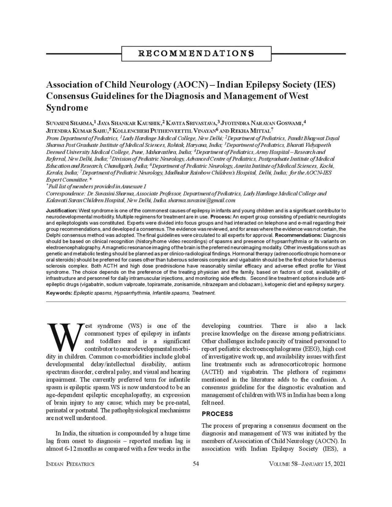
RRRRREEEEECCCCCOOOOOMMMMMMMMMMEE W developmental delayintellectual disability autismspectrum disorder cerebral palsy and visual and hearing P55V 58 15 2021AOCNIES CAnnexure 1groups definitio ID: 961568

eeg spasms treatment therapy spasms eeg therapy treatment high vgb dose etiology infantile epileptic clinical children consensus ies acth

Share:

Link:

Embed:

Download Presentation from below link

Download Pdf The PPT/PDF document "P54V 58 15 2021Association of Child Neur..." is the property of its rightful owner. Permission is granted to download and print the materials on this web site for personal, non-commercial use only, and to display it on your personal computer provided you do not modify the materials and that you retain all copyright notices contained in the materials. By downloading content from our website, you accept the terms of this agreement.


Presentation Transcript

P54V 58 15, 2021Association of Child Neurology (AOCN) – Indian Epilepsy Society (IES)Consensus Guidelines for the Diagnosis and Management of WestSyndromeUVASINIAYAAVITA RRRRREEEEECCCCCOOOOOMMMMMMMMMMEE W developmental delay/intellectual disability, autismspectrum disorder, cerebral palsy, and visual and hearing P55V 58 15, 2021.AOCN-IES CAnnexure 1groups: definitions, etiology, early diagnosis andvigabatrin and other drugs; and diet, surgery andconsensus method was adopted [6]. The writing groupgroups. These open-ended questions were administeredexperts gave their opinions to the moderator, whoexperts. A guarantor ensured that responses were blindedclose-ended responses were framed. These questionswere again sent to experts in the second round. Theirresponses were collated and presented in the meeting ofhave reached a consensus. The concerns, discrepanciesconsensus were re-polled. Any question where secondA final consensus meeting was held on 1 September,the various participating members were incorporated intothe document. The final document was prepared andThe final document was prepared andepileptic spasms, and WS were framed (Box 1differentiate spasms from myoclonic seizures [1].Differentiation from paroxysmal non-epileptic pheno-myoclonus, benign myoclonic epilepsy of infancy,concurrent surface electromyography. As per theInternational League Against Epilepsy (ILAE) 2017Against Epilepsy (ILAE) 20171. Consensus Statement: DefinitionsWS is defined as the presence of epileptic spasmsWS is defined as the presence of epileptic spasmsChildren with clinical spasms but EEG not showingovernight EEG and be referred for expert evaluation. Aalso consider treating these children similar to West P56V 58 15, 2021.AOCN-IES CChildren who have hypsarrhythmia on EEG but nocategories- known and unknown etiology. Known causessystem insult. Another classification scheme is as per theAnother classification scheme is as per theclassified into structural-metabolic, genetic, infectious,etc. However, this can be a bit confusing as there may beThe etiological profile of WS in Indi

a is different, aspresumed genetic (unknown etiology) is higher. In India,the etiology is known in 80-85% of affected childrenfected childrenneonatal hypoglycemia are the most predominant [3,4].Diagnosis of WSClinical suspicion remains the cornerstone of diagnosisof epileptic spasms. An evaluation including a thoroughhistory, examination and EEG are important in thediagnosis of infantile spasms. As etiology is the mostimportant predictor of outcome, efforts should be made toestablish the underlying etiology, as this may also affectbetter to steroids. The evaluation should follow a stepin diagnosis and management of epileptic spasms, and Diagnostic algorithm for child with West syndrome. HistorLJ and/or home ǀideos consistent ǁith epileptic spasms MRI ďrain to look for seƋuelae of perinatal insult malformations, eǀidence of genetic/metaďolic etiologLJϭ,Ϯ EEG ;ǀideo EEG preferaďleͿ recording ϯϬ minutes of sleep folloǁed ďLJ ϭϬ minutes aǁake Consider metaďolic testing if clinical and/or MRI picture suggestiǀe of IEM EEG shoǁing hLJpsarrhLJthmia or its ǀariants If no hLJpsarrhLJthmia or spasms not recorded, repeat ǀideo EEG and consider alternate diagnosis Look for deǀelopmental delaLJ, tuďerous sclerosis, dLJsmorphism, organomegalLJ etc. If MRI normal/shoǁs non- specific changes and no diagnostic clues on historLJ, consider genetic testing Blood: Glucose, ďlood gas, ďiotinidase, SGOT, serum ammonia, creatine kinase, uric acid, lactate, plasma amino acids and acLJlcarnitines ;TMSͿ, total homocLJsteine Urine: Ketones, reducing sugar, organic acids GCMS If dLJsmorphism or other sLJndromic features seen, KarLJotLJpe and chromosomal microarraLJ for micro-deletions If no dLJsmorphism, EIEE panel/Clinical or ǁhole edžome TRIO ; child and ďoth parentsͿ analLJsis to rule out point mutations. Start treatment as per Fig. Ϯ In low resource settings, CT may be considered as initial investigation, especially if the etiology is likely perinatal asphyxiconsider repeating after the age o

f 2 years, with MRS, if unknown etiology and persisting spasms. In case of refractory spasms, especially ifasymmetric, consider FDG PET to look for focal cortical dysplasia, in consultation with expert; Detailed pedigree should be drawn althoughfamily history may or may not be contributory as many variants are de novo; IEM: Inborn error of metabolism,EIEE: Early infantile epileptic encephalopathy; TMS: Tandem mass spectrometry; GCMS: Gas chromatography mass spectrometry; SGOT: Serum P57V 58 15, 2021.AOCN-IES Ccaptured during the EEG, a home video is suitable tocapture spasms. However, if inpatient facilities are not, if inpatient facilities are notEEG may also pick up other coexisting seizure types, asnoted in 22% of infants with epileptic spasms in a study;especially in those who had etiology of preterm birth orbirth asphyxia [6].Depending on the clinical and EEG findings, the levelof diagnostic certainty can be classified as confirmed,probable and possible WS according to following criteria[1,5]:a)Confirmed WS: When interictal EEG shows Whenof diagnostic certainty can be classified as confirmatory,,cases, parents should be encouraged to bring home videoof the spasms. Repeat EEG at advanced level can beplanned to record hypsarrhythmia or spasms.Following the initiation of any first line treatment, theefficacy of therapy should be assessed within 2-3 weeks;ficacy of therapy should be assessed within 2-3 weeks;treatment is seen, a repeat EEG may be considered to planthe next level of treatment. EEG should also be repeatedafter 2 weeks if the first EEG is normal or inconclusive, ornormal or inconclusive, orabnormalities are identified in infants with additionalfocal seizures, further investigations like high resolutionmulti-modality magnetic resonance imaging (MRI)/positron emission tomography (PET) can be planned tosearch for a resectable lesion [6].2. Consensus statement: EEG for Suspected WestSyndromeEEG evaluation with standard 10-20 system ofof suspected diagnosis. Video-EEG recording of atProlonged video EEG recording may be required if theSleep may be ind

uced using chloral hydrate, triclofos,diagnostic patterns include inter-ictal pattern ofRepeat EEG may be considered:-After clinical cessation of spasms, to document-If the first EEG was normal/ inconclusive;-If there is suspicion of additional/change in-If the there is no/partial clinical improvement.wakefulness and sleep. The inter-ictal patterns varyaccording to the underlying pathology, age and stage ofInter-ictal patterns:high voltage, completely disorganized and chaotic P58V 58 15, 2021.AOCN-IES Cboth in location and duration. At onset, hypsarrhythmiamay be present only during drowsiness and light sleep,causing fragmentation of the hypsarrhythmic activity,,hypsarrhythmia pattern usually attenuates in REMsleep. Capturing wakefulness after sleep is crucial todemonstrate the chaotic background activity consideredtypical of hypsarrhythmia.b.Hypsarrhythmia variants/ modified hypsarrhythmia:Up to 33% patients do not show hypsarrhythmia [7].Several variants have been described: rapid, slow,asymmetric, unilateral and even suppression burst likeasymmetric, unilateral and even suppression burst likeneuropathology. Asymmetric hypsarrhythmia constitutedfants with asymmetric spasms [9]. Also, hypsarr--Ictal EEG pattern (during spasms): The most commonpattern seen in 72% of the attacks is a brief duration (1-5sec) three phased pattern: a) diffuse high amplitude slow) low amplitude fast activity, and diffuse flattening of ongoing activity (electro-decrementalresponse) [10]. In asymmetric spasms, there may be focaldischarges preceding, during or following it; indicatingOn effective treatment, rapidnormalize. However, resolution of hypsarrhythmia withfrequently, as a reflection of underlying abnormalities. Indischarges with development of other seizure types [11].The chaotic pattern gradually becomes more organizedganizedNeuro-imaging StudiesComputed Tomography (CT) scan in view of higher yieldof abnormalities. Early MRI (T2, T1, FLAIR) withetiologic diagnosis. However, treatment should not be, treatment should not beCommon abnormalities picked up in Indian scenarioinclude

sequelae of perinatal asphyxia and hypoglycemia.Timing of MRIinitiation of therapy, as Adrenocorticotropic hormonemay be falsely misinterpreted as brain atrophy. Also,Vigabatrin (VGB) can induce T2 signal abnormalities [14].T2 signal abnormalities [14].Repeat MRI: If initial MRI is normal, and seizures persist,MRI may be repeated after 6 months, and certainly at 24-30 months age when myelination is more mature [11].Additional neuroimaging studies: Magnetic resonancespectroscopy (MRS) can help to delineate a possiblemetabolic or mitochondrial cause. Areas of hypoAreas of hypoIctal and interictal single photon emission computedtomography (SPECT) has been used to aid in thelocalization of the epileptic focus in children withasymmetric infantile spasms who are being evaluated forsurgery.MRI is the neuroimaging modality of choice, andchildren with WS. However, if it is delayed for anyIn low-resource settings, if there is clear history ofCT scan may suffice. However, if the CT is normal, anMRI must definitely be considered.If the first MRI is normal, repeat MRI (preferably 3Tesla, with epilepsy protocol incorporating 3Ddiffusion weighted MRI with post processing) shouldmetabolic etiology. P59V 58 15, 2021.AOCN-IES CIf previous MRI with epilepsy protocol is normal,However, the patient should be referred to an expertspasms estimated to be 4.7% [13]. A wide range ofsuch as Leigh’s disease, aminoacidopathies, such as non-ketotic hyperglycinemia, can present with infantile spasms.defects, pyridoxine deficiency, pyridoxal-5-phosphatedeficiency, disorders of cerebral folate metabolism,Menkes’ disease, and biotinidase deficiency. At the veryfamily history, consanguinity, previous sibling deaths,period of apparent normalcy, tone abnormalities or(for few disorders). However, if none of these features are, if none of these features are4.Consensus Statement: Metabolic TestingMetabolic evaluation should be considered if-No specific etiology can be identified on-Clinical clues to the presence of underlyingmovement disorder, failure to thrive; or systemic-There is poor response

to conventionalFirst tier investigations:-Blood: Glucose, blood gas, biotinidase, Serumglutamate oxaloactetate Transaminase (SGOT),(Tandem Mass Spectrometry) total-Urine: Ketones, reducing sugar, organic acidsSecond tier investigations: Depending on the results-Blood: Lysosomal enzymes, very long chain fatty-Urine: oligosaccharides,-CSF: glycine, lactate/pyruvate, neuro-trans-Plasma / CSF glucose ratio after a 4 hour fast is a low-epileptic encephalopathies of unexplained etiology. Apregnancies. A combination of genetic tests provided a combination of genetic tests provided aCopy number variants (CNVs) are deletions andduplications of stretches of DNA ranging from 1 kb to anentire chromosome. These CNVs can be detected byCNVs were detected in 3.6-11.8% of children withepileptic encephalopathies [17]. The yield is likely to bedisability, developmental delay disproportionate toseizure etiology/frequency, and presence of behavioralThe applications of NGS include targeted gene panel,(WGS). However, WES has shown to have higherthe entire coding genome. The current diagnostic yield ofIn one Indian study, in 36 patients with WS withpresumed genetic etiology, genetic causes were identifiedin 17 children [4]. In a multicentric study, out of 100 P60V 58 15, 2021.AOCN-IES CAOCN-IES C5. Consensus Statement: Genetic TestingGenetic testing should be considered if history, clinicalgenetic etiology.It should also be considered in children withPre and post-test counselling of parents to explainIn children with dysmorphism, karyotype and CGHWES in trios with parental samples to enable variantnegative. American College of Medical Genetics andAmerican College of Medical Genetics andIn children without dysmorphism, clinical/wholeTreatmentFirst-line Treatment Options(adrenocorticotropic hormone or oral steroids), VGB andTreatment algorithm for West syndrome.Vigabatrin dose: Start with 50 mg/kg/d and hike by 50 mg/kg every 3-7 d interval as tolerated to a maximum dose of 150 mg/kg/d; or 6 IU/Kg IM daily for 2 wk, Oral Prednisolone dose: 4 mg/kg/d for 2 wk; may be considered i

n cases with unknown etiology. Epilepticspasms;historLJ/homeǀideos/ǀideoEEGͿ and EEGshoǁing hLJpsarrhLJthmia or itsǀariants Etiolo Tuďerous sclerosis compledž Non-Tuďerous sclerosis compledž Vigaďatrin HormonaltherapLJ;high-doseACTH or high-dose PrednisoloneͿ Assess response at Ϯǁk ;clinical spasm cessation, EEG resolution of hLJpsarrhLJthmiaͿ Assess response at Ϯǁk ;clinical spasms cessation,EEGresolutionof hLJpsarrhLJthmiaͿ Clinical spasms cessationand EEGresolutionof hLJpsarrhLJthmia Continue ǀigaďatrin for 6 mo Clinicalspasmspersisting/ EEG shoǁing persistence of hLJpsarrhLJthmia ConsiderhormonaltherapLJ,other AED Refer for edžpert eǀaluation Clinical spasms cessation and EEG resolution of hLJpsarrhLJthmia Taper off hormonaltherapLJ oǀerϮ-ϰǁk ConsiderVGB,otherAED Refer foredžpert eǀaluation P61V 58 15, 2021.AOCN-IES Cfour years of age [21]. However, successful initial control, successful initial controlRecently, the effectiveness of the combination ofhormonal therapy with VGB was studied in comparisonVGB was studied in comparisondemonstrated that the combination therapy wassignificantly superior to the hormonal therapy for thecessation of epileptic spasms as a short-term response.However, the combination therapy did not significantly, the combination therapy did not significantly6. Consensus Statement: First-line treatment for WSThe first-line treatment options are hormonal therapy(adrenocorticotropic hormone or oral steroids) and VGB.than tuberous sclerosis complex and VGB should be theHormonal therapy in the form of ACTH and oral steroidshas been widely used. ACTH has disadvantages ofdifficult to summarize evidence comparing these twomodalities of treatment, as different preparations (syntheticand natural ACTH), different doses of ACTH andprednisolone, and different regimens have been used invarious studies. Also, many of the studies wereunderpowered or used varying outcomes. The mostneurodevelopmental outcomes. However, many studiesIn the 2004 United Kingdom Infantile Spasms study,children taking ACTH (40 IU/alte

rnate day) [21]. A few fewinitial management of WS, and subsequent treated failedcases with ACTH and demonstrated a response rate of40% and 33% respectively, with high-dose ACTH therapy[26,27]. Two recent systematic reviews have suggestedequivalent efficacy of high dose prednisolone as ACTHACTHDosage schedule for ACTH:Typically, in the high dosedose schedule, 20-30 IU/day has been used [30]. There isa need of high-quality studies and evidence to concludethe optimum dosing protocol of ACTH.Dosage schedule for prednisolone:mentioned UK Infantile spasms study, 40-60 mg/day of, 40-60 mg/day ofbe too high for our smaller size infants. Chellamuthu et alhave studied high dose (4 mg/kg) vs. 2 mg/kg daily oralprednisolone in a randomized controlled trial anddemonstrated the higher effectiveness (52% versus 25%)of high-dose prednisolone with similar adverse eventof high-dose prednisolone with similar adverse eventday as initial dosing protocol [26]. There are concerns ofBoth ACTH and high-dose prednisolone haveThe initial choice depends on the preference of thetreating pediatrician/neurologist and family, based onHigh-dose ACTH therapy may also be considered inHigh-dose ACTH is a preferred therapeutic option ascompared to low-dose ACTH. Suggested dose isHigh dose prednisolone (4 mg/kg/day) is recommendedIf there is clinical spasms cessation, EEG should beIn case there is no clinical spasms cessation orpersistence of hypsarrhythmia on EEG, then the firstline drug should be tapered off and second line P62V 58 15, 2021.AOCN-IES Cvariants. There is no evidence that anti-epileptic drugepilepsy.Consensus Statement: Further Treatment AfterAfter clinical cessation of spasms, if the EEG showsIf the EEG is normal, or shows backgroundtapered off and there is no need to start any other anti-If the EEG shows epileptiform abnormalities such asmultifocal or focal spike wave discharges, the patientany one of these AEDs).Treatment of Relapseslong-term epilepsy and neurological outcome [32]. Thereis a paucity of evidence on how to manage these patients.Consensus Statement: Treatmen

t of RelapsesBoth ACTH and oral steroid treatment are commonlyassociated with adverse effects such as irritability,increased appetite, hypertension, weight gain, hyper-pigmentation and risk of infections. Adverse effects areassociated with high dose and longer duration of therapy.urine sugar. There is variability in frequency ofmonitoring for these adverse effects.Screening with chest ray and Mantoux test isimmunodeficiency.Children with WS on hormonal therapy should bemonitored for adverse effects. Parents should becounseled regarding the adverse effects.Clinical surveillance for infections should be done.At least weekly blood pressure monitoring needs to bedone while the child is on therapy.No live vaccines (e.g. oral polio or measles) should bemonth after stopping the therapy.Children on systemic corticosteroids for more than 2months are at risk for adrenal insufficiency. Hence incase of inter-current illness,oral intolerance or surgical in divided doses.VigabatrinVigabatrin (VGB) is a GABA agonist that acts bycatabolism of GABA. VGB is the drug of choice forepileptic spasms in tuberous sclerosis [18]. VGB is lesseffective that hormonal therapy as first line drug inefficacy, cessation of spasm ranges from 27.3-55.3% withthe use of VGB [21]. However, there is a considerablevarious studies. Apart from TSC, there are few etiologicalpredictors of favorable response to VGB. Data from theInternational Collaborative Infantile Spasms Studytudyinfarcts responded well to combination of VGB andsteroids when compared to those with other etiology.However, there is limited data on predictors of clinicalresponse to VGB among non-TSC patients with epilepticefficacy of VGB in infantile spasms. The UnitedKingdom Infantile Spasms Study (UKISS) included non- P63V 58 15, 2021.AOCN-IES Cmean Vineland adaptive behaviour scales (VABS) scoreand VGB group [22].used by many authors for infantile spasm. The dose isThe dose isVisual field defects have been reported with use ofVGB [34]. Other side effects reported with use of VGBinclude sedation, irritability, and hypotonia. There a

re nochildren. A 30 HZ flicker electroretinography (ERG) hasbeen used to monitor ocular toxicity. The proportion ofreview of 1678 exposed patients to VGB [34].Consensus Statement: Vigabatrin in WSVGB is the first line treatment among infantile spasmAmong patients with infantile spasm (non-tuberousVGB is started at a dose of 50 mg/kg/day and hiked byhas been achieved, whichever is earlier.If effective, the duration of therapy should generally beParents should be explained the risk of possible visualinfants and among those with less than six months ofBaseline and six-monthly ophthalmological evaluationElectroretinography (ERG) is optional at 6 monthlyintervals in case of prolonged VGB therapy.Treatment After Failed Hormonal Therapy andand VGB include topiramate, sodium valproate,been approved by Central Drugs standard Organization(India) for its use in epilepsy. There have also been smallmagnesium sulphate. However, considering limitedliterature with insufficient evidence, none of these drugsare deemed effective in treatment of infantile spasmsfective in treatment of infantile spasms12.Consensus Statement: Treatment After Failure ofHormonal Therapy and VigabatrinAmong children who failed hormonal therapy andA trial of pyridoxine, pyridoxal phosphate, folinic acidDietary Therapiesthe management of WS [36]. KD administration in infantsproteins for growth [37]. This may be difficult to achievein a low resource setting. Also, ketogenic formula, whichavailable in India, and is costly. The modified Atkins diet,KD, has also been found to be effective and well-fective and well-13.Consensus Statement: Dietary therapies for WSDietary therapy should be considered in children withvigabatrin and one more AED, provided a centerproviding this therapy is accessible.KD is preferable, but in low resource settings, themodified Atkins diet may be used.Considerations while starting the diet include familyFor infants fine tuning of the diet is needed to maintain growth P64V 58 15, 2021.AOCN-IES CDietary therapy should be tried for at least 3 monthsIf effective in controll

ing spasms, it should beIf effective in controlling spasms, it should besurgeries reported include total hemispherectomy, subtotalhemispherectomy, multilobar resection, lobectomy andtuberectomy. Curative epilepsy surgery has the bestgery has the bestmonitoring have facilitated patient selection, presurgicalevaluation, and ultimately, resection planning [39].14. cephaly, Sturge Weber syndrome, tuberous sclerosis, andof spasms and EEG.As mentioned earlier, the diagnosis of WS is significantlyWS is significantlystudy, the mean age at diagnosis was 13.1 months and the, the mean age at diagnosis was 13.1 months and thePre-Symptomatic Diagnosis and Treatment ofsymptomatic stage using serial surveillance EEGs. All pre-epileptiform abnormalities detected on serial surveillanceepileptiform abnormalities detected on serial surveillanceepilepsy in tuberous sclerosis is an important associationfor mental retardation/intellectual disability. AntiepilepticAntiepilepticThere are also a few retrospective studies with smallnumbers of patients on the EEG findings preceding theonset of epileptic spasms and hypsarrhythmia in infantswith perinatal asphyxia and periventricular leukomalacia.More evidence is needed before routine serial surveillanceEEGs are recommended for presymptomatic diagnosis ofWS in at-risk infants with perinatal brain injury.Health professionals dealing with follow up and earlyParents of high-risk infants should be made awarecase spasms occur.In every infant with developmental delay, the presenceof epileptic spasms must be enquired for, especially atIf spasms are suspected, an EEG should be urgentlyInfants diagnosed with tuberous sclerosis complextreatment with VGB.pertaining to nutrition, sleep, dental care, behavior,management of West syndrome are provided. These areoutcomes of children with West syndrome are likely tomanagement. As there is on-going research on the many P65V 58 15, 2021.AOCN-IES C Dr Sushma Goyal and Dr Chaitanya Datar forrespectively.KS, JS, JNG, RM, KPV: constituted the writing committee andprocess. All the authors approved the final ve

rsion of the The logistics of faculty travel for the West syndromeLtd. and Ferring Pharmaceuticals. The venue support wasprovided by Army Hospital, Research and Referral, Dhaulamanuscript preparation. They did not have access to any version None stated.1.Lux AL, Osborne JP. A Proposal for Case Definitions andSyndrome: Consensus Statement of the West Delphi Group.2.Scheffer IE, Berkovic S, Capovilla G, et al. ILAE Classifica-3.Kaushik JS, Patra B, Sharma S, Yadav D, Aneja S. Clinical4.Surana P, Symonds, Srivastava P, (In press)5.Koutroumanidis M, Arzimanoglou A, Caraballo R, et al. The6.Kim H, Lee JH, Ryu HW, et al. Coexisting seizures in patients7.Hrachovy RA, Frost JD, Jr. Infantile epileptic encephalopa-8.Hrachovy RA, Frost JD, Jr., Kellaway P. Hypsarrhythmia:9.Drury I, Beydoun A, Garofalo EA, Henry TR. Asymmetric10.Iype M, Kunju PA, Saradakutty G, Mohan D, Khan SA. The11.Hayashi Y, Yoshinaga H, Akiyama T, Endoh F, Ohtsuka Y,12.Kotagal P. Multifocal independent spike syndrome: Relationshipsyndrome. Clin Electroencephalogr. 1995;26:23-9.13.Wirrell EC, Shellhaas RA, Joshi C, Keator C, Kumar S,14.Pellock JM, Hrachovy R, Shinnar S, et al. Infantile Spasms: A15.Sakaguchi Y, Kidokoro H, Ogawa C, et al. Longitudinal find-16.Chugani HT, Ilyas M, Kumar A, et al. Surgical treatment for17.Patel J, Mercimek-Mahmutoglu S. Epileptic encephalo-pathy18.Wilmshurst JM, Gaillard WD, Vinayan KP, et al. Summary ofTask Force Report for the ILAE commission of Pediatrics.19.Yuskaitis CJ, Ruzhnikov MRZ, Howell KB, et al. Infantile20.Richards S, Aziz N, Bale S, et al. Standards and guidelines for21.Lux AL, Edwards SW, Hancock E, et al. The United Kingdom22.Lux AL, Edwards SW, Hancock E, et al. The United Kingdomcomes to age 14 months: A multicentre randomised trial.23.O’Callaghan FJ, Edwards SW, Alber FD, et al. Safety and ef-randomised, multi- centre, open-label trial. Lancet Neurol.24.O’Callaghan FJK, Edwards SW, Alber FD, et al. Vigabatrin25.Kunnanayaka V, Jain P, Sharma S, Seth A, Aneja S. Addition26.Hussain SA, Shinnar S, Kwong G, et al. Treatment of infantile27.Eliyan Y, Heesch J,

Alayari A, Rajaraman RR, Sankar R,28.Li S, Zhong X, Hong S, Li T, Jiang L. Prednisolone/prednisone29.Chang YH, Chen C, Chen SH, Shen YC, Kuo YT. Effective- P66V 58 15, 2021.AOCN-IES C30.Hrachovy RA, Frost JD, Jr., Glaze DG. High-dose, long-dura-31.Chellamuthu P, Sharma S, Jain P, Kaushik JS, Seth A, Aneja S.32.Matsumoto A, Watanabe K, Negoro T, et al. Prognostic fac-33.Ko A, Youn SE, Chung HJ, et al. Vigabatrin and high-dose34.Maguire MJ, Hemming K, Wild JM, Hutton JL, Marson AG.35.Go CY, Mackay MT, Weiss SK, et al. Evidence-Based Guide-36.Kossoff EH, Hedderick EF, Turner Z, Freeman JM: A case-37.van der Louw E, van den Hurk D, Neal E, et al. Ketogenic diet38.Sharma S, Sankhyan N, Gulati S, Agarwala A. Use of the modi-39.Abel TJ, Losito E, Ibrahim GM, Asano E, Rutka JT.40.Whitney R, Jan S, Zak M, McCoy B. The utility of surveil- Razia Adam (Hyderabad, Telengana); Satinder Aneja (Noida,Uttar Pradesh); Biswaroop Chakrabarty (Delhi); ArijitSheffali Gulati (Delhi); Aparajita Gupta (Kolkata, West Bengal);Rachana Dubey Gupta (Indore, Madhya Pradesh); VineetBhushan Gupta (Delhi); Anaita Udwadia Hedge (Mumbai,Maharashtra); Mary Iype (Trivandrum, Kerala); Vivek Jain(Jaipur, Rajasthan); Prashant Jauhari (Delhi); Veena Kalra(Delhi); Mahesh Kamate (Belgavi, Karnataka);Lakshminarayanan Kannan (Chennai, Tamil Nadu); AnupriyaRamesh Konanki (Hyderabad, Telangana); Ajay Kumar (Patna,Bihar); PAM Kunju (Trivandrum, Kerala); Lokesh Lingappa(Hyderabad, Telengana); Priyanka Madaan (Chandigarh);Ranjith Kumar Manokaran (Chennai, Tamil Nadu); MMMehendiratta (Delhi); Ramshekhar Menon (Trivandrum, Kerala);Devendra Mishra (Delhi); Debasis Panigrahi (Bhubneshwar,Tamil Nadu); KVN Raju (Bangalore, Karnataka); Arushi GehlotSankhyan (Chandigarh); Rachna Sehgal (Delhi); Anita Sharma(Delhi); Pratibha Singhi (Gurgaon, Haryana); Vishal Sondhi(Pune, Maharasthra); Renu Suthar (Chandigarh); Sanjeev V Tho-mas (Trivandrum, Kerala); Manjari Tripathi (Delhi); VrajeshUdani (Mumbai, Maharasthra); Prashant Utage (Hyderabad,Telengana); Nitish Vora (Ahmedabad, Gujarat); SangeetaYoganathan (Vellore, Tamil Nadu).