/
Dens Invaginatus  a radiographic analysis Dens Invaginatus  a radiographic analysis

Dens Invaginatus a radiographic analysis - PDF document

olivia-moreira
olivia-moreira . @olivia-moreira
Follow
432 views
Uploaded On 2017-04-06

Dens Invaginatus a radiographic analysis - PPT Presentation

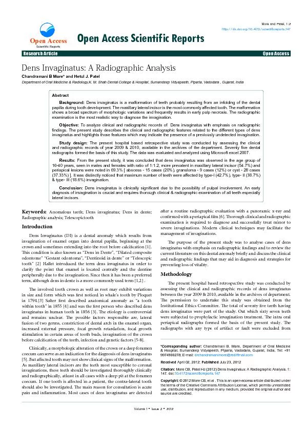
Open Access Anomalous teeth Dens invaginatus Dens in dente Dens Invaginatus DI is a dental anomaly which results from invagination of enamel organ into dental papilla beginning at the crown and ID: 336712

Open Access Anomalous teeth; Dens invaginatus;

Share:

Link:

Embed:

Download Presentation from below link

Download Pdf The PPT/PDF document "Dens Invaginatus a radiographic analysi..." is the property of its rightful owner. Permission is granted to download and print the materials on this web site for personal, non-commercial use only, and to display it on your personal computer provided you do not modify the materials and that you retain all copyright notices contained in the materials. By downloading content from our website, you accept the terms of this agreement.


Presentation Transcript

Open Access Research ArticleOpen AccessOpen Access Scientific Reports Anomalous teeth; Dens invaginatus; Dens in dente; Dens Invaginatus (DI) is a dental anomaly which results from invagination of enamel organ into dental papilla, beginning at the crown and sometimes extending into the root before calcication [1]. is condition is also known as “Dens in Dente”, “Dilated composite odontome” “Gestant odontoma”, “Dentinoid in dente” or “Telescopic tooth” [2] Hallet introduced the term dens invaginatus in order to clarify the point that enamel is located centrally and the dentine peripherally due to the invagination. Since then it has been a preferred preferred &#x/MCI; 29;&#x 000;&#x/MCI; 29;&#x 000;e involved tooth crown as well as root may exhibit variations in size and form which was rst noticed in whale’s tooth by Ploquet in 1794.[3] Salter rst described anatomical anomaly as “a tooth within tooth” in 1855 [4] and was the rst person who described dens invaginatus in human tooth in 1856 [3]. e etiology is controversial and remains unclear. e possible factors responsible are, lateral fusion of two germs, constriction of dental arch in the enamel organ, increased external pressure, focal growth retardation, focal growth stimulation in certain areas of tooth buds, invagination of the crown crown &#x/MCI; 39;&#x 000;&#x/MCI; 39;&#x 000;Clinically, a morphologic alteration of the crown or a deep foramen coecum can serve as an indication for the diagnosis of dens invaginatus [5]. But aected tooth may not show clinical signs of the malformation. As maxillary lateral incisors are the teeth most susceptible to coronal invaginations, these teeth should be investigated thoroughly clinically and radiographically, atleast in all cases with a deep pit at the foramen coecum. If one tooth is aected in a patient, the contra-lateral tooth should also be investigated. e main reason for consultation is acute pain and inammation. Most cases of dens invaginatus are detected aer a routine radiographic evaluation with a panoramic x-ray and conrmed with a periapical lm [6]. orough clinical and radiographic examination is required to diagnose and successfully treat minor to severe invaginations. Modern clinical techniques may facilitate the e purpose of the present study was to analyse cases of dens invaginatus with emphasis on radiographic ndings and to review the current literature on this dental anomaly briey and discuss the clinical and radiographic ndings that may aid in diagnosis and strategies for e present hospital based retrospective study was conducted by assessing the clinical and radiographic records of dens invaginatus between the year 2009 & 2010, available in the archives of department. e permission to undertake this study was obtained from the Institutional Ethics Committee. e total of seventy ve teeth having dens invaginatus were part of the study. Out which sixty seven teeth were subjected to prophylactic invagination treatment. e intra oral periapical radiographs formed the basis of the present study. e radiographs with any type of artifact or fault were excluded from *Corresponding author: Chandramani B. More, Department of Oral Medicine & Hospital, Sumandeep Vidyapeeth, Piparia, Vadodara, Gujarat, India, Tel: +91 PatelDens Invaginatus: A Radiographic Analysis 1: © 2012 , et al This is an open-access article distributed under the terms of the Creative Commons Attribution License, which permits unrestricted use, distribution, and reproduction in any medium, provided the original author and Dens invaginatus is a malformation of teeth probably resulting from an infolding of the dental papilla during tooth development. The maxillary lateral incisor is the most commonly affected tooth. The malformation shows a broad spectrum of morphologic variations and frequently results in early pulp necrosis. The radiographic To analyze clinical and radiographic records of Dens invaginatus with emphasis on radiographic �ndings. The present study describes the clinical and radiographic features related to the different types of dens Study design: The present hospital based retrospective study was conducted by assessing the clinical and radiographic records of year 2009 & 2010, available in the archives of the department. Seventy �ve dental radiographs formed the basis of this study. The data was evaluated and analyzed using Microsoft excel 2007.From the present study, it was concluded that dens invaginatus was observed in the age group of 16-40 years, seen in males and females with ratio of 1:1.2, more prevalent in maxillary lateral incisor (54.7%) and periapical lesions were noted in 69.3% [ abscess - 15 cases (20%), granuloma - 9 cases (12%) or cyst - 28 cases (37.33%) ]. It was distinctly noticed that maximum number of teeth were affected by type-I (42.7%), type- II (38.7%) Dens invaginatus is clinically signi�cant due to the possibility of pulpal involvement. An early diagnosis of invagination is crucial and requires thorough clinical & radiographic examination of all teeth especially Department of Oral Medicine & Radiology,K. M. Shah Dental College & Hospital, Sumandeep Vidyapeeth, Piparia, Vadodara , Gujarat, India Scientific Reports Citation: . 1: 147. doi:7Page 2 of 4 the study. All the radiographs were taken by using bisecting angle technique, were developed under standardized conditions and viewed on a standard illuminated screen by three oral radiologists to prevent inter observer bias. To determine the type of invagination, Ohlers’ system of classication for Dens invaginatus was used. e descriptive Data analysis was performed using Microso Oce Excel 2007. e study variables included age, gender, aected tooth, any associated In the present study we observed that, the patient’s aected with dens invaginatus belonged to the age group of 16-40 years with the mean age of 28 years (Graph 1). e male to female ratio was 1:1.2 (Graph 2). All the aected teeth were from permanent dentition. Out of total seventy ve teeth, maxillary teeth (88%) were most aected as compared to the mandibular teeth (12%). It was also observed that, the most common tooth aected by dens invaginatus is maxillary lateral incisors (54.7%), followed by maxillary central incisor (21.33%), maxillary canine (12%), mandibular lateral incisors (5.33%), mandibular canine (4%) and mandibular central incisor (2.67%) (Graph 3). It was also noted that periapical lesions were present in 52 cases (69.3%) & absent in 23 cases (30.7%) (Graph 4). e periapical lesions were abscess (20%), granuloma (12%) or cyst (37.33%) (Graph 5). According to Ohler’s system of classication, the type- I pattern was seen in 32 cases (42.7%), type- II in 29 cases (38.7%) & type- III in 14 Dens Invaginatus (DI) occur rarely in primary but frequently in permanent dentition and has a general prevalence of 0.04–10%. In our study all the seventy ve teeth belonged to permanent dentition. Literature study shows that, DI is commonly seen in maxillary teeth and involvement in mandibular teeth is rare [9]. is observation is consistent with the ndings of the present study. We noted that 88% of Maxillary lateral incisors are the teeth most susceptible to coronal 5 016-20 21-25 26-30 31-35 36-40 showing Age wise distribution. 5 0CENTRALLATERALCANINEMANDIBULARMAXILLARY Graph 3: Showing tooth involved. 0WITH WITHOUT Graph 4: Showing involvement of periapical lesions. 5 0PERIAPICALPERIAPICALGRANULOMAPERIAPICALCYST Graph 5: Showing type of Periapical lesion. MALEFEMALE Graph 2: Showing Gender wise distribution. Citation: . 1: 147. doi:7Page 3 of 4 invaginations [6]. Central incisors, premolars, canines and molars are in decreasing order of frequency. In our study it was observed that, the most common tooth aected by dens invaginatus is maxillary lateral incisors (54.7%), followed by maxillary central incisor (21.33%), maxillary canine (12%), mandibular lateral incisors (5.33%), mandibular canine (4%) and mandibular central incisor (2.67%). e reason behind this is thought to be the unfavorable position of maxillary lateral incisor during its formative stages and it is the last last &#x/MCI; 20; 00;&#x/MCI; 20; 00;Bilateral occurrence of DI is not uncommon and occurs in 43% of all cases with a high degree of inheritance [3]. But our study did not e gender predilection for DI i.e. male to female ratio is 1:1.7, with a slight female predilection [11]. Our study co-related this nding, wherein we observed that the male to female ratio was 1:1.2, with Dens invaginatus is clinically signicant due to the possibility of the pulp being aected. As pulpal involvement of teeth with coronal invaginations may occur shortly aer tooth eruption, an early diagnosis is mandatory to instigate preventive treatment. Clinical examination may reveal a deep ssure or pit on the surface of an anterior tooth. Due to the tortuous lingual anatomy, it is possible for caries to develop inside the invagination without any clinically detectable lesion. Since the enamel lining is thin and in close proximity to the pulp chamber, a carious lesion could easily perforate the pulp chamber. Further, there are sometimes thin canals within the enamel of the dens invaginatus, forming a direct communication with the pulp. Hence pulpitis and necrotic pulps are oen associated with this anomaly. e other reported sequelae of undiagnosed and untreated coronal invaginations are retention of neighboring teeth, displacement of teeth, cysts and internal resorption. In the present study we observed that, out of seventy ve cases, 52 cases (69.33%) had periapical lesions either abscesses - 15 cases (20%), granuloma - 9 cases (12%) or cyst - 28 cases Dens invaginatus is oen a surreptitious nding. Upon radiographic evidence of a dens invaginatus, the apical periodontium should be examined. If the radiographic appearance is unremarkable, pulp sensitivity testing should be performed. If the results suggest vital and unaected pulpal tissue, then the tooth should be promptly restored to curtail access of the dens invaginatus to the oral environment [5] e Ohlers’s system is the most popularly used classication for DI. According to this system, invaginations are classied into the following three types based on the depth of the invagination and the degree of communication with the periodontal ligament or the peri-radicular peri-radicular &#x/MCI; 24; 00;&#x/MCI; 24; 00;Type I: e invagination ends in a blind sac, limited to the dental Type II: e invagination extends to the cemento–enamel junction Type III: e invagination extends to the interior of the root, providing an opening to the periodontium, sometimes presenting presenting &#x/MCI; 25; 00;&#x/MCI; 25; 00;In this study, we distinctly observed that type I pattern was seen in 32 cases (42.7%), type II in 29 cases (38.7%) & type III in 14 cases Dens invaginatus are known to be associated with other abnormalities such as taurodontism, microdontia, talon cusp (Figure 2), gemination, supernumerary tooth, dentinogenesis imperfecta, short roots and with some medical–dental syndromes[12,14,15]. H. M. Worth has described two types of dens in dente: rst is a root dilation resembling an open umbrella and other one is eur-de-lys which resembles the French emblem [16]. e treatment of dens invaginatus ranges from preventive and restorative treatment procedures to non-Dens invaginatus is clinically signicant due to the possibility of pulpal involvement, pulpitis, necrotic pulps and chronic periapical lesions. An early diagnosis is crucial and requires thorough clinical Graph 6: Showing types of Dens Invagination. ABC Figure 1: A showing Type I Dens Invaginatus in lateral incisors. Note- periapical cyst. B Type II dens invaginatus in second premolar and C Type III dens A B C Figure 2: A. showing Type I Dens Invaginatus in lateral incisors. Note- Talons cusp. B. Type II dens invaginatus in lateral incisor and C. Type III dens invaginatus in mandibular central incisor. Note internal resorption. Citation: . 1: 147. doi:7Page 4 of 4 examination of all teeth especially lateral incisors. ese invaginations act as niche for bacterial growth and may jeopardize the status of main canal. An early detection and sealing of its opening with acid etch resin can eectively prevent these complications. e ndings stress the importance of a follow-up program for teeth in order to avoid serious peri-radicular complications that could inuence the outcome of the endodontic treatment. Because dens invaginatus is diagnosed as an incidental radiographic nding, radiographic examination is a valuable way of diagnosis in conjunction with clinical examination according to 1. Zengin AZ, Sumer AP, Celenk P (2009) Double dens invaginatus: report of 2. Sedano HO, Ocampo-Acosta F, Naranjo-Corona RI, Torres-Arellano ME (2009) Multiple dens invaginatus, mulberry molar and conical teeth. Case report and 3. Hülsmann M (1997) Dens invaginatus: aetiology, classi�cation, prevalence, 4. Silberman A, Cohenca N, Simon JH (2006) Anatomical redesign for the treatment of dens invaginatus type III with open apexes: a literature review and case presentation. J Am Dent Assoc 137: 180-185. 5. Mupparapu M, Singer SR (2004) A rare presentation of dens invaginatus in a mandibular lateral incisor occurring concurrently with bilateral maxillary dens invaginatus: case report and review of literature. Aust Dent J 49: 90-93. 6. Suprabha BS (2005) Premolarized double dens in dente in albinism--a case 7. Pandey SC, Pandey RK (2005) Radicular dens invaginatus--a case report. J 8. Regezi JA, Sciubba JJ. Oral pathology clinical pathologic correlations. In: 9. Carvalho-Sousa B, Almeida-Gomes F, Gominho LF, Albuquerque DS (2009) Endodontic treatment of a periradicular lesion on an invaginated type III mandibular lateral incisor. Indian J Dent Res 20: 243-245. 10. Tiku A, Nadkarni UM, Damle SG (2004) Management of two unusual cases of dens invaginatus and talon cusp associated with other dental anomalies. J 11. White SC, Pharoah MJ. Oral radiology principles and interpretation. In: Dental 12. Kerebel B, Kerebel LM, Daculsi G, Doury J (1983) Dentinogenesis imperfecta 13. Gupta R, Tewari S (2005) Nonsurgical management of two unusual cases of 14. de Lima MV, Bramante CM, Garcia RB, Moraes IG, Bernardineli N (2007) Endodontic treatment of dens in dente associated with a chronic periapical lesion using an apical plug of mineral trioxide aggregate. Quintessence Int 38: 15. Canger EM, Kayipmaz S, Celenk P (2009) Bilateral dens invaginatus in the 16. Worth HM, Principles and Practice of Oral radiologic interpretation. In: Odontomes and cyst. Yearbook Medical Publishers, 1975. Page 411-13. OMICS Journal of Radiology- Open Access using online manuscript submission, review and tracking systems of Editorial tracking system for quality and quick review processing. Submit your manuscript at http://www.omicsonline.org/submission/ OMICS Publishing Group 5716 Corsa Ave., Suite 110, Westlake, Los Angeles, CA 91362-7354, USA, Phone: +1- 650-268-9744, Fax: +1-650-618-1414, Toll free: +1-800-216-6499 http://omicsgroup.org/journals/roahome.php OMICS Journal of Radiology Abass Alavi Department of Radiol - ogy/Nuclear Medicine Philadelphia Donghoon Lee University of Washington, USA C L James CHOW Princess Margaret Hospital, Canada Rashid A. Fawwaz Columbia University USA Bensheng Qiu University of Washing - ton, USA Quek Swee Tian National University Hospital, Singapore Yutong Liu University of Ne - braska, USA Clark Zhe Wu University of California USA R Paulmurugan Stanford University USA Hundt Walter Philipp University Germany T Soldatos National and Capodes - trian University, Greece Kaan Orhan Near East University Cyprus Jie Zhang University of Kentucky USA Jiang Du University of Califor - nia, USA Xiaoliang Zhang University of Califor - nia, USA Sun Phillip Zhe Harvard Medical School, USA J Michael Mountz University of Pittsburgh, USA Kenji Suzuki University of Chicago USA Fangyu Peng UT Southwestern Medical Center, USA Zhao Dawen UT Southwestern Medical Center, USA Zerrin F. Yetkin UT Southwestern Medical Cente, USA Michael N. Patlas McMaster University Canada R Subramaniam Boston University USA Hao Huang University of Texas USA T Paraskevi Panteion University Greece Aruna Turaka Fox Chase Cancer Center, USA Jeon-Hor Chen China Medical University, Taiwan William T. Delfyett University of Pittsburgh USA Heng Li University of Texas USA T Bosemani Johns Hopkins University, USA Siva P Raman Johns Hopkins University, USA Richard Duszak Mid-South Imaging and Therapeutics, USA Harris L. Cohen University of Tennessee, USA R adiology is a medical specialty that employs the use of imaging to both diagnose and treat disease visualized within the human body. Radiologists use an array of imaging technologies such as x-ray radiography, ultrasound, computed tomography, nuclear medicine, positron emission tomography and magnetic resonance imaging to diagnose or treat diseases. Radiology is an Open Access, peer-reviewed journal which aims to provide the most rapid and reliable source of information on current developments in the �eld of Radiology. The emphasis will be on publishing quality papers quickly and freely available to researchers worldwide. ISSN: 2167-7964