PPT-Epidermal tissue system and its diagnostic features
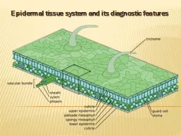
Epidermal tissue system Epidermis forms the outer most boundary of primary plant body Structures associated with the epidermis Stomata Cuticle Trichomeshairs Glands
Download Presentation
"Epidermal tissue system and its diagnostic features" is the property of its rightful owner. Permission is granted to download and print materials on this website for personal, non-commercial use only, provided you retain all copyright notices. By downloading content from our website, you accept the terms of this agreement.
Presentation Transcript
Transcript not available.