/
Urinary  S ystem Development Urinary  S ystem Development

Urinary S ystem Development - PowerPoint Presentation

patricia
patricia . @patricia
Follow
27 views
Uploaded On 2024-02-02

Urinary S ystem Development - PPT Presentation

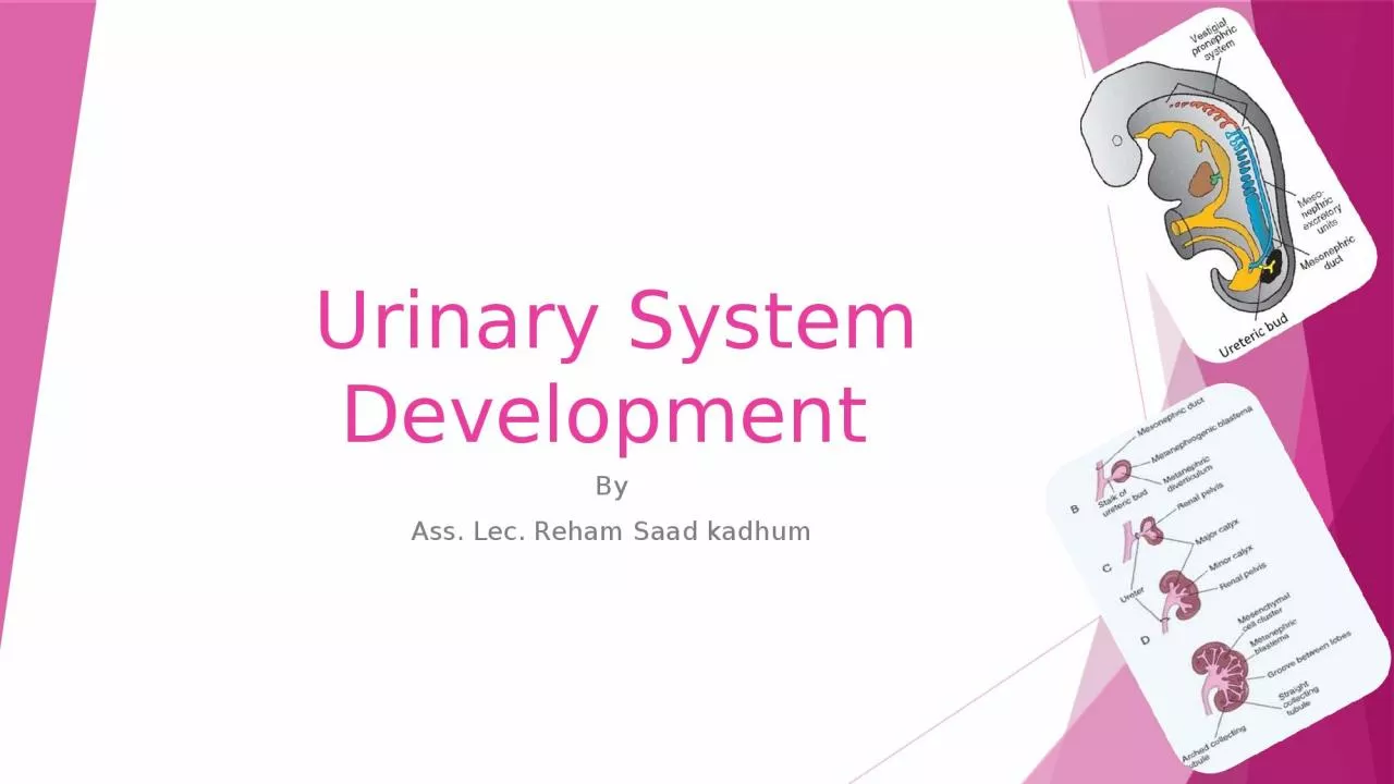
By Ass Lec Reham Saad kadhum Urogenital System Functionally the urogenital system can be divided into two entirely different compounds 1 The urinary system 2 The genital system ID: 1043878

system tubules urogenital form tubules system form urogenital bladder excretory kidney mesonephric collecting bud renal duct part ureteric sinus

Share:

Link:

Embed:

Download Presentation from below link

Download Presentation The PPT/PDF document "Urinary S ystem Development" is the property of its rightful owner. Permission is granted to download and print the materials on this web site for personal, non-commercial use only, and to display it on your personal computer provided you do not modify the materials and that you retain all copyright notices contained in the materials. By downloading content from our website, you accept the terms of this agreement.


Presentation Transcript

1. Urinary System Development By Ass. Lec. Reham Saad kadhum

2. Urogenital System Functionally the urogenital system can be divided into two entirely different compounds: 1. The urinary system. 2. The genital system. • (After the folding of the embryonic disc, the intermediate mesoderm forms a bulging on the posterior abdominal wall, called the nephrogenic cord/ urogenital ridge, it extend from the cervical to sacral region of the embryo) Both develop from a common mesodermal ridge or a urogenital ridge on either side of the aorta (along the posterior wall of the abdominal cavity).✔ Initially the excretory ducts of both system enter a common cavity, the cloaca.

3. Urinary System/Kidney System: There are three systems developing from the intermediate mesoderm, from anterior to posterior these systems are:

4. The Pronephros Pronephros is most anterior system, its excretory units (called the Nephrotomes 􀀀 At the beginning of the 4th week. 􀀀 The pronephros is represented by 7 to 10 solid cell groups in the cervical region. 􀀀 These groups form vestigial excretory units.

5. The Pronephros Nephrotomes, that regress before more caudal ones are formed. By the end of the 4th week, all indication of the pronephric system have disappears in a craniocaudally sequences at the cervical region.

6. The MesonephrosThe mesonephros & mesonephric ducts are derived from intermediate mesoderm from upper thoracic to upper lumbar (L3) segments Early in the fourth week of development, during regression of the pronephric system, the first excretory tubules of the Mesonephros appear.The excretory tubules (the Nephrotomes).lengthen to form S-shaped loop andacquire a tuft of capillaries that willform a glomerulus at their medial endfrom the dorsal aorta

7. The Mesonephros Around the glomerulus the tubules form Bowman’s capsule and together these structures constitute a renal corpuscle or called excretory unit. Laterally these tubule enters the longitudinal collecting duct known is called the mesonephric or wolffian duct that is connected caudally with the cloaca.

8. The MesonephrosIn the middle of the second month, the mesonephros forms a large ovoid organ on each side of the midline. Since the developing gonad is on its medial side, the ridge formed by both organs is known as the urogenital ridge. While caudal tubules are still differentiating, cranial tubules and glomeruli show degenerative changes. By the end of the 2nd month, the majority have disappeared.

9. The Mesonephros:In the male a few of the caudal tubules and mesonephric duct persist and participate in formation of the genital system, but they disappear in the femal.

10. MetanephrosThe definitivekidneyThe 3rd urinary organ, the metanephros, or permanent kidney, appears in the 5th week.• its excretory units develop from metanephric mesoderm in the same manner as in the mesonephric system.• The development of the duct system differs from that the other kidney system.

11. TheDefinitive KidneySystemThe kidneydevelops fromtwo sources:• The ureteric bud, whichgives rise to collectingsystem.• Metanephric mesoderm(metanephric blastema),which provides excretoryunits.

12. The Metanephros Its develops from an outgrowth of the caudal mesonephric duct, the ureteric bud at the fifth week , and from a condensation of nearby intermediate mesoderm ,the metanephric blastema Its excretory tubules (nephrotomes) form renal corpuscles in the same manner as the mesonephros; the collecting mesonephric duct is replaced by the ureteric bud.

13. The Metanephros The collecting duct of the metanephros develop from ureteric bud. The ureteric bud projects from the mesonephric duct (near its cloacal end); the bud penetrates the metanephros blastema and dilated to form the primitive renal pelvis, and splits into cranial and caudal portions, the future major calyces. Each calyx forms two new buds while penetrating the metanephric tissue .

14. Collecting system These buds continue to subdivided into 12 or more generation of tubules have formed. Meantime, at the periphery more tubules form until the end of the 5th month. The tubules of 2nd, 3rd, and 4th subdivisions fused together to form the minor calyces of the renal pelvis. 􀀀 Collecting tubules of the fifth and successive generation form the renal pyramid. The stalk of the ureteric bud becomes the ureter. The ureteric bud gives rise to the; ▪ ureter, ▪ renal pelvis, ▪ major and minor calyces, ▪ 1 to 3 million collecting tubules.

15. Execratory System Each newly formed collecting tubules is covered at distal end by metanephric tissue cap. 􀀀 The collecting tubules induce cells of the tissue cap form the renal vesicle that elongate to form the S-shaped excretory tubule. 􀀀 Capillaries grow into the pocket at one of the S and differentiate into glomeruli. 􀀀 This excretory tubule, together with their glomeruli, form excretory unites called the nephrons (including; the proximal and distal convoluted tubules and the loop of Henle) that are connecting distally with the adjacent collecting tubules. 􀀀 The proximal end of each nephron form Bowman’s capsules. 􀀀 Continuous lengthening of the execratory tubules result in formation of the proximal convoluted tubules, loop of Henle, and distal convoluted tubules.

16.

17.

18. Anatomy of Developing kidneys The excretory units of the nephrons are continuously developed till birth to be about 1 million in number.The postnatal growths in size of the nephrons contribute to the disappearance of the fetal lobulation of the kidneys.The kidney, initially in the pelvic region,Later shifts to a more cranial position in the abdomen.This ascent of the kidney is caused by diminution of the body curvature and growth of the body in the lumber and sacral regions to pass in between the arterial fork of the umbilical arteries.The kidneys are vascularized during their ascend by new branches from the aorta at higher level, the lower vascular branches degenerate.

19.

20. Clinical correlate Willm’s tumor; a cancer of the child kidney due to gene mutation.Congenital polycystic kidney; either an autosomal recessive or autosomal dominant types. It occurs due to obstruction in the urine path from the nephrons to the collecting tubules.

21. Clinical correlate Renal agenesis or dysplasia; occurs due failure of induction by the ureteric bud to the metanephros either unilateral or bilateral Duplication of ureter; due to early splitting of the ureteric bud. Ectopic ureter opening; rarely one of the ureters open into the vagina, urethra, or the vestibules.

22. Pelvic kidneys; occurring due to failure of the ascending kidney.• Horseshoe kidney; occurring by fusion of the lower poles of the kidneys as the poles are pushed close together during the passage of the kidneys through the fork of the umbilical arteries. The ascending horseshoe kidney stops at the root of the inferior mesenteric artery (IMA) at L3. the ureters pass anterior to the isthmus of this kidney.• Accessory renal arteries; occurring due to failure of degeneration of the embryonic arteries during ascend of the kidneys.

23. The Urinary bladder and UrethraDuring the 3-7 weeks of development the cloaca divides into:1. Urogenital sinus anteriorly.2. Primitive anal canal posteriorly. ✔ The mesodermal layer between primitive anal canal & urogenital sinus is called urorectal septum.

24. The Urinary bladder and UrethraThe urogenital sinus consist ofthree parts:A. The urinary bladder( the upper portion and largest part).B. The pelvic part of the urogenital sinus (narrow canal).C. The phallic part of the urogenital sinus (flattened).

25. Urogenital sinusUrinary bladder The large upper part and is connected with the allantois (allantois is obliterated later on forming fibrous cord between the bladder and the umbilicus called the urachus or the median umbilical ligament).The mesonephric ducts are opened into this part of the cloaca, the caudal part of the mesonephric duct is absorbed in the wall of the bladder to form the trigone of the bladder.

26. Urinary Bladder As a result of this change, the uretericbuds originating from the mesonephricducts became connected with thetrigone of the bladder. Then after, theopenings of the mesonephric ductsmove together and become connectedto the prostatic urethra forming themale ejaculatory ducts.The trigone acquires a transient mesodermal lining from the mesonephric tissue which is replaced later on by the endodermal lining, the same of the whole bladder.

27. B. The pelvic part of the urogenital sinus Is a narrow canal, that will form the prostaticand membranous urethra in the male.•The lining of the urethra is endodermal origin,while the connective tissue and musclessurrounding it are derived from its surroundingsplanchnic mesoderm.• At the end of the 3rd month theprostatic urethra shows manybudding forming the male prostate.• In female, budding from the cranial partof the urethra forms the urethral andparaurethral glands.

28. C. The last part is the phallic part of the urogenital sinus.• It is flattened from side to side , Note:The tip of the urorectal septum will form the perineal body.

29. Clinical correlatesBladder defectsUrachal fistula: it occurs due to failure of the obliteration of the urachus. The urine will out flow from the umbilicus. Partial failure will results in urachal sinus or urachal cyst.

30. Clinical correlatesBladder defects2. Exstrophy of the bladder Is a ventral body wall defect in which the bladder mucosa is exposed outside.This anomaly is rare, occurring in 2/100,000 live births.

31.