/
Dermatophytosis Dermatophytosis

Dermatophytosis - PDF document

payton
payton . @payton
Follow
342 views
Uploaded On 2022-08-16

Dermatophytosis - PPT Presentation

Center for Food Security and Public Health 2011 1 S l i d e 1 S l i d e 2 
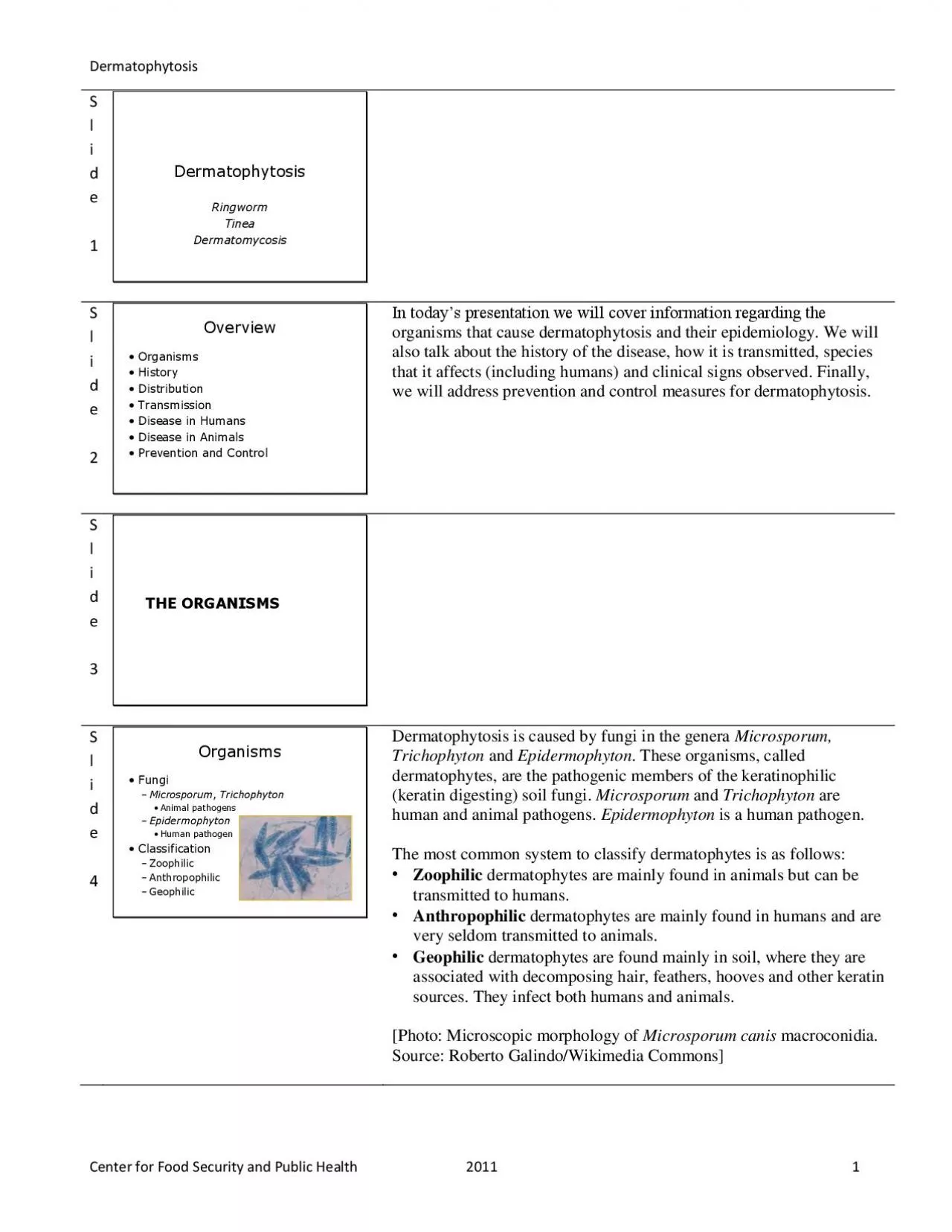
In todays presentation we will cover information regarding the organisms that cause dermatophytosis and their epidemiology ID: 937286

dermatophytes lesions common tinea lesions dermatophytes tinea common dermatophytosis skin animals mentagrophytes public health dermatophyte areas infections photo canis

Share:

Link:

Embed:

Download Presentation from below link

Download Pdf The PPT/PDF document "Dermatophytosis" is the property of its rightful owner. Permission is granted to download and print the materials on this web site for personal, non-commercial use only, and to display it on your personal computer provided you do not modify the materials and that you retain all copyright notices contained in the materials. By downloading content from our website, you accept the terms of this agreement.


Presentation Transcript

Dermatophytosis Center for Food Security and Public Health 2011 1 S l i d e 1 S l i d e 2 In today’s presentation we will cover information regarding the organisms that cause dermatophytosis and their epidemiology. We will also talk about the history of the disease, how it is transmitted, species that it affects (including humans) and clinical signs observed. Finally, we will address prevention and control measures for dermatophytosis. S l i d e 3 S l i d e 4 Dermatophytosis is caused by fungi in the genera Microsporum, Trichophyton and Epidermophyton . These organisms, called dermatophytes, are the pathogenic members of the keratinophilic (keratin digesting) soil fungi. Microsporum and Trichophyton are human and animal pathogens. Epidermophyton is a human pathogen. The most common system to classify d ermatophytes is as follows: • Zoophilic dermatophytes are mainly found in animals but can be transmitted to humans. • Anthropophilic dermatophytes are mainly found in humans and are very seldom transmitted to animals. • Geophilic dermatophytes are found mainly in soil, where they are associated with decomposing hair, feathers, hooves and other keratin sources. They infect both humans and animals. [Photo: Microscopic morphology of Microsporum canis macroconidia . Source: Roberto Galindo/Wikimedia Commons] Dermatophytosis Center for Food Security and Public Health 2011 2 S l i d e 5 Zoophilic species (found in animals) include: • Microsporum canis • M. gallinae • M. gypseum • M. equinum • M. nanum • M. persicolor • Trichophyton equinum • T. mentagrophytes Several varieties of T. mentagrophytes exist. Some are important pathogens in both animals and humans; others are mainly human pathogens. They include:

• T. simii • T. verrucosum S l i d e 6 S l i d e 7 Dermatophytes have likely existed for millions of years; however, the first historical reference did not come unt o 30 A.D., when Roman encyclopedist Aulus Cornelius Celsus described a “suppurative infection of the scalp” that was attributed to a dermatophyte. In that era, dermatophytes were described as “tineas.” The term “ringworm” emerged later, probably around the 16 th century. In the 19 th century, the mycotic etiology of these skin infections was finally described. Dermatophytes remain the most common fungal pathogens (except finger onychomycosis due to Candida) in the U.S. Sources: Libero A. Natural history of t he dermatophytes and related fungi . Mycopathologia. 1974;53(1 - 4): 93 - 110. Seebacher C., Bouchara J.P., Mignon B. Updates on the epidemiology of dermatophyte infections. Mycopathologia. 2008;166(5 - 6): 335 - 352. Dermatophytosis Center for Food Security and Public Health 2011 3 S l i d e 8 S l i d e 9 Dermatophytes grow best in warm and humid en vironments and are, therefore, more common in tropical and subtropical regions. The geographic distribution varies with the organism. M. canis , M. nanum , T. mentagrophytes , T. verrucosum and T. equinum occur worldwide. T. simii (found in monkeys) occurs only in Asia, and T. mentagrophytes var. erinacei is limited to France, Great Britain, Italy and New Zealand. Photo: Tropical and subtropical regions of the world shown in dark green and medium green colors. [Photo: Tropical and subtropical desert climates. Source: Encyclopedia Britannica Online.] S l i d e 1 0 S l i d e 1 1 Infection occurs by contact with arthrospores (asexual spores formed in the hyphae of the parasitic stage) or conidia (sexual or asexual spores formed in the “f

ree living” environment al stage). [Photo: macroconidia of Microsporum canis . J. Michael Miller/CDC via American Society for Microbiology at http://www.asm.org/division/c/fungi.htm ] Dermatophytosis Center for Food Security and Public Health 2011 4 S l i d e 1 2 Infection usually begins in a growing hair or the stratum corneum of the skin. Dermatophytes do not generally invade resting hairs, since the essential nutrients they need for growth are absent or limited. Hyphae spread in the hairs and keratinized skin, eventually developing infectious arthrospores. Transmission between hosts usually occurs by dire ct contact with a symptomatic or asymptomatic host, or direct or airborne contact with its hairs or skin scales. Infective spores in hair and dermal scales can remain viable for several months to years in the environment. Fomites such as brushes and clippe rs can be important in transmission. Geophilic dermatophytes are usually acquired directly from the soil rather than from another host. S l i d e 1 3 S l i d e 1 4 Dermatophytes generally grow only in keratinized tissues such as hair, nails and the outer layer of skin; the fungus usually stops spreading where it contacts living cells or areas of inflammation. Mucus membranes are not affected. The clinical signs may v ary, depending on the region affected. In humans, pruritus is the most common symptom. The skin lesions are usually characterized by inflammation that is most severe at the edges, with erythema, scaling and occasionally blister formation. Central clearing is sometimes seen, particularly in tinea corporis; this results in the formation of a classic “ringworm” lesion. On the scalp and facial hair, there may be hair loss. In humans, dermatophytoses are referred to as “tinea” infections, and are named with refe rence to the area of the body i

nvolved. S l i d e 1 5 Although dermatophyte infections are known to be common, their prevalence is unknown as this disease is not notifiable and many infections are treated with over - the - counter drugs. Infections are more common in chil dren than adults. Most dermatophyte infections are not serious in healthy persons; however, opportunistic bacteria can cause cellulitis in skin damaged by interdigital fungal infections. These infections are a particular concern in diabetics. Dermatophytos is is more serious in those who are immunosuppressed. These individuals may have atypical and locally aggressive dermatophyte infections, including extensive skin disease, subcutaneous abscesses, and disseminated disease. Dermatophytosis Center for Food Security and Public Health 2011 5 S l i d e 1 6 Tinea capitis, most often seen in children, is a dermatophyte infection of the hair and scalp. Tinea capitis begins with a small papule, which spreads to form scaly, irregular or well - demarcated areas of alopecia. The cervical and occipital lymph nodes may be enlarged. A kerion, a boggy, inflammatory mass, may also be seen; this reaction is usually followed by healing. Suppurative lesions are often seen when the infection is caused by zoophilic dermatophytes. Both anthropophilic and zoophilic dermatophytes can cause tinea capitis. In the U .S., it is most often caused by the anthropophilic dermatophyte T. tonsurans . Most common agents*: M. audouinii , M. canis . Other agents: M. ferrugineum , M. gypseum , M. nanum , M. persicolor , T. megninii , T. mentagrophytes , T. schoenleinii , T. soudanense , T. verrucosum , T. violaceum . [Photo: An individual with “ringworm”, or tinea capitis of the scalp caused by Microsporum gypseum . Source: CDC Public Health Image Library] S l i d e 1 7 Tinea corporis, or ringwor

m, occurs on the trunk, extremities and face. It is chara cterized by single or multiple scaly annular lesions with a slightly elevated, scaly and or erythematous edge, sharp margin and central clearing. Follicular papules, pustules or vesicles may be found on the borders of the lesion. Lesions may be variably pr uritic. Both zoophilic and anthropophilic dermatophytes are common in children, and on the neck and wrists of adults in contact with the child. In other adults, tinea corporis is often the result of chronic infection with T. rubrum , an anthropophilic derma tophyte. In many people, untreated tinea corporis resolves within a few months, particularly if it is caused by a zoophilic or geophilic organism. Most common agents*: T. rubrum , M. canis , M. tonsurans , T. verrucosum . Other agents: E. floccosum , M. audouin ii , M. gypseum , M. nanum , M. persicolor , T. equinum , T. mentagrophytes , T. raubitschekii , T. schoenleinii , T. violaceum . [Photo: A ringworm lesion on the arm, or tinea corporis, due to Trichophyton mentagrophytes . Source: CDC Public Health Image Library] S l i d e 1 8 Tinea barbae is an infection of the hairs and skin in the beard and mustache area, and is usually seen in men. The lesions may include scaling, follicular pustules and erythema. Tinea barbae can be caused by zoophilic or anthropophilic dermatophytes. Far m workers are often affected. Most common agents*: T. verrucosum . Other agents: M. canis , T. megninii , T. mentagrophytes , T. rubrum , T. violaceum . [Photo: Ringworm on the bearded areas of face and neck, known as “tinea barbae”, or “barber’s itch”. Source : CDC Public Health Image Library] Dermatophytosis Center for Food Security and Public Health 2011 6 S l i d e 1 9 Tinea faciei is seen on the non - bearded parts of the face. The lesions are u

sually pruritic; itching and burning may become worse after exposure to sunlight. Some lesions may resemble those of tinea corporis; others may have little or no scaling or raised edges. In some cases, the areas of erythema are indistinct. Due to the atypical presentation, tinea faciei is often confused with other skin diseases that affect the face. Most common agents*: T. tonsurans in North Ameri ca; T. mentagrophytes and T. rubrum in Asia. [Photo: A child with a ringworm (tinea) fungal infection on the left side of his face and left ear. Source: CDC Public Health Image Library] S l i d e 2 0 Tinea cruris is an infection of the groin, usually caused by anthro pophilic dermatophytes. The symptoms include burning and pruritus. Pustules and vesicles at the active edge of the infected area, along with maceration, are found in a background of red, scaling lesions with raised borders. Most common agents*: E. floccosu m , T. rubrum . Other agents: M. nanum , T. mentagrophytes , T. raubitschekii . S l i d e 2 1 Tinea pedis (Athlete’ s foot) is an infection of the foot, characterized by fissures, scales and maceration in the toe web, or scaling of the soles and lateral surfaces of the feet. Erythema, vesicles, pustules and bullae may also be present. It is usually caused by anthropophi lic dermatophytes. Most common agents*: T. rubrum , T. mentagrophytes var interdigitale , E. floccosum . Other agents: M. persicolor , T. raubitschekii , T. violaceum . [Photo: A patient with ringworm of the foot (tinea pedis) due to the dermatophytic fungus, Trichophyton rubrum . Source: CDC Public Health Image Library] S l i d e 2 2 Tinea manuum is a dermatophyte infection of one or, occasionally, both hands. In this form, the palms become diffusely dry, scaly and erythematous. It is most often caused by anthropophilic dermato

phytes (cases may be an extension of Athlete’s foot) but is occasionally caused by zoophilic organisms. Most common agent*: T. rubrum . Other agents: E. floccosum , M. canis , M. gypseum , T. mentagrophytes , T. verrucosum . Dermatophytosis Center for Food Security and Public Health 2011 7 S l i d e 2 3 Tinea unguium is a dermatophyte infection of the nail. It is characterized by thickened, discol ored, broken and dystrophic nails. The nail plate may be separated from the nail bed. It can be caused by anthropophilic or zoophilic dermatophytes. Most common agents*: T. rubrum , T. mentagrophytes var mentagrophytes . Other agents: E. floccosum , T. tonsurans , T. violaceum . [Photo: Onychomycosis due to Trichophyton rubrum , right and left great toe. Source: CDC Public Health Image Library] S l i d e 2 4 S l i d e 2 5 All domestic animals are susceptible to dermatophytes. The most common fungi vary with the host. • Dogs and cats: M. canis is the most common species, particularly in cats. M. gypseum and T. mentagrophytes are found occasionally. Other species are rare. • Cattle: T. verrucosum is the most important species. Species found occasionally include T. mentagrophytes , T. equinum , M. gypseum , M. nanum and M. canis . • Sheep and goats: T. verrucosum is the most common species but M. canis outbreaks have also been reported. • Horses: T. equinum and M. equinum are the most important species. M. gypseum , M. canis and T. verrucosum are seen occasionally. • Swine: M. nanum is the most important agent. This dermatophyte is rarely zoonotic. • Rodents: Varieties of T. mentagrophytes are common in rodents. Microsporum species, including M. persicolor , are seen occasionally . • Rabbits: T. mentagrophytes is the most important spec

ies. • Birds: T. gallinae is the usual agent in birds, including poultry, canaries and pigeons. This dermatophyte is rarely zoonotic. M. gypseum and T. simii infections are seen occasionally. Dermatophytosis Center for Food Security and Public Health 2011 8 S l i d e 2 6 Dermatophytes usually grow only in keratinized tissues such as hair, nails and the outer layer of skin; the fungus usually stops spreading where it contacts living cells or areas of inflammation. Mucus membranes are not affected. Dermatophyte lesions are characterized by areas of alopecia, scaling, crusts, erythema and pruritus, present to varying degrees. Occasionally, the dermatophytes die at the center of a lesion and that area resolves, leaving a circular “rin gworm” lesion. In animals, this pattern is relatively uncommon. Hairs in the affected area are usually brittle and break near the skin surface, often giving the lesion a “shaved” appearance; truncated hair shafts may be seen through the scales and crusts. S l i d e 2 7 Among small animals, the prevalence rates reported in various studies vary widely. In general, dermatophytes are thought to be carried asymptomatically more often by cats than dogs. Infection rates between 6% and 88% have been reported in felines. Others feel that subclinical dermatophyte infections are very common, particularly in cats. Among livestock, dermatophytoses are particularly common in cold climates where animals are stabled for long periods of time. Whether an animal becomes infected, after con tact with a dermatophyte, may depend on the animal’s age, the condition of its exposed skin, and grooming behavior. Young animals are more likely to have symptomatic infections. Dermatophytosis is also more common when animals are immunosuppressed, have po or nutrition or are kept in high density populations. Most i

nfections in healthy animals heal spontaneously within one to a few months. Infections can be more persistent or widespread in young or sick animals, and in some longhaired cats. S l i d e 2 8 In dogs, dermato phytosis is seen most often in puppies. It is uncommon in adult dogs unless they are immunosuppressed. The lesions may appear on any part of the body and usually consist of small circular areas of alopecia; the hairs are typically broken at the base, givin g the appearance of the area having been shaved. The center of the lesion usually contains pale skin scales, giving it a powdery appearance, and the edges are generally erythematous. In later stages, the lesion is often covered by a crust and the edges are swollen. Onychomycosis can occur concurrently. Dermatophytosis is usually self - limiting in dogs. [Photo: (Top) A dog with a ringworm lesion on its muzzle. Source: Clearly Lake Veterinary Hospital; (Bottom) A dog with multiple areas of alopecia due to de rmatophytosis. Source: University of Tennessee College of Veterinary Medicine ( www.vet.utk.edu )] Dermatophytosis Center for Food Security and Public Health 2011 9 S l i d e 2 9 Many infected cats have few or no lesions. Longhaired adult cats, in particular, can be subclinical carriers; in some cases, the cat may have minimal lesions consisting of patchy areas of short stubble, alopecia, scaly patches or erythematous plaques, visible only on close inspection. Symptomatic cases tend to be seen in kittens, with the early lesions found on the face, ears and paws. Generally, the lesions co nsist of areas of focal alopecia, with scaling and crusting containing only a few broken hairs. The cat’s grooming behavior can spread the infection to the entire body. Onychomycosis may also be seen concurrently; the nails may be opaque, with whitish mott ling, and shredding of th

e nail surface. The lesions are usually self - limiting within a few weeks to a few months in short - haired cats but may persist, either symptomatically or asymptomatically, in long - haired cats. [Photo: A kitten with alopecia (hair loss) on its ears due to dermatophytosis. Source: Dr. James Noxon, Iowa State University/CFSPH] S l i d e 3 0 In cattle, the severity of disease varies, from small focal 1 cm lesions to extensive generalized skin involvement. Most often, the disease appears as nonpruritic periocular lesions in calves. Cows and heifers may have lesions more often on the chest and l imbs, and bulls on the dewlap and intermaxillary skin. The initial lesions are discrete, grayish - white, crusty dry areas with a few brittle hairs. Some areas may become suppurative and thickly crusted. Lesions resembling light brown scabs may also be seen; when these scabs fall off, they leave an area of alopecia. The lesions usually resolve spontaneously in 2 to 4 months. [Photo: Multiple raised pale tan crusted lesions due to dermatophytosis on the skin of a cow. Source: Armed Forces Institute of Pathol ogy/CFSPH] S l i d e 3 1 In horses, most dermatophyte lesions are found in areas of contact with saddles or other tack. T. equinum lesions are usually pruritic, with exudative lesions and areas of hairless, thickened skin. M. equinum lesions are usually less severe an d consist of small scaly areas with brittle hairs. Early dermatophyte lesions may resemble papular urticaria. Dermatophytosis Center for Food Security and Public Health 2011 10 S l i d e 3 2 Dermatophytosis is common in show lambs, but uncommon in production flocks. The most noticeable lesions are usually circular, alopecic areas with thick scabs on the head and face; however, widespread lesions may be found under the wool when animals are sheare

d. In healthy lambs, the lesions are usually self - limiting. [Photo: A sheep with ringworm (or club lamb fungus). Source: Virginia Cooperativ e Extension] S l i d e 3 3 Pigs develop a wrinkled lesion covered by a thin, brown, easily removed scab, or a spreading ring of inflammation. Dermatophyte infections are often asymptomatic in adult swine. [Photo: A sow with multiple ringworm lesions. Source: American Association of Swine Veterinarians] S l i d e 3 4 Most rodents infected with T. mentagrophytes are asymptomatic or have few clinical signs. In mice, partial or complete areas of alopecia, erythema, scales, and scabs may be seen, often on the tail. In rats, the lesions are usually found on the back. Guinea pigs usually develop pruritic, ovoid, hairless, raised areas, with crusts or scales; these lesions first appear on the face then spread to the back and lim bs. In rabbits, dermatophytosis most often occurs in young, newly weaned animals. Focal alopecia, with erythema, crusts and scabs, is seen around the eyes, nose and ears, with secondary lesions appearing on the feet. The disease is usually self - limiting. S l i d e 3 5 In birds, there may be alopecia, particularly on the face and neck, scaling, auto - mutilation and feather plucking. Some lesions may be ring - shaped or pruritic. Dermatophytosis Center for Food Security and Public Health 2011 11 S l i d e 3 6 Post - mortem lesions are identical to those in live animals; dermatophytes are restricted to hai r, nails and superficial skin. S l i d e 3 7 S l i d e 3 8 A Wood’s lamp examination can detect fluorescence in some dermatophytes, including some strains of the zoophilic organisms M. canis and M. equinum and some anthropophilic dermatophytes such as M. audouinii . Potassium hydroxi de (KOH) microscopy can detect hyp

hae and conidia in skin scrapings or hair. Fungal cultures are necessary for the identification of the organism. Skin or nail biopsies are also used in humans. S l i d e 3 9 Treatment of dermatophytosis may include topical antifungal c reams or shampoos, and/or systemic antifungals. In humans, tinea capitis, tinea barbae and tinea faciei are generally treated with systemic antifungals. Tinea corporis can usually be treated with nonprescription antifungals. Animals often have self - limitin g infections that resolve within a few months, but treatment can speed recovery, decrease the spread of lesions on the animal, and decrease the risk of transmission. Onychomycosis can be very difficult to cure; long term treatment or surgical declawing may be necessary. Dermatophytosis Center for Food Security and Public Health 2011 12 S l i d e 4 0 Control of the disease in animals can prevent some cases of dermatophytosis in humans. Infected animals should be treated and the premises and fomites should be disinfected. New animals should be cultured when introduced. Gloves and protec tive clothing should be used during contact with infected animals. Such contact should be avoided as much as possible. Vaccines are available in some countries for T. verrucosum in cattle, T. equinum in horses, and M. canis in cats (does not eliminate the fungus). S l i d e 4 1 Dermatophyte spores are susceptible to common disinfectants such as benzalkonium chloride, dilute (1:10) chlorine bleach, or strong detergents. Chlorhexidine is no longer considered to be a good environmental decontaminant for these fungi. The mechanical removal of any material containing keratin, such as shed skin and hairs, facilitates disinfection. Vacuuming is considered to be the best method in many cases. S l i d e 4 2 S l i d e 4 3 Last updated: May

Related Contents


Next Show more