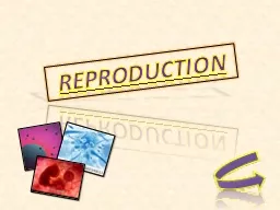
/
Figure 26.0-1 Why Reproduction Matters Figure 26.0-1 Why Reproduction Matters

Figure 26.0-1 Why Reproduction Matters - PowerPoint Presentation

phoebe-click
phoebe-click . @phoebe-click
Follow
343 views
Uploaded On 2018-10-27

Figure 26.0-1 Why Reproduction Matters - PPT Presentation

Figure 2601a Figure 2601b Figure 2601ba Figure 2601bb Figure 2601c Figure 2602 Chapter Thread HighTech Babies LM Figure 261 LM Figure 262 Figure 263 Eggs Two offspring ID: 698109

meiosis figure oocyte follicle figure meiosis follicle oocyte sperm secondary diploid cells cell uterus primary ovulation embryo estrogen days

Share:

Link:

Embed:

Download Presentation from below link

Download Presentation The PPT/PDF document "Figure 26.0-1 Why Reproduction Matters" is the property of its rightful owner. Permission is granted to download and print the materials on this web site for personal, non-commercial use only, and to display it on your personal computer provided you do not modify the materials and that you retain all copyright notices contained in the materials. By downloading content from our website, you accept the terms of this agreement.


Presentation Transcript

Slide1

Figure 26.0-1

Why Reproduction MattersSlide2

Figure 26.0-1aSlide3

Figure 26.0-1bSlide4

Figure 26.0-1baSlide5

Figure 26.0-1bbSlide6

Figure 26.0-1cSlide7

Figure 26.0-2

Chapter

Thread:

High-Tech

Babies

LMSlide8

Figure 26.1

LMSlide9

Figure 26.2Slide10

Figure 26.3

Eggs

Two

offspring

arising from

a

sexual

r

eproduction

by

fissionSlide11

Figure 26.4

EggsSlide12

Figure 26.5

Rectum

Vas deferens

Epididymis

Testis

Urinary bladder

Pubic bone

Erectile

tissue

Penis

Testicle

Glans

Prostate gland

Vas deferens

Epididymis

Seminal

v

esicle

(behind

bladder

)

Erectile

tissue

of

penis

Urethra

Glans of penis

Scrotum

Testis

Testicle

Urinary

bladder

Side view

Front

view

Urethra

Prepuce

Scrotum

Seminal

vesicle

Vas

deferens

Sperm

duct

Prostate

gland

AnusSlide13

Figure 26.5-1

Side

view

Prostate

gland

Rectum

Seminal

vesicle

Vas

deferens

Sperm

duct

Anus

Vas deferens

Epididymis

Testis

Urinary bladder

Pubic bone

Erectile

tissue

Urethra

Penis

Testicle

Glans

Prepuce

ScrotumSlide14

Figure 26.5-2

Prostate gland

Vas deferens

Epididymis

Seminal

v

esicle

(behind

bladder

)

Erectile

tissue

of

penis

Urethra

Glans of penis

Scrotum

Testis

Testicle

Urinary

bladder

Front

viewSlide15

Figure 26.6

Side view

Vaginal opening

Rectum

Cervix

Vagina

Anus

Pubic bone

Urethra

Shaft

Glans

Prepuce

Clitoris

Vulva

Labia

minora

Labia

majora

Front

view

Oviduct

Ovary

Uterus

Urinary bladder

Ovaries

Oviduct

Follicles

Corpus luteum

Wall of uterus

Vagina

Uterus

Endometrium

(lining of uterus

)

Cervix

(“neck” of uterus

)Slide16

Figure 26.6-1

Side view

Rectum

Cervix

Oviduct

Ovary

Uterus

Urinary bladder

Pubic bone

Urethra

Shaft

Glans

Prepuce

Clitoris

Vulva

Vagina

Anus

Vaginal opening

Labia minora

Labia majoraSlide17

Figure 26.6-2

Ovaries

Oviduct

Follicles

Corpus luteum

Wall of uterus

Vagina

Uterus

Front

view

Endometrium

(lining of uterus

)

Cervix

(“

neck” of uterus)Slide18

Figure 26.7

Epididymis

Cell division continuously

produces new cells

2

n

Testis

Scrotum

Penis

Diploid cell

Differentiation and

onset of meiosis

I

2

n

Primary spermatocyte

Meiosis

I

completed

(diploid)

Testis

Seminiferous

tubule

Cross

section of

seminiferous

tubule

n

n

Secondary spermatocyte

(haploid)

Meiosis

II

n

n

n

n

Developing sperm cells

Cellular differentiation

(cells become specialized)

n

Sperm cells

(haploid)

Mature sperm released into

center of seminiferous tubule

n

n

nSlide19

Figure 26.7-1

Epididymis

Testis

Scrotum

Penis

Testis

Seminiferous

tubule

Cross

section of

seminiferous

tubuleSlide20

Figure 26.7-2-s1

2

n

Diploid cellSlide21

Figure 26.7-2-s2

2

n

Diploid cell

Differentiation and

onset of meiosis

I

2

n

Primary spermatocyte

(diploid)Slide22

Figure 26.7-2-s3

2

n

Diploid cell

Differentiation and

onset of meiosis

I

2

n

Primary spermatocyte

(diploid)

Meiosis

I

completed

Secondary spermatocyte

(haploid)

n

nSlide23

Figure 26.7-2-s4

Diploid cell

Differentiation and

onset of meiosis

I

Primary spermatocyte

(diploid)

Meiosis

I

completed

Secondary spermatocyte

(haploid)

Meiosis

II

Developing sperm cells

2

n

2

n

n

n

n

n

n

nSlide24

Figure 26.7-2-s5

Diploid cell

Differentiation and

onset of meiosis

I

Primary spermatocyte

(diploid)

Meiosis

I

completed

Secondary spermatocyte

(haploid)

Meiosis

II

Developing sperm cells

Cellular differentiation

(cells become specialized)

Sperm cells

(haploid)

Mature sperm released into

center of seminiferous tubule

2

n

n

n

n

n

n

n

n

n

n

n

2

nSlide25

Figure 26.8

Primary

oocyte

within

follicle

Before birth

2

n

Differentiation

and onset of

meiosis

I

2

n

Primary oocyte

(diploid)

n

First

polar

body

n

Entry of sperm

triggers

completion

of meiosis

II

Secondary oocyte

(haploid)

n

Second

polar body

Mature egg (ovum)

(haploid)

Diploid cell

in embryo

Growing

follicle

Completion of

meiosis

I

and

onset of

meiosis

II

Mature follicle

Ruptured

follicle

Secondary

oocyte

Ovary

A secondary oocyte being

released during ovulation

n

Corpus

luteum

LM

Ovulated

secondary oocyte

arrested at prophase

of meiosis

I

; present

at birth

arrested at metaphase

of meiosis

IISlide26

Figure 26.8-1

Primary

oocyte

within

follicleSlide27

Figure 26.8-2-s1

Before birth

2

n

Differentiation

and onset of

meiosis

I

2

n

Primary oocyte

(diploid)

Diploid cell

in embryo

Primary

oocyte

within

follicle

arrested at prophase

of meiosis

I

; present

at birthSlide28

Figure 26.8-2-s2

Before birth

2

n

Differentiation

and onset of

meiosis

I

2

n

Primary oocyte

(diploid)

Diploid cell

in embryo

Primary

oocyte

within

follicle

arrested at prophase

of meiosis

I

; present

at birth

n

n

Secondary oocyte

(haploid)

arrested at metaphase

of meiosis

II

Growing

follicle

Completion of

meiosis

I

and

onset of

meiosis

II

Mature

follicle

First

polar

bodySlide29

Figure 26.8-2-s3

Before birth

2

n

Differentiation

and onset of

meiosis

I

2

n

Primary oocyte

(diploid)

Diploid cell

in embryo

Primary

oocyte

within

follicle

arrested at prophase

of meiosis

I

; present

at birth

n

n

Secondary oocyte

(haploid)

arrested at metaphase

of meiosis

II

Growing

follicle

Completion of

meiosis

I

and

onset of

meiosis

II

Mature

follicle

First

polar

body

Ruptured

follicle

Ovulated

secondary

oocyteSlide30

Figure 26.8-2-s4

Before birth

2

n

Differentiation

and onset of

meiosis

I

2

n

Primary oocyte

(diploid)

Diploid cell

in embryo

Primary

oocyte

within

follicle

arrested at prophase

of meiosis

I

; present

at birth

n

n

Secondary oocyte

(haploid)

arrested at metaphase

of meiosis

II

Growing

follicle

Completion of

meiosis

I

and

onset of

meiosis

II

Mature

follicle

First

polar

body

Ruptured

follicle

Ovulated

secondary

oocyte

Entry of sperm

triggers

completion

of meiosis

II

n

Second

polar body

Mature egg (ovum)

(haploid)

nSlide31

Figure 26.8-3

Secondary

oocyte

Ruptured

follicle

Ovary

A secondary oocyte being

released during ovulation

Corpus luteum

LM

Ovulated

secondary oocyteSlide32

Figure 26.8-4

Secondary

oocyte

Ovary

A secondary oocyte being

released during ovulation

LMSlide33

Figure 26.9

Growing

follicle

Mature

follicle

Ovulation

Corpus

luteum

Degenerating

corpus

luteum

Progesterone and

estrogen secreted

by remnant of follicle

20

25

28

Estrogen

secreted

by growing

follicle

0

5

10

14

Days

0

5

10

14

Days

20

25

28

Menstruation

(e

)

Endometrium

Menstrual

cycle

Estrogen

Progesterone

Low levels trigger

menstruation

7

Progesterone

and estrogen

promote

thickening of

endometrium

Levels of ovarian

hormones in blood

Peak causes

LH surge

Peak triggers

ovulation and

corpus

luteum

formation

Levels of pituitary

hormones in blood

LH

FSH

FSH stimulates

follicle to grow

LH peak triggers

ovulation

Ovarian

cycle

Hypothalamus

Releasing

hormone

Anterior pituitary

FSHLH

Stimulated by high levelsof estrogenInhibited by combination of

estrogen and progesteroneControl by

hypothalamus(d)

(a)(b)

(c)

1

4

3

6

7

2

5

15

15Slide34

Figure 26.9-1

(a

)

Hypothalamus

Releasing

hormone

Anterior pituitary

FSH

LH

Stimulated by high levels

of estrogen

Levels of pituitary

hormones in blood

Peak triggers

ovulation and

corpus

luteum

formation

LH

FSH

0

FSH stimulates

follicle to grow

5

10

LH peak triggers

ovulation

14

Days

20

25

28

Inhibited by combination of

estrogen and progesterone

Control

by

hypothalamus

(b)

1

4

2

15Slide35

Figure 26.9-2

Peak triggers

ovulation and

corpus

luteum

formation

LH

FSH

FSH stimulates

follicle to grow

LH peak triggers

ovulation

Levels of pituitary

hormones in blood

(b)

0

5

10

14

Days

20

25

28

Growing

follicle

Mature

follicle

Ovulation

Corpus

luteum

Progesterone and

estrogen secreted

by remnant of follicle

Estrogen secreted

by growing follicle

Degenerating

corpus

luteum

Ovarian

cycle

(c)

2

5

4

15Slide36

0

5

10

14

Days

20

25

28

Growing

follicle

Mature

follicle

Ovulation

Corpus

luteum

Progesterone and

estrogen secreted

by remnant of follicle

Estrogen secreted

by growing follicle

Degenerating

corpus

luteum

Ovarian

cycle

(c)

Peak causes

LH surge

Estrogen

Progesterone

Low levels trigger

menstruation

Progesterone

and estrogen

promote

thickening of

endometrium

Levels of ovarian

hormones

in blood

(d)

3

6

7

5

Figure 26.9-3

15Slide37

Figure 26.9-4

Peak causes

LH surge

Estrogen

Progesterone

Low levels trigger

menstruation

Progesterone

and estrogen

promote

thickening of

endometrium

Levels of ovarian

hormones

in blood

(d)

Menstrual

cycle

Endometrium

0

5

10

14

Days

20

25

28

Menstruation

(e)

3

6

7

15Slide38

Figure 26.10Slide39

Figure 26.11

Head

(carries genetic material)

Plasma membrane

Mitochondria

(produce energy

)

Acrosome

Flagellum

(used for swimming)

NucleusSlide40

Figure 26.12-1

n

n

n

2

n

Plasma

membrane

Sperm

Nucleus

Egg

nucleus

Zygote

nucleus

Sperm

nucleus

Jelly

coat

Cytoplasm

Acrosome

Follicle

cell

Acrosomal

enzymes

The sperm

squeezes through

cells

left over

from

the

follicle.

The

plasma

membranes

of

the

sperm

and egg fuse.

The

sperm nucleus

enters

the

egg

cytoplasm

.

The

sperm

and egg

chromosomes

intermingle

.

The sperm’s

acrosomal

enzymes

digest the

jelly

coat

surrounding the egg.

1

3

4

5

2

n

nSlide41

Figure 26.12-2

Colorized

S

EMSlide42

Figure 26.13

Cross

section

of

blastocyst

8

cells

Blastocyst

(hollow

ball)

Gastrula

(cross

section)

4 cells

Cleavage

2 cells

Zygote

Gastrulation

Ectoderm

Mesoderm

Endoderm

Inner cell

mass

Many cells

(solid ball)Slide43

Figure 26.13-1

Cross

section

of

blastocyst

Blastocyst

(hollow

ball)

4 cells

Cleavage

2 cells

Zygote

8

cells

Inner cell

mass

Many cells

(solid ball)Slide44

Figure 26.13-2

Cross

section

of

blastocyst

Inner

cell

mass

Blastocyst

(hollow

ball)

Endoderm

Gastrulation

Ectoderm

Mesoderm

Gastrula

(cross

section)Slide45

Figure 26.14Slide46

Figure 26.14-1Slide47

Figure 26.14-2Slide48

Oviduct

Ovulation

releases

secondary

oocyte

.

Endometrium

Ovary

Uterus

1

Figure 26.15-s1Slide49

Figure 26.15-s2

Oviduct

Ovulation

releases

secondary

oocyte

.

Endometrium

Ovary

Uterus

Sperm

fertilizes

ovum

,

forming

a

zygote.

2

1Slide50

Figure 26.15-s3

Oviduct

Ovulation

releases

secondary

oocyte

.

Endometrium

Ovary

Uterus

Sperm

fertilizes

ovum

,

forming

a

zygote.

Cleavage

of

embryo

starts.

3

1

2Slide51

Figure 26.15-s4

Oviduct

Ovulation

releases

seconda

r

y

oo

c

yt

e

.

Endometrium

O

v

a

r

y

Uterus

Sperm

fertilizes

ovum

,

forming

a

zygote.

Cleavage

of

embryo

starts.

The

emb

r

y

o

d

e

velops into

a

b

lasto

c

yst

.

3

4

1

2Slide52

Figure 26.15-s5

Oviduct

Ovulation

releases

seconda

r

y

oo

c

yt

e

.

O

v

a

r

y

The

emb

r

y

o

d

e

velops into

a

b

lasto

c

yst

.

The

b

lasto

c

yst

i

mplants

in

the

e

ndometrium

.

Cleavage

of

embryo

starts.

Sperm

fertilizes

ovum

,

forming

a

zygote.

3

4

5

1

2

Endometrium

UterusSlide53

Figure 26.16

Endometrium

Inner cell mass

January

0

Cavity

Uterine cavity

(a) Day 6: Before implantation

Endometrium

Embryo

Blood

vessel

(maternal)

Multiplying cells

(future placenta)

Placenta

Developing

umbilical

cord

Mother’s

blood

vessels

Amnion

Embryo

Chorionic

villi

Uterine cavity

(b) Day 10: Implantation under way

(c) Day 31: The embryo and its life-support

equipmentSlide54

Figure 26.16-1

Endometrium

Cavity

Uterine

cavity

(a) Day 6: Before

implantation

Inner

cell

massSlide55

Figure 26.16-2

Endometrium

Embryo

Blood

vessel

(maternal)

Multiplying cells

(future placenta)

Uterine cavity

(b) Day 10: Implantation under waySlide56

Figure 26.16-3

Placenta

Developing

umbilical

cord

Mother’s

blood

vessels

Amnion

Embryo

Chorionic

villi

(c) Day 31: The embryo and its life-support

equipmentSlide57

Figure 26.17-1

January

0

February

March

April

Gill pouches

Amnion

Limb

buds

Tail

Placenta

Umbilical cord

5 weeks (35 days)

9 weeks (63 days)

14 weeks (98 days)Slide58

Figure 26.17-1a

Gill pouches

Limb

buds

Tail

5 weeks (35 days)Slide59

Placenta

Figure 26.17-1b

Amnion

Umbilical cord

9 weeks (63 days)Slide60

Figure 26.17-1c

14 weeks (98 days)Slide61

Figure 26.17-2

May

June

July

August

September

October

20 weeks (140 days)

At birth (280 days)Slide62

Figure 26.17-2a

20 weeks (140 days)Slide63

Figure 26.17-2b

At birth (280 days)Slide64

Figure 26.18Slide65

Figure 26.18-1Slide66

Figure 26.18-2Slide67

Figure 26.19

Placenta

Umbilical

cord

Cervix

Uterus

Placenta

(detaching)

Umbilical

cord

Dilation of the

cervix

Expulsion: delivery of the

infant

Delivery of the

placenta

UterusSlide68

Figure 26.19-1

Placenta

Umbilical

cord

Uterus

Cervix

Dilation of the cervixSlide69

Figure 26.19-2

Expulsion: delivery of the infantSlide70

Figure 26.19-3

Uterus

Umbilical

cord

Placenta

(detaching)

Delivery of the placentaSlide71

Figure 26.20

Collected egg

Zygote

Collected

sperm

In vitro fertilization

Early embryo

Implantation

Colorized SEMSlide72

Figure 26.20-1

Colorized SEMSlide73

Figure 26.21

50

45

40

35

30

25

20

15

10

5

0

46.3

39.8

Cases per 100,000

person-years

19.0

15.6

Autism

Low IQ

Diagnoses

of

developmental disabilities

IVF

Non-IVFSlide74

Figure 26.22Slide75

Table 26.1Slide76

Table 26.2Slide77

Figure 26.UN01

Chlamydia

trachomatis

Colorized TEM

Neisseria

gonorrhoeae

Colorized TEM

Treponema

pallidum

Colorized SEM

Herpes

simplex

virus

Colorized TEM

Papillomaviruses

Colorized TEM

Trichomonas

vaginalis

Colorized SEM

Candida albicans

Colorized SEMSlide78

Figure 26.UN01a

Colorized

T

EM

Chlamydia

trachomatisSlide79

Figure 26.UN01b

Colorized

T

EM

Neisseria

gonorrhoeaeSlide80

Figure 26.UN01C

Colorized

S

EM

Treponema

pallidumSlide81

Figure 26.UN01d

Colorized

T

EM

Herpes simplex

virusSlide82

Figure 26.UN01e

Colorized

T

EM

PapillomavirusesSlide83

Figure 26.UN01f

Colorized

S

EM

Trichomonas

vaginalisSlide84

Figure 26.UN01g

Colorized

S

EM

Candida

albicansSlide85

Figure 26.UN02

n

Lone parent

Asexual

reproduction

Haploid

egg

n

Haploid

sperm

Sexual

reproduction

2

n

Genetically identical

offspring

Genetically unique

diploid zygoteSlide86

Figure 26.UN03

Secondary

oocyte

Oogenesis

2

n

Primary

oocyte

Spermatogenesis

Primary

spermatocyte

2

n

Once per month

n

n

Continuously

Secondary

spermatocyte

n

n

Developing sperm cells

n

n

n

n

n

n

n

Sperm

FERTILIZATION

Polar

body

Mature egg

(ovum)

n

n

2

n

Zygote

Polar

body

n

nSlide87

Figure 26.UN04

Hypothalamus sends signal about every 28 days.

Anterior pituitary secretes FSH and LH.

FSH stimulates follicle growth, and LH triggers ovulation.

Estrogen and progesterone stimulate thickening of the endometrium.

If egg is not fertilized, a drop in

estrogen and progesterone

triggers menstruation.

If egg is fertilized, a hormone

from the embryo prevents

menstruation.

Hypothalamus and pituitary

stimulate a new cycle.Slide88

Figure 26.UN05

Cleavage

Gastrulation

Ectoderm

Mesoderm

Endoderm

Zygote

2-cell

embryo

Many-celled

solid ball

Blastocyst

(cross

section)

Gastrula

(cross

section)Slide89

Figure 26.UN06

Implantation

Early

embryo

Zygote

Collected

egg

In vitro fertilization

Collected

sperm