/
Getting Truculent with Getting Truculent with

Getting Truculent with - PDF document

phoebe-click
phoebe-click . @phoebe-click
Follow
393 views
Uploaded On 2016-08-16

Getting Truculent with - PPT Presentation

Healiocom Pediatrics 311 Truncal Rashes Stan L Block MD FAAP O 
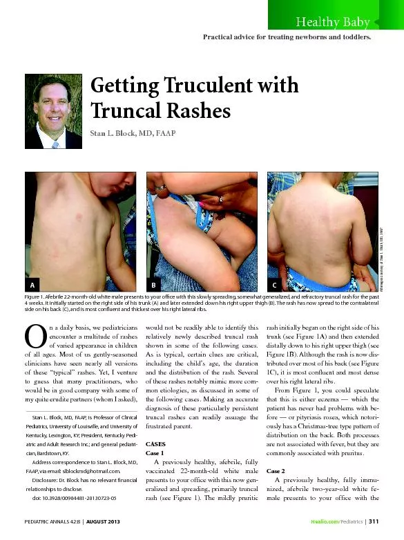
n a daily basis we pediatricians encounter a multitude of rashes of varied appearance in children clinicians have seen nea ID: 448850

Healio.com /Pediatrics 311 Truncal

Share:

Link:

Embed:

Download Presentation from below link

Download Pdf The PPT/PDF document "Getting Truculent with" is the property of its rightful owner. Permission is granted to download and print the materials on this web site for personal, non-commercial use only, and to display it on your personal computer provided you do not modify the materials and that you retain all copyright notices contained in the materials. By downloading content from our website, you accept the terms of this agreement.


Presentation Transcript

Healio.com /Pediatrics | 311 Getting Truculent with Truncal Rashes Stan L. Block, MD, FAAP O n a daily basis, we pediatricians encounter a multitude of rashes of varied appearance in children clinicians have seen nearly all versions of these “typical” rashes. Yet, I venture to guess that many practitioners, who would be in good company with some of my quite erudite partners (whom I asked), would not be readily able to identify this relatively newly described truncal rash shown in some of the following cases. As is typical, certain clues are critical, including the child’s age, the duration of these rashes notably mimic more com - mon etiologies, as discussed in some of the following cases. Making an accurate diagnosis of these particularly persistent truncal rashes can readily assuage the frustrated parent. CASES Case 1 A previously healthy, afebrile, fully vaccinated 22-month-old white male presents to your ofce with this now gen - eralized and spreading, primarily truncal rash (see Figure 1). The mildly pruritic rash initially began on the right side of his trunk (see Figure 1A) and then extended distally down to his right upper thigh (see Figure 1B). Although the rash is now dis - tributed over most of his back (see Figure 1C), it is most conuent and most dense From Figure 1, you could speculate that this is either eczema — which the patient has never had problems with be - fore — or pityriasis rosea, which notori - ously has a Christmas-tree type pattern of distribution on the back. Both processes are not associated with fever, but they are commonly associated with pruritus. Case 2 A previously healthy, fully immu - nized, afebrile two-year-old white fe - male presents to your ofce with the Stan L. Block, MD, FAAP, is Professor of Clinical Pediatrics, University of Louisville, and University of Kentucky, Lexington, KY; President, Kentucky Pedi - atric and Adult Research Inc.; and general pediatri - Address correspondence to Stan L. Block, MD, FAAP, via email: slblockmd@hotmail.com. Disclosure: Dr. Block has no relevant nancial relationships to disclose. doi: 10.3928/00904481-20130723-05 Figure 1. Afebrile 22-month-old white male presents to your ofce with this slowly spreading, somewhat generalized, and refractory truncal rash for the past 4 weeks. It initially started on the right side of his trunk (A) and later extended down his right upper thigh (B). The rash has now spread to the contralateral PEDIATRIC ANNALS 42:8 | AUGUST 2013 Healthy Baby Practical advice for treating newborns and toddlers. B C All images courtesy of Stan L. Block, MD, FAAP. A C 312 | Healio.com /Pediatrics Healthy Baby rash seen in Figure 2. The rash is mostly unilateral, quite pruritic, and has been distributed mostly on the left lateral trunk for 3 weeks. At previous physician visits elsewhere, because she is such an outdoorsy girl, she was diagnosed with poison ivy contact dermatitis. She has been unsuccessfully treated with steroid creams and diphenhydramine. You now observe some petechiae under her left axilla and left mid-section, but you attri - bute this to the scratching that you have witnessed in the ofce. She is ingesting no other medications and has no history of tick bites. You are perplexed by the truly unilateral nature of the rash. Case 3 A previously healthy afebrile 6-year- old white female presents to your ofce with a primarily unilateral, non-descript pruritic rash on the entire right side of her body (see Figure 3). The mother is quite frustrated that the child’s rash has not resolved in over 2 weeks, two physician ofce visits, and a multitude of ointments and oral medications, in - cluding mometasone cream, emollients, diphenhydramine, ranitidine (H2 hista - mine blocker) and oral prednisone for possible poison ivy contact dermatitis. Once again, you are perplexed by the nearly complete unilateral characteris - tics of the rash. Case 4 A healthy 8-month-old white female who is fully vaccinated and afebrile pres - ents with an abrupt onset rash spreading over 2 days. It is a fairly heavy crop of ovoid, round and linear pruritic eczema - toid-looking lesions distributed mostly on her right trunk and arm regions.(see Figure 4A) It is also a bit more diffuse on her back, and her mother says that the rash started with three of the small round lesions seen now on her upper back (see Figure 4B). This is her rst time ever diagnosed with “eczema.” Her previous treatments from an earlier physician visit have consisted of hydrocortisone valerate 0.2% ointment, emollients, and diphenhydramine. How - ever, something peculiar about the appear - ance and history of the rash sends alarm bells to your cerebral cortex. But she is too young to have that type of rash? Case 5 A 6-year-old white male presents to your ofce with low-grade fever, mild pharyngitis for 2 days and a 24-hour his - tory of the rash seen in Figure 5 (see page 313). He describes the rash as not painful, but very itchy. It is distributed over his en - tire anterior trunk and even up to his axilla Figure 2. Afebrile 2-year-old white female who pre - sented with a mostly unilateral, lichenoid, pruritic rash on the left lateral trunk for 3 weeks. Previous treatment with steroid ointments for presumed poison ivy as diagnosed by another physician, has not ameliorated the condition at all, and in fact, the rash is now spreading. The rash also has some pe - techiae in the axilla and in the lateral rib areas (see arrrows), which you presume are due to excoriation. She has been outdoors for many days during the summer, but denies any tick bites or recent medica - tions other than diphenhydramine for pruritus. Figure 3. Afebrile 6-year-old white female who presented with a mostly unilateral rash on the lat - eral abdomen, ribs, and back for 2 weeks. The rash was pruritic, non-tender and unresponsive to ste - roid creams and two separate previous physician ofce visits. Figure 4. (A) Afebrile 8-month-old white female presents with a 3-week, slowly spreading, heavy crop of ovoid, round, and linear pruritic eczematoid lesions over mostly the right trunk and arm, but which is more diffuse on her back. (B) The mother recalls that the rash started with three of the more central round lesions on her upper back 3 weeks ago. A B Healio.com /Pediatrics | 313 Healthy Baby but not on his back, legs or face. What ad - ditional examination should be undertak - en, and how should you proceed? Case 6 You are seeing this 2-year-old white female during the summer for these very discreet isolated but quite pruritic macule- papules on both of her sides and a few on her upper thighs for the last 24 hours. She is afebrile, fully vaccinated, and has been playing outside or swimming most days this summer. When you mention the words “insect bites” during this Saturday morn - ing’s visit, the mother adamantly states that she always sprays her children with insect repellant whenever they proceed outside, particularly the DEET 24% family version. She had read about DEET in Consumer Reports magazine as being the only effec - tive insect repellant. What additional his - tory might have been important to know when trying to identify the etiology? Case 7 A 4-year-old white female has devel - oped a fever to 100.5°F, headache, and this pruritic truncal morbillifom rash for 3 days. She denies recent travel, tick bites, sore throat, diarrhea, or joint aches. During your examination you notice the ushed cheeks immediately, along with the reticular mor - billiform rash localized mostly on the bilat - eral anterior trunk and upper extremities (see Figure 7, page 314). Once you presume the correct diagnosis, you must warn the family that the rash may persist for a few weeks — so that they do not get frustrated — and that treatment is merely for the symptoms. They also have a teenage daughter who has never had this type of rash. DISCUSSION Cases 1, 2 and 3 Each of these three children presented with a rash history, pattern and distribution consistent with “unilateral latero-thoracic exanthem” (ULE), otherwise known as “asymmetric periexural exanthem” of childhood. However, if you do not recog - nize these terms, do not be alarmed. The most recent edition of the Nelson Textbook of Pediatrics (19th edition) 1 still does not include this disorder in my search of its in - dex. In each of these patients, the rash be - gan on the lateral trunk area , and then later spread distally or across the midline over a few weeks. The rst series of children with ULE was described by Bodemer and de Prost in 1992. 2 ULE is depicted in the Hurwitz Pedi - atric Clinical Dermatology textbook, 3rd edition 3 as most commonly occurring in children between ages 1 and 5 years. The lesions begin on the lateral trunk area and then may spread centrifugally up to the axilla or inguinal area or posteriorly in up to 50% of children. The lesions often dis - play as maculo-papules, eczematoid, li - chenied, morbilliform, or even in scarla - tiniform and reticular morphologies. Half of children have pruritus, and desqua - mation is common as the rash resolves. Occasionally the patient may have some low-grade constitutional symptoms, just as with any other low-grade viral infec - tion. In general, most experts postulate that this illness is likely caused by a host of multiple viral etiologies. Some have speculated that it may occasionally evolve into the papular acrodermatitis of childhood (Gianotti-Crosti syndrome, which I presented in the July 2013 Pedi - atric Annals issue), and may be caused by those same potential pathogens. 4 One of the critical features of ULE that practitio - ners must be aware of and must be able to reassure frustrated parents of — the rash usually resolves over 3 to 4 weeks, but it Figure 5. During August, this 6-year-old white male presented with this diffuse, pruritic, ne mac - ulopapular rash over the entire anterior trunk and axilla. No rash was noted on the back. He has also complained of fever to 101°F and mild sore throat. Figure 6. (A and B) The afebrile 2-year-old white female presented this summer with these discreet very itchy maculopapular scattered lesions over her trunk and upper thighs for the last 24 hours. Although she has been swimming frequently, her mother usually applies DEET insect repellant to her when she is outdoors. Note that the rash is bilaterally distributed. She is fully vaccinated for her age. A B 314 | Healio.com /Pediatrics Healthy Baby may last as long as 8 weeks. Treatment is symptomatic only. Case 4 The 8-month-old female in Figure 4 has typical pityriasis rosea. Ovoid ecze - matoid “herald” lesion(s) initiated the bilateral truncal rash, along with the al - most Christmas-tree-like bilateral pat - tern on the back. The rash can sometimes be pruritic; constitutional symptoms are uncommon and low grade when they do occur. And again like ULE, the rash may persist for 8 to 12 weeks; its etiology is unknown. Case 5 Although Group A streptococcal phar - yngitis is uncommon in the summer, this young boy in Figure 5 (see page 313) pre - sented with the classic bilateral abdominal truncal rash of scarlet fever. The rash may be pruritic, mostly involves the anterior trunk and axilla, and most importantly on physical examination, it nearly always starts in the groin region. Thus you must always partially pull the pants or under - wear down to examine the skin below the pant-line. At 7 to 10 days later, the rash often desquamates in a similar manner as ULE, but only on the hands, feet and groin area, not on the trunk region. A rapid anti - gen detection test of the throat for Group A strep will nearly always conrm the eti - ology, except in the rare case of “surgical scarlet fever” caused by Staphylococcus aureus . Treatment is usually with beta- lactam antibiotics, such as amoxicillin. Case 6 The rash of the afebrile 2-year-old in Figure 6 (see page 313) upon rst glance could easily be mistaken for multiple in - sect bites from chiggers or mosquitoes, especially with her presenting in the rst 24 hours of eruption during the swim - ming season. However, further question - ing revealed that the child, although vac - cinated against varicella, was exposed earlier for an extended period of time to an aunt with known herpes zoster. About 20% of children who have received a sin - gle dose of varicella vaccine will develop breakthrough varicella over a period of 10 years. However, these cases are often “mild and clinically modied,” according to the 2012 AAP Red Book , 5 as in our pa - tient here. Over the ensuing next few days, some of the lesions crusted over, appeared to be in different stages, and disappeared to - tally in about 5 days, instead of the usual 7 to 10 days in unvaccinated chickenpox. Therapy can consist of oral acyclovir, or merely symptomatic treatment with anti-pruritics and daily oatmeal baths. An important point to remember is the avoidance of steroid creams or oral ste - roids when the diagnosis of varicella is considered likely. Case 7 The truncal rash in our 4-year-old girl from Figure 7 is distinctly different than the previous rashes displayed. It is re - ticulated, morbilliform, and heavily dis - tributed bilaterally over the abdomen and upper extremities, with none on the back or legs. This is the 2 to 3 week rash of Fifth disease, caused by Parvovirus B19. Although the rash here is not the typical reticular-lacy appearance of Fifth disease, the clinical diagnosis is usually clinched by the appearance of ushed cheeks bilat - erally, as seen in Figure 7. This infection may cause some low- grade fever in 15% to 30% of patients, headaches and other mild constitutional symptoms. It most commonly occurs in the spring. Once infected, immunity seems to be permanent. Importantly, Fifth disease may cause fairly severe arthral - gias for days to months in more than 50% of older adolescents, particularly females (so warn the sibling). 4 In addition, a po - tential hemolytic anemia from this infec - tion can be quite dangerous for the fetus in the rst two trimesters, and for those with chronic anemia. 4 CONCLUSION Clinicians should familiarize them - selves with the characteristics of this novel rash (ULE) along with the possible atypical presentations of these other com - mon unique rashes, which are primarily distributed on the trunk. Because many of these rashes may persist for weeks, if misdiagnosed they will become a source of much cost, consternation and frustra - tion for you and the family. REFERENCES Kleigman RM, Stanton B, St. Geme J, Schor NF, Behrman RE, eds. Nelson Textbook of Pediatrics. Philadelphia, PA: WB Saunders; 2011. Bodemer C, de Prost Y. Unilateral laterotho - racic exanthem in children: a new disease? J Am Acad Dermatol. 1992;27(5 Pt 1):693-696. Paller AS, Mancini AJ, eds. Hurwitz Clinical Pediatric Dermatology: A Textbook of Skin Disorders of Childhood and Adolescence. 3rd ed. Philadelphia, PA: Elsevier/Saunders; 2006. Feigin RD, Cherry J, Demmler-Harrison GJ, Kaplan SL. Feigin and Cherry’s Textbook of Pediatric Infectious Diseases. 6th ed. Philadel - phia, PA: WB Saunders; 2009. 5. AAP Committee on Infectious Diseases; Pick - ering LK, Baker CJ, Kimberlin DW, Long SS, eds. Red Book: 2012. Report of the Committee on Infectious Diseases (Red Book Report of the Committee on Infectious Diseases) . 29th ed. Elk Grove Village, IL. American Academy of Pediatrics; 2012. Figure 7. The 4-year-old white female presented to your ofce with a fever to 100.5°F, headache, and this pruritic generalized truncal morbillifom rash for 3 days. Note the red cheeks. PEDIATRIC ANNALS 42:8 | AUGUST 2013