/
Harvard Medical School, Boston, Massachusetts, USA.Infantilehemangioma Harvard Medical School, Boston, Massachusetts, USA.Infantilehemangioma

Harvard Medical School, Boston, Massachusetts, USA.Infantilehemangioma - PDF document

phoebe-click
phoebe-click . @phoebe-click
Follow
415 views
Uploaded On 2015-10-05

Harvard Medical School, Boston, Massachusetts, USA.Infantilehemangioma - PPT Presentation

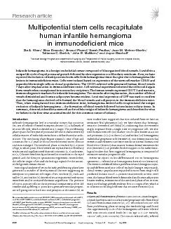
endothelial progenitor cells HemEPCs Our demonstration that HemECs and HemEPCs are more similar to normal human cord blood EPCs cbEPCs than mature human dermal microvascular ECs HDMECs 13 ID: 150506

endothelial progenitor cells (HemEPCs). Our

Share:

Link:

Embed:

Download Presentation from below link

Download Pdf The PPT/PDF document "Harvard Medical School, Boston, Massachu..." is the property of its rightful owner. Permission is granted to download and print the materials on this web site for personal, non-commercial use only, and to display it on your personal computer provided you do not modify the materials and that you retain all copyright notices contained in the materials. By downloading content from our website, you accept the terms of this agreement.


Presentation Transcript

Harvard Medical School, Boston, Massachusetts, USA.Infantilehemangiomaisbenignendothelialtumorcomposedofdisorganizedbloodvessels.Itexhibitsuniquelifecycleofrapidpostnatalgrowthfollowedbyslowregressiontofibrofattyresiduum.Here,wehavereportedtheisolationofmultipotentialstemcellsfromhemangiomatissuethatgiverisetohemangioma-likelesionsinimmunodeficientmice.Cellswereisolatedbasedonexpressionofthestemcell endothelial progenitor cells (HemEPCs). Our demonstration that HemECs and HemEPCs are more similar to normal human cord blood EPCs (cbEPCs) than mature human dermal microvascular ECs (HDMECs) (13) provides further support to the earlier histo - logical studies that first suggested hemangioma is composed of immature vascular progenitor cells. Herein we report on less-differentiated SCs in hemangioma specimens — referred to as hemangioma-derived SCs (HemSCs). The HemSCs exhibited robust proliferative and clonogenic capac - ity and differentiated into cells of multiple lineages. Importantly, clonal HemSC populations expanded in vitro from single cells produced human GLUT-1–positive microvessels in vivo. Over time, the human blood vessels diminished and human adipocytes became evident, reminiscent of the involuted phase of IH. In sum - mary, our findings provide evidence for a SC origin of IH, which is in contrast to the long-held view that IH arises from ECs (14). Isolation and characterization of HemSCs . Anti-CD133–coated magnet - ic beads have been used to isolate stem/progenitor cells from blood (15) and from human tumors (16, 17). We isolated CD133 + cells from proliferating phase IH using this methodology and found SFTFBSDIBSUJDMF TheJournalofClinicalInvestigation http://www.jci.org Volume 118 Number 7 July 2008  seen in the scant cytoplasm of the lipid-filled adipocytes (Figure 5B). Controls to rule out nonspecific staining with the anti-GFP and anti–perilipin A antibodies are shown in Supplemental Fig - ure 6. As an alternative approach to follow HemSC adipogen - esis in vivo, sections were immunostained with an anti-human nuclear antigen antibody (Figure 5C). The human nuclear antigen was detected in the nuclei of adipocytes in the HemSC Matrigel implants. Mouse heart tissue sections served as a negative control, and human hemangioma tissue sections served as a positive con - trol (Figure 5C). Some adipocytes negative for the human nuclear antigen were evident, suggesting that recruitment of murine cells into the Matrigel microenvironment may have occurred. These results with tagged, GFP-labeled HemSCs and the anti-human nuclear antigen antibody indicate that HemSCs form adipocytes at time points subsequent to vessel formation. The signals that determine whether and when a HemSC becomes an EC or an adi - pocyte are unknown. For what we believe is the first time, we have shown that CD133- selected HemSCs recapitulate human IH in a murine in vivo model. Clonal HemSCs produced human GLUT-1–positive microvessels at 7–14 days; after 28 days in vivo, human adipocytes were also detected. HemSCs that had differentiated into endothelium in vivo, as deter - mined by cell-surface CD31 expression, could be retrieved and implanted into secondary recipients, where the cells formed blood vessels once more. This indicates the robust vasculogenic poten - tial of these hemangioma-derived cells. GFP-labeled HemSCs were shown to form vessels and adipocytes at 14- and 56-day time points, respectively, confirming that the vessels and adipocytes were not host derived. Using in vitro cellular activity assays, we showed that HemSCs are highly proliferative and able to differentiate into mul - tiple cell types, including adipocytes. These results demonstrate that HemSCs are the cellular precursors of IH. CD133 has been used to identify tumor-initiating cells from medulloblastomas, glioblastomas, and prostate and colon car - cinomas (16, 17, 39–42). Xenografting CD133 + tumor cells in immune-compromised mice has also been shown to generate a primary tumor (16, 17). In a similar fashion, we demonstrated that the CD133-selected cells from IH formed GLUT-1–positive blood vessels and, at later time points, differentiated into adipocytes, the prominent cellular compartments of proliferating and involuting phase hemangioma, respectively. For the secondary transplan - tation experiment (Figure 3, C and D), we reimplanted human CD31-selected cells because (a) these cells had shown vasculo - genic activity in the primary implant and (b) the level of CD133 expression in the CD31-positive and CD31-negative populations recovered from the primary implant was very low, which would have made reselecting CD133-positive cells difficult. The human CD31-selected cells showed a robust degree of blood vessel forma - tion (78 vessels/mm 2 ) upon secondary transplantation, a finding that supports the vasculogenic potential of these cells. To date, there are no animal models that truly reflect the human hemangioma phenotype and life cycle. Although murine heman - gioendothelioma cells have been studied (43), these do not pheno - copy IH or provide a hemangioma model because the tumors do not regress and are fatal within 3 months. In addition, polyoma middle T antigen–transformed cells used in animal models result in “cavernous hemangioma” lesions, more akin to malignant vas - cular tumors, which are also fatal within 3–10 months (44). Hem - angioma tissue fragments xenografted into nude mice have been shown to undergo an initial period of necrosis followed by approx - imately 2-fold enlargement and then subsequent regression (45), which may provide a model for testing new therapies for IH. Our in vivo model is unique in that we are able to produce IH lesions with a small number of purified primary cells suspended in Matrigel but without exogenous growth factors or supplements. These find - ings implicate a preset genetic or epigenetic program in the Hem - SCs that directs the cells to recapitulate the IH tumor, including endothelial and adipocytic differentiation. An important future experiment will be to implant hemangioma-derived CD133 + cells that have not undergone in vitro expansion to determine whether HemSCs lose or gain hemangioma-forming potential during the in vitro culture. It is still not understood what triggers the adipo - genesis that characterizes the involuting and involuted phases of IH. It is likely that changes in the microenvironment, such as levels of circulating EPCs, immune cells, and growth factors, initiate the regression of the tumor. It is well established that hemangioma endothelium expresses GLUT-1, merosin, Lewis Y antigen (CD174), and Fc receptor II (CD32) in all 3 phases of the life cycle (34). These markers are also expressed by placental microvessels. This immunohistochemical similarity suggested the hypothesis that IH is formed from embol - ic placental ECs (34). Microarray analyses have supported this link between placenta and hemangioma (46). It is also possible that the progenitor cells reside in fetal and neonatal tissues or come from the BM and express a transcriptional program similar to that of placenta. To date, placenta-derived cells with in vitro clonogenic and differentiation abilities similar to HemSCs have not been described. In conclusion, we have identified the hemangioma-initiating cell. We show that a single multipotential cell (HemSC) produces GLUT-1–positive microvessels and that this lesion undergoes an involutive process by differentiation into adipocytes. This could be a result of a somatic mutation within a stem or progenitor cell and subsequent expansion/differentiation. It is also plausible that a normal SC in the context of the postnatal infantile microenviron - ment becomes disrupted or delayed in its differentiative program, resulting in an accumulation of early progenitor cells. Such a clon - al population could then undergo ill-timed or abnormal differen - tiation, which subsequently resolves. Understanding the cellular origin of IH provides an avenue for deciphering the mechanisms of EC differentiation and delineating targets for adjuvant therapy. Our in vitro and in vivo models of IH offer powerful tools to evalu - ate potential therapeutic modalities and insight into the biology of de novo formation of blood vessels. Cell isolation and culture . Specimens of proliferating IH were obtained from the Department of Plastic Surgery at Children’s Hospital Boston under a human subject protocol that was approved by the Committee on Clini - cal Investigation. The clinical diagnosis was confirmed in the Department of Pathology at Children’s Hospital Boston. Informed consent was pro - vided for the specimens, according to the Declaration of Helsinki. Single- cell suspensions were prepared from the proliferating phase specimens as described previously (13). HemSCs were selected using anti-CD133–coated magnetic beads (Miltenyi Biotec). CD133-selected cells were cultured on fibronectin-coated (1 g/cm 2 ) plates with EBM-2 (CC-3156; Cambrex). EBM-2 was supplemented with 20% FBS, endothelial growth media-2 SingleQuot (CC-4176; Cambrex), and 1 PSF (100 U/ml penicillin, SFTFBSDIBSUJDMF TheJournalofClinicalInvestigation http://www.jci.org Volume 118 Number 7 July 2008  0.25 mM L -glutamine, and 25 ng/ml nerve growth factor) was then used for 14 days. Hematopoietic activity was assessed in MethoCult medium (StemCell Technologies) at 1 10 3 , 1 10 4 , 1 10 5 , and 1 10 6 cells per 35-mm dish. Mesenchymal differentiation was assessed by oil red O (lipid droplets), alkaline phosphatase induction (substrate-based activity) and Von Kossa (mineralization), and collagen type 2 staining. Immunofluores - cent analysis of cells was carried out using goat anti-human CD31 (Santa Cruz Biotechnology Inc.), mouse anti-human VE-cadherin (Immunotech), mouse anti-human tubulin -tubilin III (Millipore), and goat anti-human GFAP (Dako) antibodies. FITC- and PE-conjugated secondary antibodies (Vector Laboratories) were used for antigen detection. Images were taken with a Nikon Eclipse TE300 (Nikon) using SPOT Advanced 3.5.9 software (Diagnostic Instruments Inc.) and a 20/0.45 objective lens. Statistics . The data were expressed as means ± SEM and analyzed by ANOVA followed by Student’s two-tailed t test where appropriate. Differ - ences were considered significant at P 0.05. This work was supported by an NIH grant (P01 AR048564 to J. Bischoff). We thank Jill Wylie-Sears for her technical assistance, Elke Pravda for confocal microscopic imaging, and Kristin John - son for the preparation of the figures. Received for publication August 2, 2007, and accepted in revised form April 23, 2008. Address correspondence to: Joyce Bischoff, Vascular Biology Program and Department of Surgery, Children’s Hospital Bos - ton, Harvard Medical School, Boston, Massachusetts 02115, USA. Phone: (617) 919-2192; Fax: (617) 730-0231; E-mail: joyce. bischoff@childrens.harvard.edu. Zia A. Khan and Elisa Boscolo are co–first authors.1.Mulliken, J.B., Fishman, S.J., and Burrows, P.E. 2000. Vascular anomalies. Curr. Probl. Surg. 37 :517–584.Bischoff, J. 2002. Monoclonal expansion of endothelial cells in hemangioma: an intrinsic defect with extrinsic consequences? Trends Cardio - vasc. Med. 12 Boye, E., et al. 2001. Clonality and altered behav - ior of endothelial cells from hemangiomas. J. Clin. Invest. 107 Bielenberg, D.R., et al. 1999. Progressive growth of infantile cutaneous hemangiomas is directly corre - lated with hyperplasia and angiogenesis of adjacent epidermis and inversely correlated with expression of the endogenous angiogenesis inhibitor, IFN- beta. Int. J. Oncol. 14 Berard, M., et al. 1997. Vascular endothelial growth factor confers a growth advantage in vitro and in vivo to stromal cells cultured from neonatal hem - angiomas. Am. J. Pathol. 150 Virchow, R. 1863. Die Krankhaften Geschwulste . August Hirschwald. Berlin, Germany. 306 pp.7.Pack, G., and Miller, T.R. 1950. Hemangiomas; classification, diagnosis, and treatment. Angiology. 1 Malan, E. 1974. Vascular malformations (angiodys - plasias). In Carlo Erba Foundation . Milan, Italy. 4.Smoller, B.R., and Apfelberg, D.B. 1993. Infan - tile (juvenile) capillary hemangioma: a tumor of heterogeneous cellular elements. J. Cutan. Pathol. 20 Dosanjh, A., et al. 2000. In vitro characteristics of neonatal hemangioma endothelial cells: similari - ties and differences between normal neonatal and fetal endothelial cells. J. Cutan. Pathol. 27 11.Shmelkov, S.V., St. Clair, R., Lyden, D., and Rafii, S. 2005. AC133/CD133/Prominin-1. Int. J. Biochem. Cell Biol. 37 Yu, Y., Flint, A.F., Mulliken, J.B., Wu, J.K., and Bischoff, J. 2004. Endothelial progenitor cells in infantile hemangioma. Blood. 103 Khan, Z.A., et al. 2006. Endothelial progenitor cells from infantile hemangioma and umbilical cord blood display unique cellular responses to end - ostatin. Blood. 108 :915–921.Bauland, C.G., et al. 2006. The pathogenesis of hemangiomas: a review. Plast. Reconstr. Surg. 117 Wu, X., et al. 2004. Tissue-engineered microves - sels on three-dimensional biodegradable scaffolds using human endothelial progenitor cells. Am. J. Physiol. Heart Circ. Physiol. 287 :H480–H487.O’Brien, C.A., Pollett, A., Gallinger, S., and Dick, J.E. 2007. A human colon cancer cell capable of ini - tiating tumour growth in immunodeficient mice. Nature. 445 17.Ricci-Vitiani, L., et al. 2007. Identification and expansion of human colon-cancer-initiating cells. Nature. 445 Melero-Martin, J.M., et al. 2007. In vivo vasculogen - ic potential of human blood-derived endothelial progenitor cells. Blood. 109 Baal, N., et al. 2004. Expression of transcription factor Oct-4 and other embryonic genes in CD133 positive cells from human umbilical cord blood. Thromb. Haemost. 92 Okuda, T., van Deursen, J., Hiebert, S.W., Gros - veld, G., and Downing, J.R. 1996. AML1, the tar - get of multiple chromosomal translocations in human leukemia, is essential for normal fetal liver hematopoiesis. Cell. 84 21.Li, Z., Chen, M.J., Stacy, T., and Speck, N.A. 2006. Runx1 function in hematopoiesis is required in cells that express Tek. Blood. 107 Reyes, M., et al. 2002. Origin of endothelial pro - genitors in human postnatal bone marrow. J. Clin. Invest. 109 Jiang, Y., et al. 2002. Pluripotency of mesenchymal stem cells derived from adult marrow. Nature. 418 Egusa, H., Schweizer, F.E., Wang, C.C., Matsuka, Y., and Nishimura, I. 2005. Neuronal differentiation of bone marrow-derived stromal stem cells involves suppression of discordant phenotypes through gene silencing. J. Biol. Chem. 280 :23691–23697.Tondreau, T., et al. 2004. Bone marrow-derived mesenchymal stem cells already express specific neural proteins before any differentiation. Differen - tiation. 72 Pittenger, M.F., et al. 1999. Multilineage potential of adult human mesenchymal stem cells. Science. 284 :143–147.27.Ritter, M.R., Reinisch, J., Friedlander, S.F., and Friedlander, M. 2006. Myeloid cells in infantile hemangioma. Am. J. Pathol. 168 Choi, K., Kennedy, M., Kazarov, A., Papadimitriou, J.C., and Keller, G. 1998. A common precursor for hematopoietic and endothelial cells. Development. 125 Ito, Y., Iwamoto, Y., Tanaka, K., Okuyama, K., and Sugioka, Y. 1996. A quantitative assay using base - ment membrane extracts to study tumor angiogen - esis in vivo. Int. J. Cancer. 67 Hoang, M.V., and Senger, D.R. 2005. In vivo and in vitro models of Mammalian angiogenesis. Methods Mol. Biol. 294 31.Yu, Y., et al. 2006. Mesenchymal stem cells and adi - pogenesis in hemangioma involution. Stem Cells. 24 Parums, D.V., et al. 1990. JC70: a new monoclonal antibody that detects vascular endothelium associ - ated antigen on routinely processed tissue sections. J. Clin. Pathol. 43 :752–757.North, P.E., Waner, M., Mizeracki, A., and Mihm, M.C., Jr. 2000. GLUT1: a newly discovered immunohistochemical marker for juvenile heman - giomas. Hum. Pathol. 31 North, P.E., et al. 2001. A unique microvascular phenotype shared by juvenile hemangiomas and human placenta. Arch. Dermatol. 137 Kawaguchi, N., et al. 1998. De novo adipogenesis in mice at the site of injection of basement membrane and basic fibroblast growth factor. Proc. Natl. Acad. Sci. U. S. A. 95 36.Kimura, Y., Ozeki, M., Inamoto, T., and Tabata, Y. 2002. Time course of de novo adipogenesis in matri - gel by gelatin microspheres incorporating basic fibroblast growth factor. Tissue Eng. 8 :603–613.37.Tabata, Y., et al. 2000. De novo formation of adi - pose tissue by controlled release of basic fibroblast growth factor. Tissue Eng. 6 Greenberg, A.S., et al. 1991. Perilipin, a major hor - monally regulated adipocyte-specific phosphopro - tein associated with the periphery of lipid storage droplets. J. Biol. Chem. 266 Singh, S.K., et al. 2003. Identification of a can - cer stem cell in human brain tumors. Cancer Res. 63 40.Salmaggi, A., et al. 2006. Glioblastoma-derived tumorospheres identify a population of tumor stem- like cells with angiogenic potential and enhanced multidrug resistance phenotype. Glia. 54 :850–860.41.Collins, A.T., Berry, P.A., Hyde, C., Stower, M.J., and Maitland, N.J. 2005. Prospective identification of tumorigenic prostate cancer stem cells. Cancer Res. 65 :10946–10951.Richardson, G.D., et al. 2004. CD133, a novel mark - er for human prostatic epithelial stem cells. J. Cell Sci. 117 Obeso, J., Weber, J., and Auerbach, R. 1990. A hemangioendothelioma-derived cell line: its use as a model for the study of endothelial cell biology. Lab. Invest. 63 Bautch, V.L., Toda, S., Hassell, J.A., and Hanahan, D. 1987. Endothelial cell tumors develop in trans - genic mice carrying polyoma virus middle T onco - gene. Cell. 51 :529–537.45.Tang, Y., et al. 2007. A novel in vivo model of human hemangioma: xenograft of human hemangioma tis - sue on nude mice. Plast. Reconstr. Surg. 120 :869–878.46.Barnes, C.M., et al. 2005. Evidence by molecular pro - filing for a placental origin of infantile hemangio - ma. Proc. Natl. Acad. Sci. U. S. A. 102 :19097–19102.47.Tai, M.H., et al. 2005. Oct4 expression in adult human stem cells: evidence in support of the stem cell theory of carcinogenesis. Carcinogenesis. 26 :495–502.Choi, S.J., et al. 2003. AML-1A and AML-1B regula - tion of MIP-1alpha expression in multiple myelo - ma. Blood. 101 :3778–3783.